. 2024 Mar 9;8(3):298-311.
doi: 10.7150/ntno.93064. eCollection 2024.

Adipose-derived stem cell exosomes act as delivery vehicles of microRNAs in a dog model of chronic hepatitis

Affiliations

Adipose-derived stem cell exosomes act as delivery vehicles of microRNAs in a dog model of chronic hepatitis

Ilaria Zanolla et al. Nanotheranostics. .

Abstract

Exosomes are nanosized extracellular vesicles secreted by all cell types, including canine adipose-derived stem cells (cADSCs). By mediating intercellular communication, exosomes modulate the biology of adjacent and distant cells by transferring their cargo. In the present work after isolation and characterization of exosomes derived from canine adipose tissue, we treated the same canine donors affected by hepatopathies with the previously isolated exosomes. We hypothesize that cADSC-sourced miRNAs are among the factors responsible for a regenerative and anti-inflammatory effect in the treatment of hepatopathies in dogs, providing the clinical veterinary field with an effective and innovative cell-free therapy. Exosomes were isolated and characterized for size, distribution, surface markers, and for their miRNomic cargo by microRNA sequencing. 295 dogs affected with hepatopathies were treated and followed up for 6 months to keep track of their biochemical marker levels. Results confirmed that exosomes derived from cADSCs exhibited an average diameter of 91 nm, and positivity to 8 known exosome markers. The administration of exosomes to dogs affected by liver-associated inflammatory pathologies resulted in the recovery of the animal alongside the normalization of biochemical parameters of kidney function. In conclusion, cADSCs-derived exosomes are a promising therapeutic tool for treating inflammatory disorders in animal companions.

Keywords: exosomes; extracellular nanovesicles; liver diseases; miRNA; regenerative medicine..


Conflict of interest statement

Competing Interests: The authors have declared that no competing interest exists.

Figures

Figure 1
Figure 1
Canine ADSC characterization. cADSCs showed their typical elongated morphology. Scale bar is 20 µm. Images were taken with the Evos XL Core microscope (Invitrogen, Waltham, Massachusetts, USA) equipped with a 4x objective.
Figure 2
Figure 2
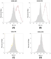
Flow cytometry analysis of surface markers in cADSCs. (A-B) cADSCs are positive for CD90-APC and CD44-FITC. (C-D) cADSCs are negative for CD14-PE and CD45-FITC. Measurements were taken with the Attune™ NxT Acoustic Focusing Cytometer (Life Technologies, Carlsbad, California, USA).
Figure 3
Figure 3
cADSC-exo characterization. (A) The histogram shows the size distribution of cADSC-exos evaluated by Tunable resistive pulse sensing. The mean diameter is 91 nm (St. Dev ± 28.7), while the average concentration is 6.62x10⁸ particles/mL. (B) SEM images confirm the exosome diameter. Images were taken with the SEM Zeiss EVO 40 microscope (Zeiss, Oberkochen, Germany) at a 20.000x magnification. (C) cADSC-exos show a protein concentration of 483.9µg/mL (St. Dev ± 18.3). Results are the mean of 3 replicates. (D) cADSC-exos are positive for the specific markers MSC-derived exosomes CD81 and CD63. Results are expressed as percentages compared to positive control. Analyses were performed in triplicate.
Figure 4
Figure 4
Internalization of cADSC-exos by cADSCs. PKH26-stained exosomes have been taken up by cADSCs and appeared in the cytoplasmic region of cells as red spots. Images were acquired with the Zeiss Axiovert 200M Fluorescence Microscope (Zeiss, Oberkochen, Germany) equipped with a 63x oil objective. Cell nuclei are stained in blue; actin filaments are stained in green; exosomes are stained in red. Scale bar is 20 µm.
Figure 5
Figure 5
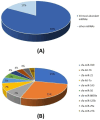
miRNA cargo of cADSC-exos. (A) The 10 most abundant miRNAs represent the 83% of the total miRNA cargo in cADSC-exos. (B) Distribution of the 10 top-ranked miRNAs. Analysis was performed in triplicate.
Figure 6
Figure 6
Diff-quick stain of cytological samples before and 30 days after the second exosome injection after 30 days. (A) Before exosome treatment, inflammatory processes are occurring with foamy macrophages and hepatic debris; red cells are also visible. (B) After exosome treatment, fibrosis and inflammation are strongly reduced.
Figure 7
Figure 7
Sonoelastography of the canine liver before injection of autologous exosomes. Fibrotic tissue and altered portal vein blood flow are shown.
Figure 8
Figure 8
Sonoelastography of the canine liver 180 days after injection of autologous exosomes. No fibrotic tissue is visible, and the portal vein blood flow is restored.

References

    1. Diomaiuto E, Principe V, Luca AD, Laperuta F, Alterisio C, Loria AD. Exosomes in dogs and cats: an innovative approach to neoplastic and non-neoplastic diseases. Pharmaceuticals. 2021. 14. - PMC - PubMed
    1. Kalluri R, LeBleu VS. The biology, function, and biomedical applications of exosomes. Science. 2020. 367. - PMC - PubMed
    1. Brunello G, Zanotti F, Trentini M, Zanolla I, Pishavar E, Favero V. et al. Exosomes derived from dental pulp stem cells show different angiogenic and osteogenic properties in relation to the age of the donor. Pharmaceutics. 2022;14:908. - PMC - PubMed
    1. Li J, Zheng S, Ma C, Chen X, Li X, Li S, Research progress on exosomes in podocyte injury associated with diabetic kidney disease. Front Endocrinol. 2023. 14. - PMC - PubMed
    1. Zanotti F, Zanolla I, Trentini M, Tiengo E, Pusceddu T, Licastro D. et al. Mitochondrial metabolism and EV cargo of endothelial cells is affected in presence of EVs derived from MSCs on which HIF is activated. Int J Mol Sci. 2023;24:6002. - PMC - PubMed