

Facile preparation of a CoNiS/CF electrode by SILAR for a high sensitivity non-enzymatic glucose sensor
- PMID: 38577432
- PMCID: PMC10993041
- DOI: 10.1039/d3ra08154k
Facile preparation of a CoNiS/CF electrode by SILAR for a high sensitivity non-enzymatic glucose sensor
Abstract
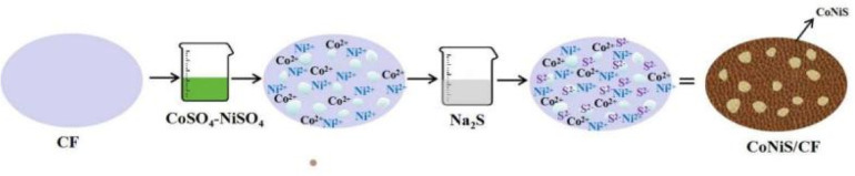
The nanomaterials for non-enzymatic electrochemical sensors are usually pre-synthesized and coated onto electrodes by ex situ methods. In this work, amorphous cobalt-nickel sulfide (CoNiS) nanoparticles were facilely prepared on copper foam (CF) by the in situ successive ionic layer adsorption and reaction (SILAR) method, and as-prepared CoNiS/CF was studied as an electrode for non-enzymatic glucose sensing. It was analyzed by field emission scanning electron microscopy (FESEM), energy dispersive X-ray analysis (EDAX) and X-ray photoelectron spectroscopy (XPS). The electrochemical performance was investigated by cyclic voltammetry (CV) and chronoamperometry (CA). This binary sulfide electrode showed better performance toward glucose oxidation compared to the corresponding single sulfide and showed a wide linear range of 0.005 to 3.47 mM, a high sensitivity of 2298.7 μA mM-1 cm-2 and a low detection limit of 2.0 μM. The sensor exhibited high sensitivity and good repeatability and stability and was able to detect glucose in an actual sample. This work provides a simple and fast in situ electrode preparation method for a high-sensitivity glucose sensor.
This journal is © The Royal Society of Chemistry.
Conflict of interest statement
There are no conflicts to declare.
Figures


References
-
- Zhou Y. Chen J. P. Gan L. Xu W. Liu Y. Zhao Y. G. Zhu Y. J. Chromatogr. 2022;1685:463564. - PubMed
-
- Srivastava M. Srivastava S. K. Ojha R. P. Prakash R. Microchem. J. 2022;182:107850.
-
- Vaishanav S. K. Korram J. Nagwanshi R. Ghosh K. K. Satnami M. L. Sens. Actuators, B. 2017;245:196–204.
-
- Sheibani N. Kazemipour M. Jahani S. Foroughi M. M. Microchem. J. 2019;149:103980.
LinkOut - more resources
Full Text Sources