. 2024 Mar 11;15(9):2518-2537.
doi: 10.7150/jca.92578. eCollection 2024.

circular RNA circ-231 promotes protein biogenesis of TPI1 and PRDX6 through mediating the interaction of eIF4A3 with STAU1 to facilitate unwinding of secondary structure in 5' UTR, enhancing progression of human esophageal squamous cell carcinoma (ESCC)

Affiliations

circular RNA circ-231 promotes protein biogenesis of TPI1 and PRDX6 through mediating the interaction of eIF4A3 with STAU1 to facilitate unwinding of secondary structure in 5' UTR, enhancing progression of human esophageal squamous cell carcinoma (ESCC)

Guo-Wei Huang et al. J Cancer. .

Abstract

Background: The nuclear cap-binding complex (CBC)-dependent translation (CT) is an important initial translation pathway for 5'-cap-dependent translation in normal mammal cells. Eukaryotic translation initiation factor 4A-III (eIF4A3), as an RNA helicase, is recruited to CT complex and enhances CT efficiency through participating in unwinding of secondary structure in the 5' UTR. However, the detailed mechanism for eIF4A3 implicated in unwinding of secondary structure in the 5' UTR in normal mammal cells is still unclear. Specially, we need to investigate whether the kind of mechanism in normal mammal cells extrapolates to cancer cells, e.g. ESCC, and further interrogate whether and how the mechanism triggers malignant phenotype of ESCC, which are important for identifying a potential therapeutic target for patients with ESCC. Methods: Bioinformatics analysis, RNA immunoprecipitation and RNA pulldown assays were performed to detect the interaction of circular RNA circ-231 with eIF4A3. In vitro and in vivo assays were performed to detect biological roles of circ-231 in ESCC. RNA immunoprecipitation, RNA pulldown, mass spectrometry analysis and co-immunoprecipitation assays were used to measure the interaction of circ-231, eIF4A3 and STAU1 in HEK293T and ESCC. In vitro EGFP reporter and 5' UTR of mRNA pulldown assays were performed to probe for the binding of circ-231, eIF4A3 and STAU1 to secondary structure of 5' UTR. Results: RNA immunoprecipitation assays showed that circ-231 interacted with eIF4A3 in HEK293T and ESCC. Further study confirmed that circ-231 orchestrated with eIF4A3 to control protein expression of TPI1 and PRDX6, but not for mRNA transcripts. The in-depth mechanism study uncovered that both circ-231 and eIF4A3 were involved in unwinding of secondary structure in 5' UTR of TPI1 and PRDX6. More importantly, circ-231 promoted the interaction between eIF4A3 and STAU1. Intriguingly, both circ-231 and eIF4A3 were dependent on STAU1 binding to secondary structure in 5' UTR. Biological function assays revealed that circ-231 promoted the migration and proliferation of ESCC via TPI1 and PRDX6. In ESCC, the up-regulated expression of circ-231 was observed and patients with ESCC characterized by higher expression of circ-231 have concurrent lymph node metastasis, compared with control. Conclusions: Our data unravels the detailed mechanism by which STAU1 binds to secondary structure in 5' UTR of mRNAs and recruits eIF4A3 through interacting with circ-231 and thereby eIF4A3 is implicated in unwinding of secondary structure, which is common to HEK293T and ESCC. However, importantly, our data reveals that circ-231 promotes migration and proliferation of ESCC and the up-regulated circ-231 greatly correlates with tumor lymph node metastasis, insinuating that circ-231 could be a therapeutic target and an indicator of risk of lymph node metastasis for patients with ESCC.

Keywords: Interaction; RNA binding protein; STAU1; circ-231; eIF4A3.


Conflict of interest statement

Competing Interests: The authors have declared that no competing interest exists.

Figures
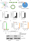
Figure 1
Figure 1
circ-231 interacts with eIF4A3. (A) The overlapped circRNAs were counted using R package gplots in seven cell lines, including A549, BJ, Hela S3, HepG2, K562, MCF7 and HEK293 cells. (B and C) The number of circRNAs in total 51 overlapped circRNAs containing eIF4A3-binding sites were analyzed and counted according to Circinteractome database. (D) circ-231, circ-069 and circ-248 were measured by qRT-PCR. β-actin used as internal control. (E) Total protein from HEK293T cells was extracted and 10% extracts were selected as input. Mouse anti-eIF4A3 or normal mouse IgG were incubated with protein extracts overnight at 4°C. RNA from the precipitated complex was extracted and circ-231, circ-069 and circ-248 were determined using qRT-PCR. Values are mean ± standard deviation (SD). *P˂0.05, **P<0.01. (F) RNA immunoprecipitation assays for eIF4A3 in HEK293T, KYSE150 cells were performed according to the method described in (E). Mouse anti-eIF4AI/II and normal IgG were selected as negative control. The relative enrichment of circ-231 was detected using qRT-PCR. Values are mean ±SD. *P˂0.05, **P<0.01, ***P<0.001. (G) circ-231 pulldown assays were performed. Biotin-labeled DNA probe complementary to junction sequence of circ-231 or random probe were incubated with HEK293T or KYSE150 cell extracts at 4°C overnight and the biotin-labeled probes were pulldown by streptavidin-coupled magnetic beads and eIF4A3 was determined with western blot.
Figure 2
Figure 2
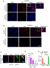
circ-231 was localized in cytoplasm and co-localized with eIF4A3 in HEK293T and ESCC cells. Fluorescence in situ hybridization assays for circ-231 was performed in HEK293T (A), KYSE150 cells (B). circ-231 antisense or sense probe (negative control) conjugated to biotin was detected using streptavidin-Cy3 (red). Cells were counterstained with DAPI (blue). Magnification, 40x. circ-231 is localized in cytoplasm in cells. circ-231 antisense or sense probe (negative control) conjugated to biotin was detected using streptavidin-Cy3 (red). Cells were counterstained with DAPI (blue). Magnification, 40 x. (C) Co-localization of circ-231 and eIF4A3 was performed in human esophageal tumor tissues. After biotin-coupled antisense probe for circ-231 was incubated with tumor tissue slides, mouse anti-eIF4A3 was simultaneously added to tissues at 37°C for 2 h and then Cy3-streptavidin and Alexa Fluor 488 dye-coupled goat anti-mouse IgG was added to tissues at room temperature for 1 h. Fluorescent signals were observed under 60 x magnification with 2 x optical magnification. (D) The relative levels of circ-231 and eIF4A3 transcripts in human esophageal tumor tissues (n=25) were detected using qRT-PCR. Values are mean ±SD. *P˂0.05, **P<0.01. β-actin used as internal control. (E) Cytoplasmic and nuclear RNAs were isolated from HEK293T cells and circ-231 was measured with qRT-PCR. A nuclear lncRNA MALAT1 transcript was used as control transcript. Values are mean ± SD.
Figure 3
Figure 3
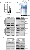
circ-231 cooperating with eIF4A3 promotes protein expression of PRDX6 and TPI1. (A) siRNAs against circ-231 or eIF4A3 were transfected into HEK293T and the relative levels of circ-231 and eIF4A3 transcripts were measured using qRT-PCR. Three independent experiments were performed. Values are mean ±SD. *P<0.05, **P<0.01. (B) After circ-231 or eIF4A3 knockdown, proteins were extracted and coomassie blue staining assays were performed and the differentially expressed protein bands were cutout and subjected to LCMS-Q-TOF analysis. Three independent experiments for coomassie blue staining were performed. (C) siRNAs against circ-231 or eIF4A3 were transfected into HEK293T or KYSE150 cells and protein levels of PRDX6, TPI1 and eIF4A3 were measured with western blot. In shRNA1-circ-231 or shRNA-Scramble stable cell lines, including KYSE150 (D) and KYSE510 (E) cells, protein levels of PRDX6, TPI1 and eIF4A3 were determined using western blot.
Figure 4
Figure 4
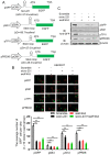
circ-231 in coordination with eIF4A3 promotes unwinding of secondary structure at 5′ end of mRNAs. (A) The schematic diagrams represent that those secondary structures with different free energy were individually inserted into Hind III sites upstream of initiation codon of pcDNA3.1-EGFP. (B) siRNAs against circ-231 or eIF4A3 were transfected or co-transfected into HEK293T cells and after 48 h, fluorescent signals of GFP were observed with fluorescent microscope. Magnification, 10x. The number of GFP+ cells from at least 20 views in each of groups were counted and analyzed. Values are mean ±SD. *P<0.05, **P<0.01. (C) siRNAs against circ-231 or eIF4A3 were transfected or co-transfected into HEK293T cells and after 48 h, the protein levels of GFP from every group were detected with western blot.
Figure 5
Figure 5
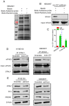
circ-231 promotes the interaction of eIF4A3 with STAU1. (A) circ-231 pulldown assays were performed. Biotin-labeled DNA probe complementary to junction sequence of circ-231 or random probe was incubated with HEK293T cell extracts at 4°C overnight and the biotin-labeled probes were pulled-down by streptavidin-coupled magnetic beads and the immunoprecipitated complex was subjected to SDS-PAGE electrophoresis and differential bands were dissected and identified by Mass analysis. (B) circ-231 pulldown was performed in HEK293T cells and STAU1 was determined with western blot. (C) RNA immunoprecipitation was performed using anti-STAU1 antibody in HEK293T and KYSE150 cells and RNA was extracted from the immunoprecipitated complex and circ-231 was determined using qRT-PCR. Values are mean ±SD. Two independent experiments were performed. (D) After circ-231 knockdown in HEK293T or KYSE150 cells, co-immunoprecipitation for STAU1 was performed and eIF4A3 was determined with western blot. (E) STAU1 was down-regulated in HEK293T or KYSE150 cells and the levels of TPI1 and PRDX6 protein were determined with western blot.
Figure 6
Figure 6
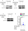
The binding of both eIF4A3 and circ-231 to secondary structure at 5′ UTR depends on STAU1. Biotinylated RNA from 5′ UTR of TPI1 mRNAs was synthesized in vitro transcription and allowed to form secondary structure and incubated with extracts from HEK293T cells with STAU1 (A), eIF4A3 (B) and circ-231 (C) knockdown, eIF4A3 or STAU1 from the immunoprecipitated complex was determined with western blot. (D) For circ-231, RNA from the immunoprecipitated complex was extracted and circ-231 was determined using qRT-PCR. Values are mean ±SD.
Figure 7
Figure 7
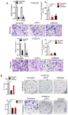
circ-231 promotes the migration and proliferation of human esophageal cancer cells. KYSE150 shRNA1-circ-231, shRNA2-circ-231 or shRNA-Scramble stable cell lines were subjected to starvation without serum culture medium for 18 h, the migration of cells was measured by transwell assays (A) and the proliferation of cells was detected by colony formation assays (B). Scale bar: 10 x. Values are mean ±SD. *P<0.05, **P<0.01. (C-F) KYSE150 Tet-on shRNA1-circ-231, shRNA2-circ-231 or shRNA-Scramble cell lines were individually subcutaneously injected into the right flank of each of six-week-old male BALB/c nude (nu/nu; n=5) mice (5.0 × 10⁶ cells / flank) and 2 mg/ml doxycycline and 10 mg/ml sucrose were added to drinking water before cell transplantation and maintained through all lifetime. The tumor volume was measured every two days and tumors were collected and weighed at three weeks after all mice being euthanized by the inhalation of CO2.
Figure 8
Figure 8
TPI1 and PRDX6 promote migration and proliferation of human esophageal cancer cells. After TPI1 or PRDX6 knockdown in KYSE510 and KYSE150 cells, the cells were subjected to starvation in serum-free culture medium for 18 h. Then, cell migration was measured by transwell assay (A), and cell proliferation was measured by colony formation assay (B). Scale bar: 10x. Values are mean ±SD. *P<0.05, **P<0.01, ***P<0.001.
Figure 9
Figure 9
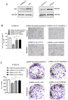
circ-231 promotes the migration and proliferation of human ESCC cells through TPI1 or PRDX6. (A) The expression vectors of pcDNA3.1, pcDNA3.1-TPI1 and pcDNA3.1-PRDX6 were individually transfected into KYSE510 cells and TPI1 and PRDX6 proteins were measured by using western blot. pcDNA3.1 was used as control. After the vectors of pcDNA3.1-TPI1 and pcDNA3.1-PRDX6 transfected into in KYSE510 shRNA-circ-231 cells, the cells were subjected to starvation in serum-free culture medium for 18 h. Then, cell migration was measured by transwell assay (B), and cell proliferation was measured by colony formation assay (C). pcDNA3.1 vectors were transfected into KYSE510 shRNA-Scramble and shRNA-circ-231 cells and used as control. Scale bar: 10x. Values are mean ±SD. *P<0.05, **P<0.01, ***P<0.001.
Figure 10
Figure 10
The schematic diagram for the interaction of circ-231 with eIF4A3 and STAU1 to promote unwinding of secondary structure in 5′ end of TPI1 and PRDX6 mRNAs, leading to ESCC progression.

References

    1. Wang Z, Murigneux V, Le Hir H. Transcriptome-wide modulation of splicing by the exon junction complex. Genome Biol. 2014;15:551. - PMC - PubMed
    1. Jiang Z, Tai Q, Xie X, Hou Z, Liu W, Yu Z. et al. EIF4A3-induced circ_0084615 contributes to the progression of colorectal cancer via miR-599/ONECUT2 pathway. J Exp Clin Cancer Res. 2021;12:227. - PMC - PubMed
    1. Li Q, Lei C, Lu C, Wang J, Gao M, Gao W. LINC01232 exerts oncogenic activities in pancreatic adenocarcinoma via regulation of TM9SF2. Cell Death Dis. 2019;10:698. - PMC - PubMed
    1. Kanellis DC, Espinoza JA, Zisi A, Sakkas E, Bartkova J, Katsori AM. et al. The exon-junction complex helicase eIF4A3 controls cell fate via coordinated regulation of ribosome biogenesis and translational output. Sci Adv. 2021;7:eabf7561. - PMC - PubMed
    1. Budiman ME, Bubenik JL, Driscoll DM. Identification of a signature motif for the eIF4a3-SECIS interaction. Nucleic Acids Res. 2011;39:7730–9. - PMC - PubMed