. 2024 Jun 5;14(6):jkae075.
doi: 10.1093/g3journal/jkae075.

Sse1, Hsp110 chaperone of yeast, controls the cellular fate during endoplasmic reticulum stress

Affiliations

Sse1, Hsp110 chaperone of yeast, controls the cellular fate during endoplasmic reticulum stress

Mainak Pratim Jha et al. G3 (Bethesda). .

Abstract

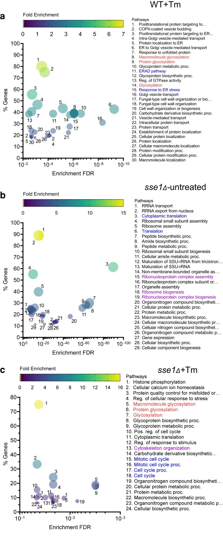
Sse1 is a cytosolic Hsp110 molecular chaperone of yeast, Saccharomyces cerevisiae. Its multifaceted roles in cellular protein homeostasis as a nucleotide exchange factor (NEF), as a protein-disaggregase and as a chaperone linked to protein synthesis (CLIPS) are well documented. In the current study, we show that SSE1 genetically interacts with IRE1 and HAC1, the endoplasmic reticulum-unfolded protein response (ER-UPR) sensors implicating its role in ER protein homeostasis. Interestingly, the absence of this chaperone imparts unusual resistance to tunicamycin-induced ER stress which depends on the intact Ire1-Hac1 mediated ER-UPR signaling. Furthermore, cells lacking SSE1 show inefficient ER-stress-responsive reorganization of translating ribosomes from polysomes to monosomes that drive uninterrupted protein translation during tunicamycin stress. In consequence, the sse1Δ strain shows prominently faster reversal from ER-UPR activated state indicating quicker restoration of homeostasis, in comparison to the wild-type (WT) cells. Importantly, Sse1 plays a critical role in controlling the ER-stress-mediated cell division arrest, which is escaped in sse1Δ strain during chronic tunicamycin stress. Accordingly, sse1Δ strain shows significantly higher cell viability in comparison to WT yeast imparting the stark fitness following short-term as well as long-term tunicamycin stress. These data, all together, suggest that cytosolic chaperone Sse1 is an important modulator of ER stress response in yeast and it controls stress-induced cell division arrest and cell death during overwhelming ER stress induced by tunicamycin.

Keywords: endoplasmic reticulum stress; endoplasmic reticulum unfolded protein response; heat shock protein 110; molecular chaperone; protein homeostasis.


Conflict of interest statement

Conflicts of interest The author(s) declare no conflict of interest.

Figures

Fig. 1.
Fig. 1.
SSE1 deletion imparts resistance to tunicamycin-induced ER stress in yeast. a) (ai) Yeast growth assay by serial drop dilutions using the strains WT (BY4741), sse1Δ, and sse2Δ in YPAD plates at permissive temperature (30°C) (left panel) and under heat stress (37°C) (right panel). The triangles above each panel indicate the increasing dilutions. The time mentioned in hours represents the time of incubation before taking the image of the plates. aii) Growth curves in liquid YPD media at permissive temperature (30°C) of the yeast strains used for drop-dilution assay in panel (ai). The normalized growth is plotted as line plot for each strain with shaded area representing the error range for the measurements. (aiii) Similar to panel (ai), a drop-dilution assay was done in presence of ER stressor Tunicamycin (Tm); in two concentrations 2.5 µg/ml and 5.0 µg/ml sufficient to elicit ER-UPR. Longer time of incubation than panel Ai is used here before capturing the pictures of plates as spots appear at later time points due to extremely slow growth of yeast in presence of Tm. (aiv) Growth curves of three strains used in (aiii) in liquid media in presence of tunicamycin (2.5 μg/ml) in YPD are shown as stated earlier in (aii). (av) The spots of the strains from the (ai) and (aiii) panel drop-dilution assays were quantified using densitometry and were plotted as a bar plot with whiskers representing SEM as shown in the right panel (n = 3). Statistical significance was calculated using unpaired t-tests and the significant pairs were plotted in the graph (all comparisons had P < 0.0001, and they were significant even after Bonferroni Correction). b) (bi) The presence of HAC1 mRNA splice variants was checked from untreated yeast strains (WT, sse1Δ) and the same strains after treatment with tunicamycin (2.5 µg/ml) by synthesizing the cDNAs followed by PCR amplifications with the help of specific primers. (bii) The band intensities were quantified using densitometry and were plotted as a bar plot with whiskers representing SEM as shown in the bottom panel (n = 3). Statistical significance was calculated using unpaired t-tests and the significant pairs were plotted in the graph (Hac1u-WT-Tm/Hac1u-WT + Tm, P = 0.0008, ***; Hac1s-WT-Tm/Hac1s-WT + Tm, P-value = 0.0002, ***; Hac1u-sse1Δ-Tm/Hac1u-sse1Δ+Tm, P = 0.0033, **; and Hac1s-sse1Δ-Tm/Hac1s-sse1Δ+Tm, P = 0.0006, ***). All the above pairwise comparisons are significant even after Bonferroni correction with the only exception being Hac1s-WT-Tm/Hac1s-sse1Δ-Tm (one-tailed P = 0.0383, *) showed marginal significance and plotted in the graph. c) (ci) Western blot showing the distinct increase in Kar2 (ER-resident Hsp70 and ER-UPR marker) levels in response to ER stress by optimum (2.5 µg/ml) concentration of Tm signifying proper mounting of ER-UPR. GAPDH was used as the loading control. cii) The bands were quantified by densitometry and were plotted as a bar plot with whiskers representing SEM in the bottom panel (n = 3). Statistical significance was calculated using unpairedt-tests and the significant pairs were plotted in the graph (WT-Tm/WT + Tm, P = 0.0112, *; sse1Δ-Tm/sse1Δ+Tm, P = 0.0142, *; and sse2Δ-Tm/sse2Δ+Tm, P = 0.0347, *). The above pairwise comparisons, except the last pair (sse2Δ-Tm/sse2Δ+Tm), are significant even after Bonferroni correction. di) Yeast growth assay by serial drop dilutions using the strains wild-type (BY4741), sse1Δ transformed with either the empty plasmid vectors (EV or vector control) or the plasmids expressing the Sse1 protein either at endogenous level (from pRS315 plasmid) or overexpressed (from pJV340 plasmid) for complementation assay. Along with Sse1, Fes1, a second cytosolic nucleotide exchange factor (NEF) is also overexpressed from the pJV340 plasmid. The drop-dilution assay was performed using the strains wild-type (BY4741) + pRS315 (empty vector, EV), sse1Δ + pRS315 (empty vector, EV), sse1Δ + pJV340 (empty vector over expression, EV-OE), sse1Δ + pRS315—Sse1, sse1Δ + pJV340—Sse1, and sse1Δ + pJV340—Fes1 in SD-Leu (synthetic media with Dextrose without leucine) agar plates at permissive temperature (30°C), and in presence of the optimal tunicamycin (2.5 µg/ml) concentrations. The triangles above each panel indicate the increasing dilutions. The time mentioned in hours represents the time of incubation before taking the image of the plates. dii) The spots of the strains from the Di panel drop-dilution assays were quantified using densitometry and were plotted as a bar plot with whiskers representing SEM as shown in the right panel (n = 3). Statistical significance was calculated using unpaired t-tests and the significant pairs were plotted in the graph (all comparisons had P < 0.0001, and they were significant even after Bonferroni Correction). ei) Similar complementation assay using the SSE2 overexpressing plasmid taken from yeast overexpression library. The strains were grown in SR-Ura + 1% galactose plates at the indicated temperatures and with tunicamycin (right panel). eii) represents the quantification of the spot densities from (ei). Bars were plotted as described in the (dii) panel.
Fig. 2.
Fig. 2.
Tunicamycin resistance of sse1Δ is dependent on IRE1-HAC1 mediated ER-UPR signaling and the fitness is attributed to the abrogation of CLIPS function of Sse1 and not to the basal high heat shock response of the strain. a) Yeast growth assay by serial drop dilutions using the strains WT, sse1Δ, hac1Δ, and ire1Δ in YPAD plates at permissive temperature (30°C) and at heat-shock condition (37°C). The time mentioned in hours represents the time of incubation before taking the image of the plates. b) Drop-dilution assay as shown in (a) using the strains WT, and single deletion strains sse1Δ, hac1Δ, ire1Δ, and double deletion strains, hac1Δ-sse1Δ and ire1Δ-sse1Δ in YPAD plates at permissive temperature (30°C) and at heat-shock condition (37°C). c) The same strains as in (b) were used for drop-dilution assay at optimal concentrations of Tm (2.5 µg/ml and 5.0 µg/ml). (d) Drop-dilution assay of WT, sse1Δ and all other single deletion strains of yeast reported to exhibit high basal Heat Shock Response (HSR), (sse2Δ, ssa1Δ, ssa2Δ, get1Δ, ino2Δ, ino4Δ, and ssb1Δ) in YPAD plates at permissive temperature (30°C) (left panel), and in presence of ER stressor Tm (2.5 µg/ml) (right panel). e) The effect of constitutive activation of heat shock response (HSR) on Tm-resistance phenotype of WT and sse1Δ strains was checked by expressing the Hsf1R206S mutant from plasmid by drop dilution assay. The strains used here are WT (BY4741) + pRS423 (empty vector, EV), WT + pRS423—Hsf1, WT + pRS423—Hsf1-R206S, sse1Δ + pRS423 (empty vector, EV), sse1Δ + pRS423—Hsf1, and sse1Δ + pRS423—Hsf1-R206S in SD-His (synthetic media with dextrose without histidine) agar plates at permissive temperature (30°C), heat stress condition (37°C), and in presence of the optimal tunicamycin (2.5 µg/ml) concentrations. f) Similar to (e), growth assay by serial drop dilutions was performed for the WT, sse1Δ and all other single deletion strains for CLIPS proteins apart from Sse1, (ssb1Δ, ssz1Δ, zuo1Δ, jjj1Δ, snl1Δ, gim2Δ, gim3Δ, gim5Δ, cpr6Δ, cct8Δ, and egd1Δ) in YPAD plates in permissive temperature (30°C) (left panel), heat shock condition (37°C) (middle panel) and in presence of an optimal concentration of tunicamycin (2.5 µg/ml) (right panel). All spots shown in (a–e) were quantified and the quantifications have been shown in Supplementary Fig. 5.
Fig. 3.
Fig. 3.
Sse1 plays an important role in modulating tunicamycin-induced ER stress-associated changes in protein translation. a) Polysome profiles are plotted for the WT and sse1Δ strains isolated from untreated cells and cells treated with Tm (2.5 µg/ml) and comparison between polysome profiles of (left panel) untreated WT vs. untreated sse1Δ, (middle panel) untreated vs. Tm-treated WT cells and (right panel) untreated vs. Tm-treated sse1Δ cells are shown. b) The rate of translation of WT, and sse1Δ strains in untreated and Tm-treated (2.5 µg/ml) conditions were analyzed using the CLICK-IT chemistry reaction using L -azido-homoalanine (AHA) and Alexa-Fluor 488 alkyne dye. The incorporated fluorescence in newly synthesized proteins was measured in each sample by flow cytometry and was plotted as a bar plot with whiskers representing SEM. The left panel shows the incorporated fluorescence following 6 hrs of Tm-stress in comparison to untreated cells of WT and sse1Δ strains. The right panel shows same after 24 hrs of Tm-treatment. Statistical significance was calculated using unpaired T-tests and the pairwise comparison outputs were plotted in the graph (Left panel: WT-Tm/WT + Tm, two-tailed P = 0.0003, ***; sse1Δ-Tm/sse1Δ+Tm, two-tailed P = 0.0367, *; WT-Tm/sse1Δ-Tm, two-tailed P < 0.0001, ****; WT + Tm/sse1Δ+Tm, two-tailed P = 0.0021, **. Right panel: WT-Tm/WT + Tm, two-tailed P < 0.0001, ****; sse1Δ-Tm/sse1Δ+Tm, two-tailed P = 0.0019, **; WT-Tm/sse1Δ-Tm, two-tailed P = 0.0052, **; WT + Tm/sse1Δ+Tm, two-tailed P < 0.0001, ****). c) Schematic of the yeast strain YMJ003 (wild type) that serves as a reporter strain for ER-UPR activation. The strain contains the GFP under unfolded protein response element (UPRE) to report for ER-UPR induction. d) Kinetics of ER-UPR activation was measured by measuring UPRE-GFP fluorescence by flow cytometry and shown as overlaid histograms at different timepoints post-Tm-treatment. Each histogram represents the data of the strains WT (YMJ003), sse1Δ, and sse2Δ (in YMJ003 strain background) at designated time points mentioned in each panel following treatment with Tm (2.5 µg/ml). The shift in the two populations, P2 (UPR-activated population) and P1 (basal state after reversal from UPR-activated state), among the strains are marked accordingly. e) The P2 population's (as shown in d) mean GFP fluorescence intensity over time is plotted in this line plot for the same set of WT, sse1Δ and sse2Δ cells. The whiskers over each time point value represents SEM.
Fig. 4.
Fig. 4.
The ER-UPR kinetics is different in absence of Sse1. a) (Left) Schematic of the yeast strains where individual UPR target genes are tagged with GFP, so that their real time translation can be monitored by measuring the GFP fluorescence. (Right) Using these strains as background, SSE1 was deleted using URA3 cassette in each of the GFP reporter strain. The left panel strains serve as the WT strain and the right panel strain serve as sse1Δ strain. b–e) The GFP-tagged UPR target protein's expression as measured by flow cytometry over time are plotted. The mean GFP fluorescence of the UPR-activated population is plotted as line plots at designated time points following treatment with Tm (2.5 µg/ml). The line plots of the expression of cellular targets of ER-UPR like Pdi1-GFP (b), Lhs1-GFP (c), Sec62-GFP (d), and Ubc7-GFP (e) strains with sse1Δ counterparts along with untreated and Tm-treated samples are shown. The whiskers at each time point value represents SEM.
Fig. 5.
Fig. 5.
The pathway enrichment analysis of the cellular transcriptome upon tunicamycin stress in WT and sse1Δ strains. a–c) Transcriptome analysis of WT and sse1Δ strains in untreated condition and after treatment with tunicamycin (2.5 μg/ml) along with other yeast strains of same genetic background as described previously (Narayana Rao et al. 2022) was done by RNA sequencing. The outputs were converted to Z scores. Transcripts above and below z-score 2 were considered as differentially upregulated or downregulated, respectively. The multivariate bubble plots showing the enriched upregulated pathways with the attributes—the enrichment false discovery rate (enrichment FDR) on the X-axis, the percentage of genes identified for each of the enriched pathway with respect to the total annotated genes on the Y-axis, the color scale represents the fold enrichment, and the larger bubble size signifies highly enriched and significant pathways. a) Bubble plot representing the pathways enriched in WT cells when treated with tunicamycin (2.5 μg/ml). Bubble plot showing the enriched pathways of the sse1Δ strain in untreated condition (b) and tunicamycin (2.5 μg/ml)-treated condition (c). The important enriched pathways in all three cases have been highlighted in different colors.
Fig. 6.
Fig. 6.
The changes in cellular proteome upon tunicamycin stress in WT and sse1Δ strains. a) The total number of proteins identified by quantitative mass spectrometry and the outputs of the statistical analysis of various pairwise comparisons are plotted as composite bar plots showing the total number of proteins that were differentially upregulated, downregulated, and insignificant for each of the comparisons between the sse1Δ and WT strain in untreated and after treatment with optimal concentration of tunicamycin (2.5 µg/ml). b–e) Volcano plots showing the differentially expressed (upregulated and downregulated) as well as proteins with insignificant changes in the expression level in the untreated sse1Δ strain with respect to the untreated WT strain (B), in the Tm (2.5 µg/ml) treated WT strain with respect to the untreated WT strain (C), in the Tm (2.5 µg/ml) treated sse1Δ strain with respect to the untreated sse1Δ strain (D) and in Tm (2.5 µg/ml) treated sse1Δ strain with respect to Tm (2.5 µg/ml) treated WT strain (E).
Fig. 7.
Fig. 7.
Sse1 controls tunicamycin-induced ER-stress-mediated cell division arrest and cell death of yeast. a) Images of WT and sse1Δ cells taken by confocal microscopy following treatment with Tm (2.5 µg/ml) along with the untreated control cells. Images were captured at 6, 12, 18, 24, and 48-h time points, respectively. b) The quantitation of the size (surface area in μm2) of the yeast cells from the previous panels, which are represented as a box and whiskers plot where the top whiskers represent the highest and the bottom whiskers represent the lowest individual cell surface area. The horizontal line within each box represents the median cell surface area. Statistical significance was calculated using unpaired T-tests and the pairwise comparison outputs were plotted in the graph (12 h: WT-Tm/WT + Tm, two-tailed P = 0.0144, *; sse1Δ-Tm/sse1Δ+Tm, two-tailed P = 0.0060, **; 18 hrs: WT-Tm/WT + Tm, two-tailed P < 0.0001, ****; sse1Δ-Tm/sse1Δ+Tm, two-tailed P = 0.0403, *; 24 hrs: WT-Tm/WT + Tm, two-tailed P < 0.0001, ****; sse1Δ-Tm/sse1Δ+Tm, two-tailed P < 0.0001, ****; 48 hrs: WT-Tm/WT + Tm, two-tailed P < 0.0001, ****; sse1Δ-Tm/sse1Δ+Tm, two-tailed P < 0.0001, ****). c–f) Cell cycle analysis was done for the WT and sse1Δ cells using the DNA binding fluorescent dye Sytox Green following treatment with Tm (2.5 µg/ml) along with untreated controls. The data were captured after 6 and 24 hrs of Tm (2.5 µg/ml) treatment. c) The overlaid histogram represents the pairwise comparison of WT-untreated/WT + Tm cell cycle pattern at 6 hrs post-Tm-treatment. d) The overlaid histogram represents the pairwise comparison of sse1Δ-untreated/sse1Δ+Tm cell cycle pattern at 6 hrs post-Tm-treatment. e) The overlaid histogram represents the pairwise comparison of WT-untreated/WT + Tm cell cycle pattern at 24 hrs post-Tm-treatment. f) The overlaid histogram represents the pairwise comparison of sse1-untreated/sse1Δ+Tm cell cycle pattern at 24 hrs post-Tm-treatment. g) Cell death percentage was analysed using propidium iodide staining through flow cytometry and was plotted as a bar plot (with whiskers representing SEM, n = 3) using the strains WT (BY4741) and sse1Δ (in BY4741 strain background) at the revival stage [after 36 hrs following optimal Tm (2.5 µg/ml) treatment for 6 hrs]. Statistical significance was calculated using unpaired T-tests and the significant pair was plotted in the graph (WT + Tm/sse1Δ+Tm, P < 0.0001, ****). h) A similar cell death percentage as shown in panel G was determined for WT (BY4741) and sse1Δ (in BY4741 strain background) after chronic ER stress of 24 hrs by Tm (2.5 µg/ml) treatment. Statistical significance was calculated using unpaired T-tests and the significant pair was plotted in the graph (WT + Tm/sse1Δ+Tm, P < 0.0001, ****).
Fig. 8.
Fig. 8.
A schematic summary of the role of Sse1 during ER stress. A schematic model summarizing the role of Sse1 during ER stress. The upper box represents the physiological condition when there is no stress to ER. The lower left box summarizes the condition of WT cells during Tm-mediated ER stress where polysomes are majorly reorganized into monosomes, and in this process Sse1 plays an important role. There is UPR induction by activation of Ire1-Hac1 pathway, which restores homeostasis up to a tolerable level of stress. The lower right box summarizes the condition of sse1Δ cells during ER stress with tunicamycin. Polysome to monosome conversion is inefficient in absence of Sse1 which leads to faster production of UPR-responsive proteins which in turn restores the homeostasis faster. Additionally, the ER stress induced cell cycle-arrest is evaded in sse1Δ cells leading to fitness advantage during tunicamycin stress and more cell viability.

Similar articles

Cited by

References

    1. Albanese V, Yam AY, Baughman J, Parnot C, Frydman J. 2006. Systems analyses reveal two chaperone networks with distinct functions in eukaryotic cells. Cell. 124(1):75–88. doi:10.1016/j.cell.2005.11.039. - DOI - PubMed
    1. Andreasson C, Fiaux J, Rampelt H, Druffel-Augustin S, Bukau B. 2008a. Insights into the structural dynamics of the Hsp110-Hsp70 interaction reveal the mechanism for nucleotide exchange activity. Proc Natl Acad Sci U S A. 105(43):16519–16524. doi:10.1073/pnas.0804187105. - DOI - PMC - PubMed
    1. Andreasson C, Fiaux J, Rampelt H, Mayer MP, Bukau B. 2008b. Hsp110 is a nucleotide-activated exchange factor for Hsp70. J Biol Chem. 283(14):8877–8884. doi:10.1074/jbc.M710063200. - DOI - PubMed
    1. Babour A, Bicknell AA, Tourtellotte J, Niwa M. 2010. A surveillance pathway monitors the fitness of the endoplasmic reticulum to control its inheritance. Cell. 142(2):256–269. doi:10.1016/j.cell.2010.06.006. - DOI - PMC - PubMed
    1. Brandman O, Stewart-Ornstein J, Wong D, Larson A, Williams CC, Li G-W, Zhou S, King D, Shen PS, Weibezahn J, et al. 2012. A ribosome-bound quality control complex triggers degradation of nascent peptides and signals translation stress. Cell. 151(5):1042–1054. doi:10.1016/j.cell.2012.10.044. - DOI - PMC - PubMed

MeSH terms

LinkOut - more resources