. 2024 May;64(5):54.
doi: 10.3892/ijo.2024.5642. Epub 2024 Apr 5.

Heterogeneity of primary and metastatic CAFs: From differential treatment outcomes to treatment opportunities (Review)

Affiliations

Heterogeneity of primary and metastatic CAFs: From differential treatment outcomes to treatment opportunities (Review)

Zixing Kou et al. Int J Oncol. 2024 May.

Abstract

Compared with primary tumor sites, metastatic sites appear more resistant to treatments and respond differently to the treatment regimen. It may be due to the heterogeneity in the microenvironment between metastatic sites and primary tumors. Cancer‑associated fibroblasts (CAFs) are widely present in the tumor stroma as key components of the tumor microenvironment. Primary tumor CAFs (pCAFs) and metastatic CAFs (mCAFs) are heterogeneous in terms of source, activation mode, markers and functional phenotypes. They can shape the tumor microenvironment according to organ, showing heterogeneity between primary tumors and metastases, which may affect the sensitivity of these sites to treatment. It was hypothesized that understanding the heterogeneity between pCAFs and mCAFs can provide a glimpse into the difference in treatment outcomes, providing new ideas for improving the rate of metastasis control in various cancers.

Keywords: heterogeneity; metastatic cancer‑associated fibroblast; primary cancer-associated fibroblast; treatment opportunities; treatment outcomes; tumor microenvironment.


Conflict of interest statement

The authors declare that they have no competing interests.

Figures

Figure 1
Figure 1
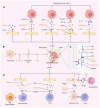
Heterogeneity of mCAFs and pCAFs. (A) mCAFs and pCAFs come from different sources. (B) mCAFs and pCAFs are activated in different ways. For example, in PDAC, the precursor of pCAFs are activated by the tumor-secreted TGF-β and in LMs, mCAFs are activated by macrophage-secreted granulin. (C) mCAFs and pCAFs differ in terms of exosomes, transcriptomes, biomarkers, matreotype and soluble molecules. CAFs, cancer-associated fibroblasts; mCAFs, metastatic CAFs; pCAFs, primary CAFs; TGF-β, transforming growth factor-β; TGF-βR, TGF-β receptor; MicRNA, microRNA.
Figure 2
Figure 2
mCAFs and pCAFs are from different sources. (A) PDAC cells can activate PSCs into pCAFs by secreting TGF-β, PDGF and FGF2. CRC cells secrete TGF-β to activate intestinal RFs into pCAFs and simultaneously secrete CCL2 and PDGF to activate intestinal MSC and a SDF-1/CXCR-4 secretory loop is formed between CRC cells and pCAFs, which not only activates pCAFs but also promotes tumor progression. Mammary resident fibroblasts can be activated by TGF-β, SDF-1 and microRNAs secreted by BC cells. SDF-1 can activate BM-MSCs and BM-MSCs are also activated by OPN and CCL5. Conditional culture medium of lung cancer can activate lung resident fibroblasts and NSCLC-derived factors can activate lung MSC into CAF. (B) HSCs can be activated by macrophage-secreted granulin, as well as by HSC-secreted CCL2, PDGF and TGF-β; at the same time, HSCs also release SDF-1 to promote the secretion of TGF-β by LM cells. Bone metastasis cells secrete IL-6 and TGF-β to induce BM-MSCs into mCAFs. microRNAs from exosomes of lung metastatic tumor cells and IL-1 secreted by neutrophils can activate lung resident fibroblasts. Lung cancer pCAF-derived EVs can activate lung resident fibroblasts within the niche. CAFs, cancer-associated fibroblasts; mCAFs, metastatic CAFs; pCAFs, primary CAFs; PDAC, pancreatic cancer; PSCs, pancreatic stellate cells; TGF-β, transforming growth factor-β; PFs, portal fibroblasts; PDGF, platelet-derived growth factor; MSC, mesenchymal stem cells; SDF-1, stromal cell-derived factor 1; CXCR, C-X-C chemokine receptor; CRC, colorectal cancer; ; BC, breast cancer; BM-MSC, bone marrow-derived MSC; OPN, osteopontin; CCL, chemokine (c-c motif) ligand; NSCLC, non-small cell lung cancer; HSCs, hepatic stellate cells; IL, interleukin; EVs, extracellular vesicles; LM, liver metastasis; RFs, resident fibroblasts; TGF-βR, TGF-β receptor; BM-macrophages, bone marrow-derived macrophages; FGF, fibroblast growth factors; FGFR, FGF receptor; CCR, C-C chemokine receptor; IL-R, interleukin receptors.
Figure 3
Figure 3
mCAFs provide an efficient protective environment for metastases. (A) The thickened ECM of metastases forms a physical barrier for drug penetration and immune cell infiltration. (B) mCAFs highly express IGF2 and Jagged1, activate downstream pathways, promote the secretion of IFN, VEGFA, CDKNIA and collagen and upregulate the expression of stem cell and mesenchymal markers in metastatic cells. (C) mCAFs secrete soluble factors to inhibit the polarization and chemotactic functions of immune cells. CAFs, cancer-associated fibroblasts; mCAFs, metastatic CAFs; pCAFs, primary CAFs; ECM, extracellular matrix; IGF, insulin like growth factor; CDKNIA, cyclin-dependent kinase; IGFR, IGF receptor; RAF, mitogen-activated protein kinase; MEK, mitogen-activated protein kinase kinase; ERK, extracellular regulated kinase; NICD, Notch intracellular domain; CSL, DNA-binding transcription factor; VEGFA, vascular endothelial growth factor A; IFN, interferon; SNAI, snail family transcriptional repressor.
Figure 4
Figure 4
Potential therapeutic opportunities by targeting mCAFs. (A) Blockage of highly expressed signaling molecules or receptors in mCAFs. FAPi can directly target the FAP on the surface of CAFs and exert cytotoxic effects on CAFs by chelating radioactive isotopes and toxins. Using inhibitors such as BI836845, AMD3100 and anakinra to block the corresponding receptors can cut off the signaling communication between metastatic tumors and mCAFs; Similarly, silencing key genes such as LTBP2 can also achieve the objective. (B) Depletion or reversal of mCAFs; curcumin can induce HSC apoptosis and ATRA administration is able to convert PSC activation. (C) Degradation or softening of the ECM of metastasis sites. Using a LOX inhibitor to block ECM crosslinking for antifibrotic and antitumor effects or degradation of hyaluronan components by the PEGPH20 is able to reduce the interstitial fluid pressure of the tumor, leading to an increase in drug delivery. Although MMP is an endogenous collagenolytic enzyme, a number of studies have demonstrated its involvement in the degradation of the ECM, which provides a migratory path for tumor metastasis. (D) Relieving the immunosuppression effect of mCAFs and increasing T cell infiltration. Tranilast reduces M2 macrophage polarization by inhibiting SDF-1 secretion. The use of AMD3100 to competitively inhibit the SDF-1 receptor can increase the infiltration of CD8+ T cells into the ECM. IL-1b secretion from neutrophils can enhance Ptgs2 expression and PGE2 secretion in fibroblasts, leading to reduced anti-tumor immunity and enhanced metastasis to the lung. Genetic loss of Ptgs2 or EP2/4 inhibiting in fibroblasts reverses the immunosuppressive phenotype in BMDCs. mCAFs, metastatic cancer-associated fibroblasts; FAPi, FAP inhibitor; FAP, fibroblast activation protein; CAFs, cancer-associated fibroblasts; IGF2, insulin like growth factor 2; IGF2R, IGF2 receptor; SDF-1, stromal cell-derived factor 1; CXCR, C-X-C chemokine receptor; TGF-β, transforming growth factor-β; TGF-βR, TGF-β receptor; LTBP2, Latent TGF-β binding protein-2; CCL, chemokine (c-c motif) ligand; CCR, C-C chemokine receptor; ATRA, all-trans retinoic acid; COL, collagen; MMP, matrix metalloproteinases; HA, hyaluronan; LOX, lysyl oxidases; Ptgs2, prostaglandin-endoperoxide synthase-2; EP2/4, E prostanoid receptors 2 and 4; PGE2, Prostaglandin E2; IL, interleukin; IL-R, IL receptors.

Similar articles

Cited by

References

    1. Garcia-Vicién G, Mezheyeuski A, Bañuls M, Ruiz-Roig N, Molleví DG. The Tumor microenvironment in liver metastases from colorectal carcinoma in the context of the histologic growth patterns. Int J Mol Sci. 2021;22:1544. doi: 10.3390/ijms22041544. - DOI - PMC - PubMed
    1. Luo F, Li J, Wu S, Wu X, Chen M, Zhong X, Liu K. Comparative profiling between primary colorectal carcinomas and metastases identifies heterogeneity on drug resistance. Oncotarget. 2016;7:63937–63949. doi: 10.18632/oncotarget.11570. - DOI - PMC - PubMed
    1. Hu Z, Li Z, Ma Z, Curtis C. Multi-cancer analysis of clonality and the timing of systemic spread in paired primary tumors and metastases. Nat Genet. 2020;52:701–708. doi: 10.1038/s41588-020-0628-z. - DOI - PMC - PubMed
    1. Korentzelos D, Clark AM, Wells A. A perspective on therapeutic pan-resistance in metastatic cancer. Int J Mol Sci. 2020;21:7304. doi: 10.3390/ijms21197304. - DOI - PMC - PubMed
    1. Hirata E, Sahai E. Tumor microenvironment and differential responses to therapy. Cold Spring Harb Perspect Med. 2017;7:a026781. doi: 10.1101/cshperspect.a026781. - DOI - PMC - PubMed