. 2024 Jun;27(2):335-354.
doi: 10.1007/s40477-023-00869-2. Epub 2024 Apr 5.

Musculoskeletal disorders in padel: from biomechanics to sonography

Affiliations

Musculoskeletal disorders in padel: from biomechanics to sonography

Giulio Cocco et al. J Ultrasound. 2024 Jun.

Abstract

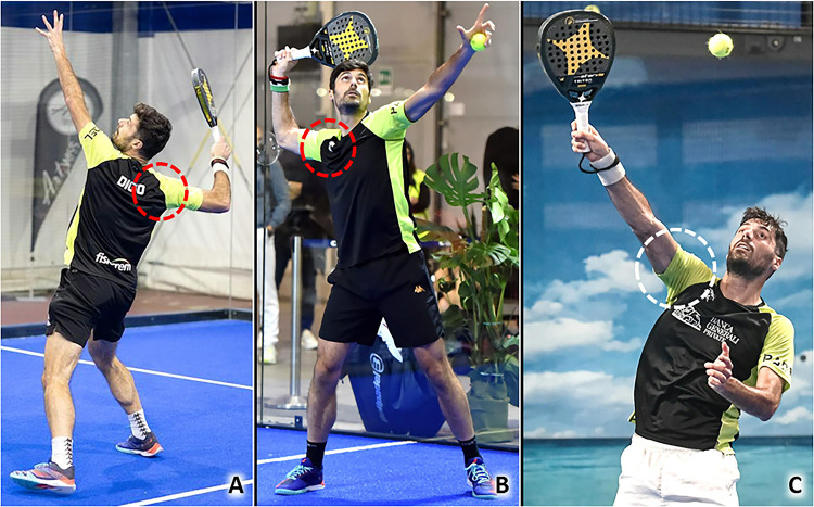
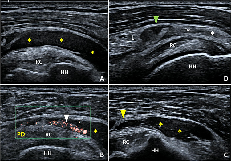
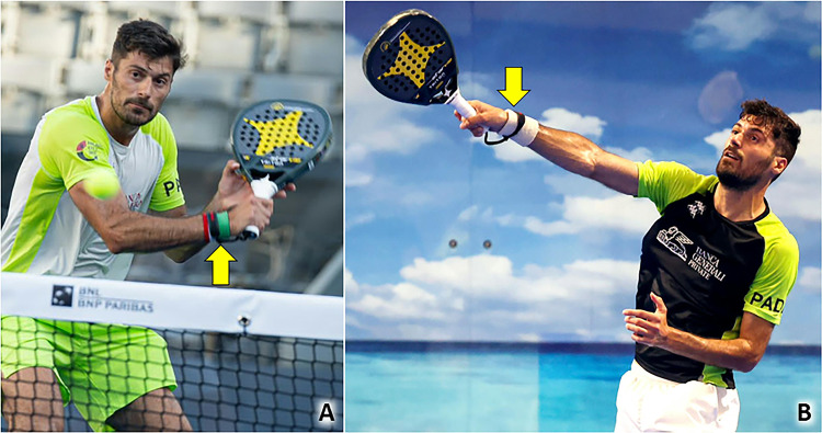
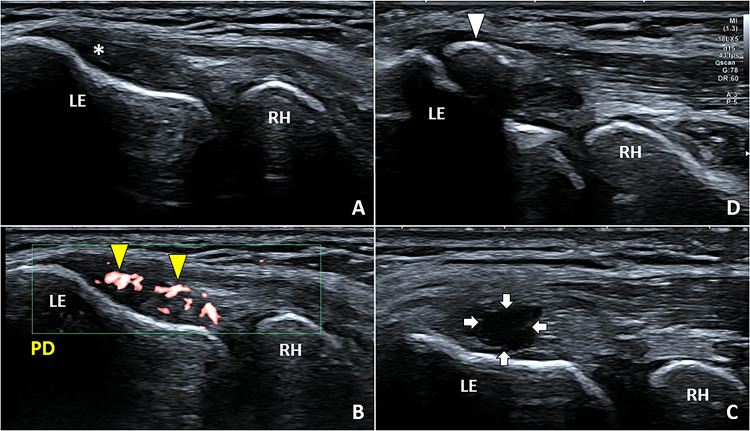
Padel is a racket sport, combining high-frequency and low-intensity athletic gestures, that has been gaining growing scientific interest in recent years. Musculoskeletal injuries are very common among padel players with an incidence rate of 3 per 1000 h of training and 8 per 1000 matches. To the best of our knowledge, a comprehensive collection describing the most common sonographic findings in padel players with musculoskeletal injuries is lacking in the pertinent literature. In this sense, starting from the biomechanical features of padel-specific gestures we have reported the ultrasonographic patterns of most frequent injuries involving the upper limb, the trunk, and the lower limb. Indeed, comprehensive knowledge of the biomechanical and clinical features of musculoskeletal injuries in padel is paramount to accurately perform a detailed ultrasound examination of the affected anatomical site. So, the present investigation aims to provide a practical guide, simple and ready-to-use in daily practice, to optimize the sonographic assessment of padel players by combining it with the clinical findings and the biomechanical features of athletic gestures.

Keywords: Biomechanics; Injuries; Musculoskeletal; Padel; Ultrasound.


Conflict of interest statement

The authors declare that they have no competing interests.

Figures

Fig. 1
Fig. 1
Combined movements of abduction (over 90 degrees) and external rotation (red dotted circle) of the shoulder (A, B), and elevations (white dotted circle) of the upper limb overhead (C), are very frequently performed during a padel match considering the court’s size and, the upward trajectory of the ball due to the multiple bounces on the perimetral walls
Fig. 2
Fig. 2
In the acute phase, the effusion (yellow asterisks) is the most common sonographic finding defining exudative bursitis (A). Progressively, synovial hypertrophy (B) with hypervascularization (white arrowhead) and fibrotic septae (yellow arrowhead) can develop within the bursal cavity (C). Adhesive bursopathy (D) shows a well-defined, hypoechoic, nodular thickening (green arrowhead) of the synovial tissue (white asterisk) close to the overlying hyperechoic coracoacromial ligament (L). HH humeral head, RC rotator cuff, PD power Doppler
Fig. 3
Fig. 3
An expert padel player starts and keeps the one-hand backhand stroke with the extended wrist (yellow arrow) to reduce the eccentric load on the common extensor tendon in the lateral compartment of the elbow both in the pre-impact phase (A) and after the impact of the ball (B)
Fig. 4
Fig. 4
Hypoechoic thickening (white asterisk) of the CET in B-Mode (A) and neo-vessels (yellow arrowheads) in color/power Doppler (B) are the most common sonographic findings in padel players with lateral elbow tendinopathy. Focal injury (white arrows) of the CET (C) and calcific depositions (white arrowheads) can also be identified especially in players with recalcitrant lateral elbow pain (D). LE lateral epicondyle, RH radial head, PD power Doppler
Fig. 5
Fig. 5
By rapidly activating the pronator teres and flexor carpi radialis muscles, a sudden flexion of the wrist (yellow arrow) coupled with complete pronation of the forearm (white curved arrow) can be performed by elite padel players—the “whipping” effect (A, B). The aforementioned gestures result in a global torsional movement of the upper limb pivotal to increase the power/velocity of the ball and control its trajectory (C)
Fig. 6
Fig. 6
Hypoechoic thickening (white arrowhead) of the conjoint tendon (CT) in B-Mode (A) with neo-vessels (yellow arrowheads) penetrating the degenerated tendon tissue in power Doppler (PD) (B) are common sonographic findings in padel players with medial elbow pain. More rarely, a small bony fragment (yellow arrow) embedded in the ulnar collateral ligament (ucl) and surrounded by an intense hypervascularization (C) can be observed close to the painful medial epicondyle (ME). Of note, a small ossicle (white arrow) within the collagen fibers of the ulnar collateral ligament (ucl), commonly rounded in shape and not encircled by neo-vessels, can be identified also in asymptomatic volunteers (D). U ulna, CD color Doppler
Fig. 7
Fig. 7
Repeated ulnar (white curved arrow) and radial (red curved arrow) deviations of the radiocarpal joint (A, B) and quick torsional movements (black curved arrow) of the wrist (C) during a padel match may progressively lead to pathologies of the extensor tendons
Fig. 8
Fig. 8
Thickening of the fibrous retinaculum (yellow arrowhead), sheath effusion (white asterisks), tenosynovial hypertrophy (white arrowhead) with hypervascularization of the first extensor compartment is visualized in a padel player with radial wrist pain (A, B). Likewise, tenosynovitis of the extensor carpi ulnaris tendon with multiple vascular signals within the hypertrophic synovial sheath (white arrowhead) can be observed on the ulnar side (C, D). Rad radius, A artery, V vein, PD power doppler, Ul ulna, Tri triquetrum, L lunate, 5M fifth metacarpal bone
Fig. 9
Fig. 9
The transition from the loading to impact phase of overhead strokes during a Padel match requires a very rapid and explosive change of the shape of the core (red dotted lines). Indeed, an eccentric contraction of abdominal muscles to control the hyperextension of the spine (A) is immediately followed by powerful concentric contractions to flex and rotate the trunk (B) transmitting the forces from the lower to upper limbs
Fig. 10
Fig. 10
Combining transverse (A, B) and longitudinal (C) scans, a focal injury (white arrowhead) involving the superficial fibers of rectus abdominis muscle (RA)—with a mild peri-lesional hypervascularization (B) in power Doppler (PD)—can be observed in a professional padel player due to a powerful overhead stroke with massive contraction of the core muscles. Instead, a full-thickness tear (yellow arrowheads) of the rectus abdominis (RA) with blood collections (white asterisks) within the muscle belly can be visualized in transverse sonograms of an amatorial padel player (D, E). Of note, the longitudinal scan (F) confirms the anatomical integrity of the deep lamina of its fascial sheath (blue arrowhead). sc subcutaneous tissue
Fig. 11
Fig. 11
Comparative ultrasound scanning, in a transverse plane, of the normal (A, B) and painful (C) side of the anterolateral abdominal wall clearly shows a massive thickening and hyperechogenicity of the left internal oblique muscle (IO) due to severe intramuscular edema with a loss of the normal echotexture. Shifting the probe distally (D), disruption of its muscle fibers with a large intramuscular hematoma (yellow asterisk) has been identified confirming the post-traumatic myofascial injury with a sparing of the overlying external oblique muscle (EO). TA transversus abdominis muscle, RA rectus abdominis muscle
Fig. 12
Fig. 12
Rapid combined knee flexion-rotation movements (white dotted circle) are often necessary to correctly orientate the trunk and hit the ball at the optimal height (A). The patellar tendon (yellow arrow) and Achilles tendon (green arrow) function as shock absorber cords during the landing phase of the jump (black dotted arrow) modulating the velocity of braking and reducing the risk of knee/ankle sprain (B)
Fig. 13
Fig. 13
Normally (A), the patellar tendon presents a fibrillar echotexture in the longitudinal scan; instead, a hypoechoic thickening of the deep and proximal fibers (white asterisk), lamellar calcifications (yellow arrowhead), and cortical irregularities (white arrowhead) of the inferior pole of the patella (Pat) can be commonly observed in padel players with patellar tendinopathy (B). The color Doppler (CD) depicts neo-vessels (C) originating from the Hoffa fat pad (FP) and infiltrating the hypoechoic degenerated tendon tissue (white asterisk). More rarely, an isolated longitudinal tear of the PT (green arrowhead) can be identified with a regular enthesis (orange arrowhead) (D)
Fig. 14
Fig. 14
Powerful contraction of the triceps surae and extension of the knee (yellow arrow) are key biomechanical factors during the take-off phase of the jump (black dotted arrow) to reach the ball (A). Likewise, during the landing phase of the jump (white dotted arrow), contraction of the gastrocnemius muscle with the knee slightly flexed (yellow arrow) allows soft braking protecting the ankle from an excessive mechanical overload (B)
Fig. 15
Fig. 15
A small injury of the anterior aponeurosis (white arrowhead) of the medial gastrocnemius (MG) is enough to allow the blood to spill within the inter aponeurotic space (green arrowhead) of the triceps surae muscle (A). Instead, post-traumatic disruption of deep fibers of its free aponeurosis (white asterisk), with sparing of superficial fibers (yellow arrowhead), should be considered such as a tendon injury (B) with a worse functional prognosis compared to the previous myoaponeurotic lesion. In padel athletes with a large hematoma (yellow asterisks) in the inter aponeurotic space of the triceps surae muscle (C) ultrasound-guided drainage is paramount to avoid progression toward an irregular mass of granulation tissue (white dotted line) that “blocks” a correct healing process (D). Sol soleus muscle
Fig. 16
Fig. 16
Fusiform hypoechoic thickening (white arrowheads) of the midportion of AT and its hypervascularization with neo-vessels (yellow arrowheads) originating from the underlying Kager fat pad (FP) can be considered the most common sonographic signs of non-insertional pathology (AC). More rarely, isolated peri tendinitis may be identified with vascular signals (blue arrowheads) confined inside the thickened paratenon and a normal thickness/echotexture of the underlying AT (D). In players with suspected partial injury of the AT, ankle dorsiflexion may be performed to dynamically and selectively glide the disrupted tendon fibers (white asterisks) confirming the diagnosis (E). CD color doppler, PD power doppler
Fig. 17
Fig. 17
Comparative scanning (A, B) shows bilaterally the posterosuperior bony spur (bS) of calcaneus but, the hypoechoic thickening of the insertional portion of AT (yellow arrowhead) only on the painful side. The color/power Doppler (CD) confirms the presence of neo-vessels (white arrowhead) originating from the subcutaneous fat and penetrating the tendon tissue selectively on the painful heel (C). In another padel player with heel pain, the B-mode scan (D) showed effusion in the deep retrocalcaneal bursa (b), and color Doppler (CD) confirmed its hypervascularization (yellow arrow) (E). Interestingly, hyperemia also involves the superficial retrocalcaneal bursa (white arrow) and the underlying tendon tissue (E). Cal calcaneus

Similar articles

Cited by

References

    1. García-Giménez A, de la Fuente FP, Otín CC, Páez LC. Performance outcome measures in padel: a scoping Review. Int J Environ Res Public Health. 2022;19:4395. doi: 10.3390/ijerph19074395. - DOI - PMC - PubMed
    1. Dahmen J, Emanuel KS, Fontanellas-Fes A, Verhagen E, Kerkhoffs GMMJ, Pluim BM. Incidence, prevalence and nature of injuries in padel: a systematic review. BMJ Open Sport Exerc Med. 2023;9(2):e001607. doi: 10.1136/bmjsem-2023-001607. - DOI - PMC - PubMed
    1. Tagliafico AS, Torri L, Righetto R. Injuries in non-professional padel tennis players. Results of a survey of the Italian Federation of Tennis in Liguria, Italy. J Sports Med Phys Fitn. 2023;63(4):566–569. doi: 10.23736/S0022-4707.22.14280-5. - DOI - PubMed
    1. Priego Quesada JI, Sanchís Almenara M, Kerr ZY, Alcantara E. Examination of the risk factors associated with injured recreational padel players in Spain. J Sports Med Phys Fitn. 2018;58(1–2):98–105. doi: 10.23736/S0022-4707.16.06729-3. - DOI - PubMed
    1. Pérez F, Rubia A, Cañadas E, Lorenzo-Calvo J, Marquina M, García-Sánchez C. Musculoskeletal injury prevalence in professional padel players. A retrospective study of the 2021 season. Phys Ther Sport. 2023;63:9–16. doi: 10.1016/j.ptsp.2023.06.003. - DOI - PubMed

MeSH terms

LinkOut - more resources