

A Shape Memory Polymeric Shield for Protecting Corneal Endothelium During Phacoemulsification
- PMID: 38578634
- PMCID: PMC11005075
- DOI: 10.1167/tvst.13.4.11
A Shape Memory Polymeric Shield for Protecting Corneal Endothelium During Phacoemulsification
Abstract
Background: The purpose of this study was to explore the protective effect of a shape memory polymeric shield on corneal endothelium during phacoemulsification in rabbits.
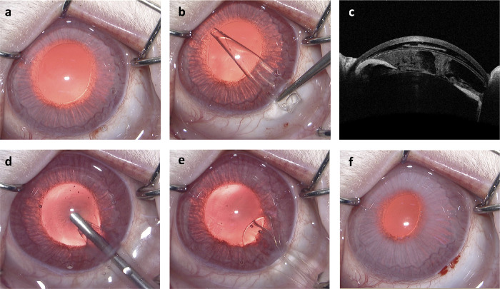
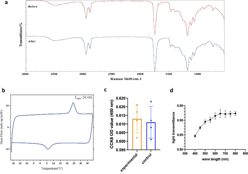
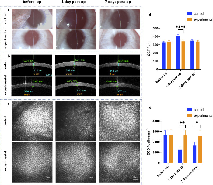
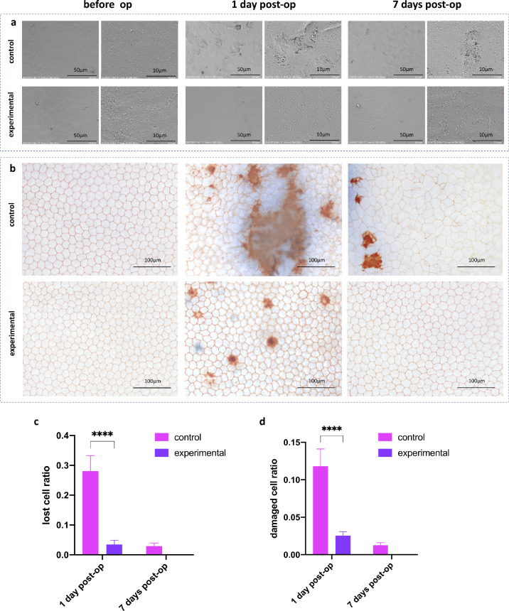
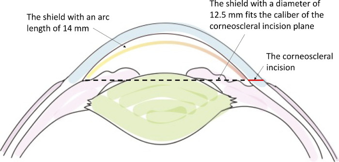
Methods: Poly-(glycerol dodecanedioate) (PGD) with a transition temperature of 24.416°C was prepared to make a shape memory shield with a thickness of 100 µm, an arc length of 14 mm, and a radius of curvature of 8.8 mm. In the control group, a phaco-tip with bevel-down was used to simulate injury to the corneal endothelium by phacoemulsification in rabbits. In the experimental group, the pre-cooled and curled shape memory shield was injected into and removed from the anterior chamber before and after phaco-power release. Anterior segment optical coherence tomography (AS-OCT), confocal microscope, trypan blue/alizarin red staining, and scanning electron microscope were performed to measure endothelial damage after surgery.
Results: One day postoperatively, the lost cell ratio of the control group and the experimental group were 28.08 ± 5.21% and 3.50 ± 1.43%, respectively (P < 0.0001), the damaged cell ratios were 11.83 ± 2.30% and 2.55 ± 0.52%, respectively (P < 0.0001), and the central corneal thicknesses (CCT) were 406.75 ± 16.74 µm and 340. 5 ±13.48 µm, respectively (P < 0.0001). Seven days postoperatively, the endothelial cell density (ECD) of the control group and the experimental group were 1674 ± 285/mm2 and 2561 ± 554/mm2, respectively (P < 0.05). The above differences were all statistically significant.
Conclusions: This PGD based shape memory shield has a protective effect on corneal endothelium during phacoemulsification. It reduces postoperative corneal edema and ECD decrease in the short term after surgery.
Translational relevance: The shape memory PGD "shield" in this study may have a use in certain human patients with vulnerable corneas of low endothelial cell count or shallow anterior chambers.
Conflict of interest statement
Disclosure:
Figures
Similar articles
-
Temporal evolution of the biological response to laser-induced refractive index change (LIRIC) in rabbit corneas.Exp Eye Res. 2021 Jun;207:108579. doi: 10.1016/j.exer.2021.108579. Epub 2021 Apr 20. Exp Eye Res. 2021. PMID: 33864783 Free PMC article.
-
Different-sized incisions for phacoemulsification in age-related cataract.Cochrane Database Syst Rev. 2017 Sep 20;9(9):CD010510. doi: 10.1002/14651858.CD010510.pub2. Cochrane Database Syst Rev. 2017. PMID: 28931202 Free PMC article.
-
The association of corneal thickness with ocular structures from corneal epithelium to optic nerve sheath diameter.Int Ophthalmol. 2025 Jul 22;45(1):306. doi: 10.1007/s10792-025-03683-3. Int Ophthalmol. 2025. PMID: 40694268
-
Non-steroidal anti-inflammatory drugs versus corticosteroids for controlling inflammation after uncomplicated cataract surgery.Cochrane Database Syst Rev. 2017 Jul 3;7(7):CD010516. doi: 10.1002/14651858.CD010516.pub2. Cochrane Database Syst Rev. 2017. PMID: 28670710 Free PMC article.
-
Prescription of Controlled Substances: Benefits and Risks.2025 Jul 6. In: StatPearls [Internet]. Treasure Island (FL): StatPearls Publishing; 2025 Jan–. 2025 Jul 6. In: StatPearls [Internet]. Treasure Island (FL): StatPearls Publishing; 2025 Jan–. PMID: 30726003 Free Books & Documents.
References
-
- Lee CM, Afshari NA.. The global state of cataract blindness. Curr Opin Ophthalmol. 2017; 28: 98–103. - PubMed
-
- Xie LX, Yao Z, Huang YS, Ying L.. Corneal endothelial damage and its repair after phacoemulsification. Zhonghua Yan Ke Za Zhi. 2004; 40: 90–93. - PubMed
-
- Gogate P, Ambardekar P, Kulkarni S, Deshpande R, Joshi S, Deshpande M.. Comparison of endothelial cell loss after cataract surgery: phacoemulsification versus manual small-incision cataract surgery: six-week results of a randomized control trial. J Cataract Refract Surg. 2010; 36: 247–253. - PubMed
Publication types
MeSH terms
LinkOut - more resources
Full Text Sources
Research Materials
Miscellaneous
