. 2024 Jul 4;144(1):84-98.
doi: 10.1182/blood.2024024072.

Loss of the stress sensor GADD45A promotes stem cell activity and ferroptosis resistance in LGR4/HOXA9-dependent AML

Affiliations

Loss of the stress sensor GADD45A promotes stem cell activity and ferroptosis resistance in LGR4/HOXA9-dependent AML

Nunki Hassan et al. Blood. .

Abstract

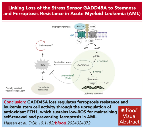
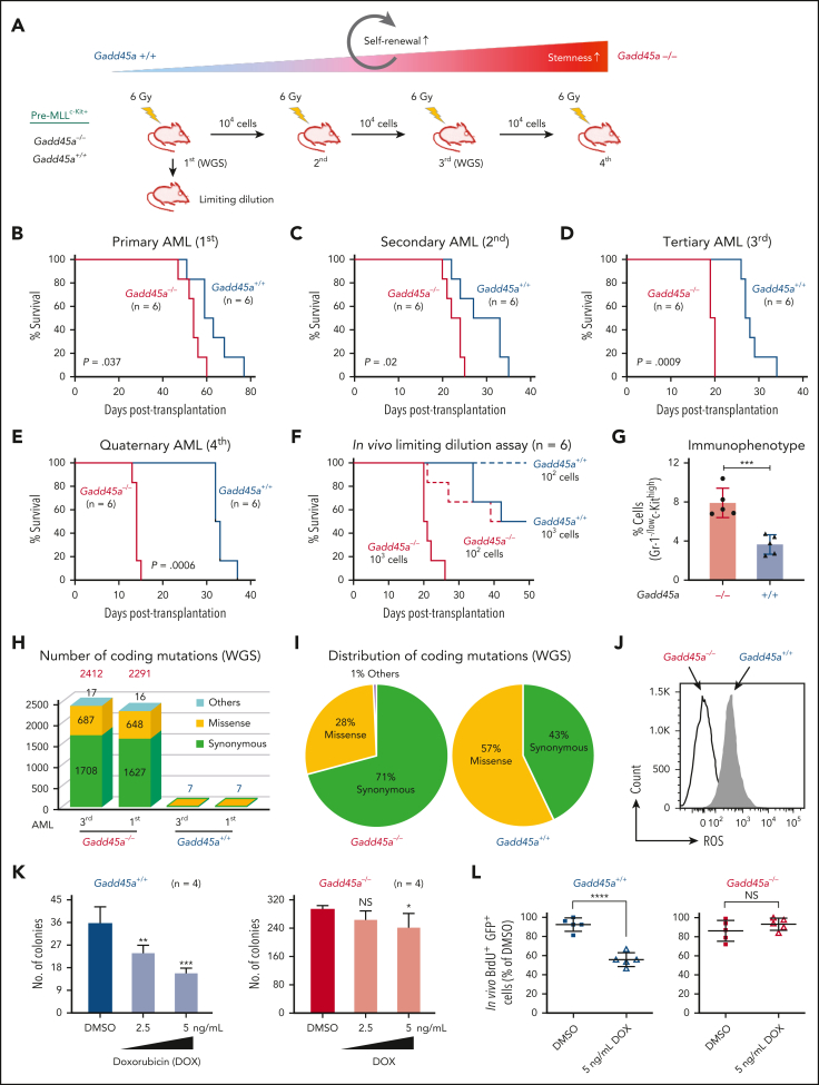
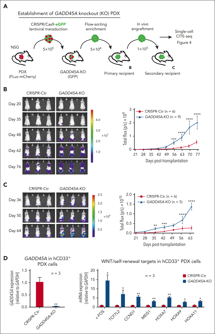
The overall prognosis of acute myeloid leukemia (AML) remains dismal, largely because of the inability of current therapies to kill leukemia stem cells (LSCs) with intrinsic resistance. Loss of the stress sensor growth arrest and DNA damage-inducible 45 alpha (GADD45A) is implicated in poor clinical outcomes, but its role in LSCs and AML pathogenesis is unknown. Here, we define GADD45A as a key downstream target of G protein-coupled receptor (LGR)4 pathway and discover a regulatory role for GADD45A loss in promoting leukemia-initiating activity and oxidative resistance in LGR4/HOXA9-dependent AML, a poor prognosis subset of leukemia. Knockout of GADD45A enhances AML progression in murine and patient-derived xenograft (PDX) mouse models. Deletion of GADD45A induces substantial mutations, increases LSC self-renewal and stemness in vivo, and reduces levels of reactive oxygen species (ROS), accompanied by a decreased response to ROS-associated genotoxic agents (eg, ferroptosis inducer RSL3) and acquisition of an increasingly aggressive phenotype on serial transplantation in mice. Our single-cell cellular indexing of transcriptomes and epitopes by sequencing analysis on patient-derived LSCs in PDX mice and subsequent functional studies in murine LSCs and primary AML patient cells show that loss of GADD45A is associated with resistance to ferroptosis (an iron-dependent oxidative cell death caused by ROS accumulation) through aberrant activation of antioxidant pathways related to iron and ROS detoxification, such as FTH1 and PRDX1, upregulation of which correlates with unfavorable outcomes in patients with AML. These results reveal a therapy resistance mechanism contributing to poor prognosis and support a role for GADD45A loss as a critical step for leukemia-initiating activity and as a target to overcome resistance in aggressive leukemia.


Conflict of interest statement

Conflict-of-interest disclosure: The authors declare no competing financial interests.

Figures

None
Graphical abstract
Figure 1.
Figure 1.
Gadd45a is negatively regulated by Lgr4 and its deletion retains β-catenin activity even in the absence of Lgr4. (A) Heat map analysis of microarray data (n = 3, P ≤ .005, fold change ≥ 1.8) showing differentially expressed genes induced by Lgr4 knockdown in MLL-AF9–induced AML cells. (B) Quantitative polymerase chain reaction (qPCR) (n = 3) and Western blots confirming upregulation of Gadd45a induced by Lgr4 knockdown in MLL-AF9 leukemic cells carrying scrambled control (Scr) vs Lgr4 shRNA1 (sh1). Data are given as mean ± SD. ∗P < .05, unpaired t-test. (C) qPCR (n = 3) and Western blots showing downregulation of Gadd45a induced by Lgr4 overexpression in HOXA9/MEIS1 leukemic cells, compared with endogenous expression of Gadd45a in MLL-AF9 leukemic cells. Data are given as mean ± SD. ∗∗∗∗P < .0001, 1-way analysis of variance (ANOVA). (D) Analysis of TCGA dataset,, revealing the correlation between LGR4 and GADD45A expression in all patients with AML (n = 244, r = −0.328, P = 1.6e-07), patients with AML with unfavorable outcome (n = 81, r = −0.482, P = 5.1e-06), and patients with AML with favorable outcome (n = 120, r = −0.155, P = .091). (E) Western blots confirming efficient Gadd45a knockout with a resultant increase in β-catenin and inactive phospho-Ser9-Gsk3β (p-Gsk3βSer9) in GFP+ pre-MLLc-Kit+ cells. qPCR showing upregulation of Wnt/self-renewal target genes induced by Gadd45a deletion (n = 3). Data are given as mean ± SD. ∗P < .05, ∗∗P < .005, unpaired t-test. (F) Colony formation of Gadd45a−/− vs Gadd45a+/+ pre-MLLc-Kit+ cells. The percentage of colonies (relative to Gadd45a−/−) at the third round of serial replating is shown. n = 4 independent experiments. Data are given as mean ± SD. ∗∗P < .005, unpaired t-test. (G) Western blots confirming efficient Lgr4 knockdown with a resultant change of endogenous β-catenin expression in response to Gadd45a knockout in LSCs (LinSca-1c-KithighCD16/32highCD34+), flow sorted from the bone marrow (BM) of AML mice following transplantation with pre-MLLc-Kit+ cells.
Figure 2.
Figure 2.
Deletion of Gadd45a enhances oncogenic potential and LSC activity on serial transplantation while inducing mutations. (A) Schematic overview of the experimental procedure. (B-E) Kaplan-Meier mouse survival curves of primary AML (B), secondary AML (C), tertiary AML (D), and quaternary AML (E). Primary AML was generated by transplanting 1 × 106Gadd45a−/− vs Gadd45a+/+ preleukemic cells, induced by MLL-AF9-GFP, into sublethally irradiated C57BL/6 (BL6) recipient mice (n = 6 for each group). Secondary AML, tertiary AML, and quaternary AML were generated by transplanting 1 × 104 GFP+ leukemic bone marrow (BM) cells flow sorted from mice with primary, secondary, and tertiary AML, respectively, into recipient mice (n = 6 for each group). P value was determined by the log-rank test. (F) In vivo limiting dilution transplantation assay showing a 10-fold increase in LSC frequency in Gadd45a−/− vs Gadd45a+/+ mice (1/142 vs 1/1650; P = .00114). LSC frequency was calculated using L-Calc software (StemCell Technologies). Kaplan-Meier survival curves of mice receiving the indicated number of GFP+ leukemic BM cells (n = 6 for each group), and P value was determined by the log-rank test. (G) Scatter plots with a bar graph depicting the percentage of Gr-1-/lowc-Kithigh population in total GFP+ leukemic BM cells from Gadd45a−/− vs Gadd45a+/+ mice (n = 5 for each group). Data are given as mean ± SD. ∗∗∗P < .0005. Unpaired t-test. (H) Stacked bar plots displaying the number of coding mutations identified by whole-genome sequencing (WGS) of Gadd45a−/− vs Gadd45a+/+ LSCs from primary (1st) and tertiary (3rd) AML. Green, yellow, and silver stacked bars represent synonymous, missense, and others (inframe insertion/deletion, frameshit, splice donor variant in coding sequence, start lost, and stop retained/gained), respectively. (I) Distribution of the coding mutations in Gadd45a−/− vs Gadd45a+/+ LSCs from primary AML. (J) Flow cytometry histograms showing decreased intracellular ROS in Gadd45a−/− LSCs compared with Gadd45a+/+ LSCs. (K) The number of colonies following treatment of Gadd45a−/− vs Gadd45a+/+ AML LSCs with dimethyl sulfoxide (DMSO) control or doxorubicin (DOX: 2.5 or 5 ng/mL) for 5 days in methylcellulose (n = 4). Data are given as mean ± SD. ∗P < .05, ∗∗P < .005, ∗∗∗P < .0005; NS, not significant (P > .05). One-way ANOVA. (L) In vivo BrdU proliferation assay with dot plots depicting BrdU+ GFP+ leukemic cells engrafted in the mouse BM at 12 days posttransplantation. Gadd45a−/− or Gadd45a+/+ AML LSCs were pretreated ex vivo with DMSO vs 5 ng/mL DOX for 48 hours, and 1 × 105 treated cells were transplanted into BL6 mice for leukemic cell engraftment and subsequent BrdU proliferation assay. Data are presented as the mean percentage relative to DMSO ± SD (n = 5 mice per group). ∗∗∗∗P < .0001; NS, not significant (P > .05). Unpaired t-test. (M) Flow cytometry histograms showing intracellular ROS levels in Gadd45a−/− vs Gadd45a+/+ AML LSCs treated with DMSO vs DOX (2.5 or 5 ng/mL) for 5 days, or pretreated with 1 mM N-acetylcysteine (NAC) for 1 hour, followed by treatment with 100 μM menadione for an additional 1 hour.
Figure 2.
Figure 2.
Deletion of Gadd45a enhances oncogenic potential and LSC activity on serial transplantation while inducing mutations. (A) Schematic overview of the experimental procedure. (B-E) Kaplan-Meier mouse survival curves of primary AML (B), secondary AML (C), tertiary AML (D), and quaternary AML (E). Primary AML was generated by transplanting 1 × 106Gadd45a−/− vs Gadd45a+/+ preleukemic cells, induced by MLL-AF9-GFP, into sublethally irradiated C57BL/6 (BL6) recipient mice (n = 6 for each group). Secondary AML, tertiary AML, and quaternary AML were generated by transplanting 1 × 104 GFP+ leukemic bone marrow (BM) cells flow sorted from mice with primary, secondary, and tertiary AML, respectively, into recipient mice (n = 6 for each group). P value was determined by the log-rank test. (F) In vivo limiting dilution transplantation assay showing a 10-fold increase in LSC frequency in Gadd45a−/− vs Gadd45a+/+ mice (1/142 vs 1/1650; P = .00114). LSC frequency was calculated using L-Calc software (StemCell Technologies). Kaplan-Meier survival curves of mice receiving the indicated number of GFP+ leukemic BM cells (n = 6 for each group), and P value was determined by the log-rank test. (G) Scatter plots with a bar graph depicting the percentage of Gr-1-/lowc-Kithigh population in total GFP+ leukemic BM cells from Gadd45a−/− vs Gadd45a+/+ mice (n = 5 for each group). Data are given as mean ± SD. ∗∗∗P < .0005. Unpaired t-test. (H) Stacked bar plots displaying the number of coding mutations identified by whole-genome sequencing (WGS) of Gadd45a−/− vs Gadd45a+/+ LSCs from primary (1st) and tertiary (3rd) AML. Green, yellow, and silver stacked bars represent synonymous, missense, and others (inframe insertion/deletion, frameshit, splice donor variant in coding sequence, start lost, and stop retained/gained), respectively. (I) Distribution of the coding mutations in Gadd45a−/− vs Gadd45a+/+ LSCs from primary AML. (J) Flow cytometry histograms showing decreased intracellular ROS in Gadd45a−/− LSCs compared with Gadd45a+/+ LSCs. (K) The number of colonies following treatment of Gadd45a−/− vs Gadd45a+/+ AML LSCs with dimethyl sulfoxide (DMSO) control or doxorubicin (DOX: 2.5 or 5 ng/mL) for 5 days in methylcellulose (n = 4). Data are given as mean ± SD. ∗P < .05, ∗∗P < .005, ∗∗∗P < .0005; NS, not significant (P > .05). One-way ANOVA. (L) In vivo BrdU proliferation assay with dot plots depicting BrdU+ GFP+ leukemic cells engrafted in the mouse BM at 12 days posttransplantation. Gadd45a−/− or Gadd45a+/+ AML LSCs were pretreated ex vivo with DMSO vs 5 ng/mL DOX for 48 hours, and 1 × 105 treated cells were transplanted into BL6 mice for leukemic cell engraftment and subsequent BrdU proliferation assay. Data are presented as the mean percentage relative to DMSO ± SD (n = 5 mice per group). ∗∗∗∗P < .0001; NS, not significant (P > .05). Unpaired t-test. (M) Flow cytometry histograms showing intracellular ROS levels in Gadd45a−/− vs Gadd45a+/+ AML LSCs treated with DMSO vs DOX (2.5 or 5 ng/mL) for 5 days, or pretreated with 1 mM N-acetylcysteine (NAC) for 1 hour, followed by treatment with 100 μM menadione for an additional 1 hour.
Figure 3.
Figure 3.
Deletion of GADD45A promotes engraftment of human AML PDX cells in NSG mice. (A) Schematic describing in vivo generation of CRISPR/Cas9-mediated knockout (KO) of GADD45A in PDX mice. Also see supplemental Figure 4 for the additional experimental detail. (B) Representative in vivo bioluminescence imaging and total flux (photons/second [p/s]) of GADD45A KO PDX mice (n = 9) vs CRISPR control (Ctr) PDX mice (n = 6) in primary NSG recipients. A total of 5 × 104 mCherry+ GFP+ PDX cells were transplanted into each of recipient mice. Scatter dot plots represent the mean ± SD. ∗∗∗P < .0005, ∗∗∗∗P < .0001, 2-way ANOVA. (C) Representative in vivo bioluminescence imaging and total flux (p/s) of GADD45A KO PDX mice (n = 5) vs CRISPR Ctr PDX mice (n = 6) in secondary NSG recipients. A total of 1 × 105 mCherry+ PDX cells from primary recipient mice were transplanted into each of secondary recipient mice. Data are given as mean ± SD. ∗∗P < .005, ∗∗∗P < .0005, ∗∗∗∗P < .0001, 2-way ANOVA. (D) Quantitative polymerase chain reaction (qPCR) confirming stable knockout of GADD45A and showing relative expression levels of WNT/self-renewal target genes in GADD45A KO vs CRISPR Ctr hCD33+ PDX bone marrow (BM) cells (n = 3). Data are given as mean ± SEM. ∗P < .05, ∗∗P < .005, unpaired t-test.
Figure 4.
Figure 4.
Coupling single cell RNA-sequencing (scRNA-seq) with cellular indexing of transcriptomes and epitopes by sequencing (CITE-seq) on AML PDX cells reveals an increased proportion of LSCs and identifies genes/pathways upregulated by GADD45A deletion at a single stem cell level. (A) t-distributed stochastic neighbor embedding (T-SNE) clustering of CITE-seq data showing human CD34+ LSC-enriched subpopulations (in red) with GADD45A KO (n = 1511 cells, 19.6%) vs CRISPR Ctr (n = 542 cells, 8.7%) in the bone marrow (BM) of PDX mice. Cutoff: minimum = q85 and maximum = q97 (q stands for quantile). (B) Heat map of integrated CITE-seq data analysis identifying 43 differentially expressed genes (DEGs) upregulated in GADD45A KO PDX LSCs (n = 233 cells), compared with CRISPR Ctr LSCs (n = 110 cells). The LSC compartment (CD34+CD38CD45RA+CD90) was defined by a stringent cutoff of CD34 > q85, CD38 < q50, CD45RA > q65, and CD90 < q65. Wilcoxon rank-sum test, fold change > 1.4, and adjusted P value with Benjamini-Hochberg method < 0.05. (C) Bar chart showing the top 9 enriched cancer-associated pathways from MSigDB oncogenic signatures, along with their corresponding P values and associated DEGs. (D) Scatter plot showing Gene Ontology (GO) function enrichments of the DEGs upregulated in GADD45A KO LSCs, compared with CRISPR Ctr LSCs. Clusters were computed using the Leiden algorithm, and similar gene sets were clustered together. Larger, black-outlined points represent significantly enriched terms. Points are plotted on the first 2 UMAP dimensions. The table lists enriched gene sets with P < 8.0e-04 and associated DEGs. (E) Fast preranked gene set enrichment analysis (FGSEA) of CITE-seq data (cutoff: adjusted P < 0.05 and NES > 1.8) identifying GADD45A loss-induced enrichment of gene sets associated with ROS sensing by NFE2L2, detoxification of ROS, cellular response to chemical stress, and iron uptake and transport, on GADD45A knockout in human AML PDX cells. (F) Kaplan-Meier curves of overall survival for 172 patients with AML, as stratified by expression levels of FTH1 (P = .0039) and PRDX1 (P = .019) in TCGA dataset.
Figure 5.
Figure 5.
Deletion of GADD45A prevents RSL3-induced ferroptosis and DNA damage through upregulation of FTH1. (A) Flow cytometry dot plots showing the percentage of nonapoptotic or ferroptotic cell death (annexin V negative, SYTOX blue positive; Q3), following treatment of Gadd45a−/− vs Gadd45a+/+ AML LSCs with dimethyl sulfoxide (DMSO) or 3 μM RSL3 for 4 days in methylcellulose. (B) Percentages of viable cells measured by trypan blue exclusion assay in Gadd45a−/− vs Gadd45a+/+ LSCs, following treatment with DMSO or 3 μM RSL3 for 4 days in methylcellulose (n = 3). Data are given as mean ± SD. ∗∗P < .005; NS, not significant (P > .05). Unpaired t-test. (C) In vivo BrdU proliferation assay with dot plots showing the percentage of BrdU+DAPI leukemic cells engrafted in the mouse bone marrow (BM) at 21 days posttransplantation. Gadd45a−/− vs Gadd45a+/+ LSCs were pretreated ex vivo with DMSO or 3 μM RSL3 for 4 days in methylcellulose, and 1 × 105 GFP+ treated cells were then transplanted into C57BL/6 (BL6) mice for the engraftment of GFP+ LSCs and subsequent in vivo BrdU cell proliferation assay. Data are presented as the mean percentage relative to DMSO ± SD. ∗∗∗P < .0005; NS, not significant (P > .05). Unpaired t-test. (D) Quantitative polymerase chain reaction (qPCR) (n = 3) showing upregulation of Gadd45a and downregulation of Fth1 induced by RSL3 treatment in Gadd45a+/+ LSCs but not in Gadd45a−/− LSCs. Data are given as mean ± SD. ∗∗P < .005; NS, not significant (P > .05), unpaired t-test. (E) qPCR (n = 3) confirming efficient knockdown of Fth1 by shRNA (Fth1-sh) in Gadd45a−/− LSCs and Gadd45a+/+ LSCs. Data are given as mean ± SD. ∗P < .05, ∗∗∗∗P < .0001, unpaired t-test. (F) Flow cytometry histograms illustrating intracellular iron (Fe2+) and ROS levels in Gadd45a−/− vs Gadd45a+/+ LSCs carrying Scr or Fth1-sh, following treatment with DMSO or 3 μM RSL3 for 4 days in methylcellulose. (G) Percentages of viable cells measured by trypan blue exclusion assay in Gadd45a−/− vs Gadd45a+/+ LSCs carrying Scr or Fth1-sh, following treatment with DMSO or 3 μM RSL3 for 4 days in methylcellulose (n = 3). Data are given as mean ± SD. ∗∗P < .005, ∗∗∗∗P < .0001; NS, not significant (P > .05). One-way ANOVA. (H) Western blots showing increased expression of γH2AX induced by RSL3 treatment in Gadd45a+/+ LSCs but not in Gadd45a−/− LSCs. (I) Alkaline comet assay used to quantify the level of DNA-strand breaks illustrating heightened DNA damage (tail moment) in Gadd45a+/+ LSCs treated in methylcellulose with 3 μM RSL3 compared with DMSO. Scatter plots with a bar graph depicting tail moments (a combined measure of tail length and the amount of migrated DNA) calculated using CometScore. Representative fluorescence images showing DAPI-stained single cells after electrophoresis (×20 magnification). During electrophoresis, damaged DNA migrated out of the nucleus toward the anode, forming a comet tail, whereas undamaged DNA remained in the comet head. The comet tail moment represents the extent of DNA damage in individual cells.
Figure 6.
Figure 6.
Lack of GADD45A influences the response of primary human AML cells to ferroptosis induction. (A) Percentages of viable cells (DAPI negative) in GADD45A-KO vs CRISPR-Ctr mCherry+ human AML PDX cells, following ex vivo treatment with 3 μM RSL3 for 24 hours. n = 3 independent experiments. Data are given as mean ± SD. ∗P < .05, ∗∗∗P < .0005, unpaired t-test. (B) Percentages of viable (DAPI-negative) mCherry+ human AML cells (n = 3, mean ± SD) and quantitative polymerase chain reaction (qPCR) of GADD45A expression (n = 6, mean ± SEM) in CRISPR-Ctr PDX bone marrow (BM) cells, pretreated ex vivo for 18 hours with 5 μM ferrostatin-1 (Fer-1; ferroptosis inhibitor) and subsequently treated with 3 μM RSL3 (ferroptosis inducer) for an additional 24 hours. ∗P < .05, ∗∗∗∗P < .0001; NS, not significant (P > .05). One-way ANOVA. (C) qPCR showing relative expression levels of GADD45A in primary specimens from patients with AML (n = 6), compared with remission samples (n = 2). Data are given as mean ± SD. n = 3 replicates. ∗∗∗P < .0005, ∗∗∗∗P < .0001, one-way ANOVA. Note: two paired diagnostic/remission samples: AML-NK_1/remission_1 and AML-NK_2/remission_2, showing higher levels of GADD45A at remission than paired diagnosis, consistent with higher expression of GADD45A in normal human BM and CD34+ cells than in MLL-rearranged patients with AML (supplemental Figure 8). (D) Schematic overview of AML patient specimens responding to ferroptosis inducer RSL3 ex vivo. (E) Intracellular ferrous iron (Fe2+) was detected using the fluorescent turn-off sensor Phen Green (PG) SK that is quenched on binding iron, while ROS levels were measured using the lipid peroxidation sensor C11-BODIPY (581/591) that shifts its fluorescence from red (∼590 nm) to green (∼530 nm) on oxidation in hCD34+ primary AML patient specimens, following ex vivo treatment with 3 μM RSL3 for 24 hours. ΔPG-SK revealed the reversed value of PG-SK fluorescence quenching, showing an increased intracellular Fe2+ in hCD34+ cells from a patient with AML with acute promyelocytic leukemia (APL) but not 9p deletion. Data are given as mean ± SD. ∗∗P < .005, ∗∗∗P < .0005; NS, not significant (P > .05). Unpaired t-test. Also see supplemental Figure 9 for additional patient samples examined. (F) Percentages of viable cells (n = 4 replicates, mean ± SD) tested using the alamarBlue assay in primary AML patient specimens pretreated ex vivo for 18 hours with 5 μM Fer-1, followed by treatment with 3 μM RSL3 for an additional 24 hours. ∗∗∗P < .0005, ∗∗∗∗P < .0001; NS, not significant (P > .05). One-way ANOVA. (G) qPCR of GADD45A expression (mean ± SEM) in primary AML patient specimens pretreated ex vivo for 18 hours with 5 μM Fer-1, followed by treatment with 3 μM RSL3 for an additional 24 hours. ∗∗∗P < .0005; NS, not significant (P > .05). One-way ANOVA.

Comment in

References

    1. Krivtsov AV, Twomey D, Feng Z, et al. Transformation from committed progenitor to leukaemia stem cell initiated by MLL-AF9. Nature. 2006;442(7104):818–822. - PubMed
    1. Stavropoulou V, Kaspar S, Brault L, et al. MLL-AF9 expression in hematopoietic stem cells drives a highly invasive AML expressing EMT-related genes linked to poor outcome. Cancer Cell. 2016;30(1):43–58. - PubMed
    1. George J, Uyar A, Young K, et al. Leukaemia cell of origin identified by chromatin landscape of bulk tumour cells. Nat Commun. 2016;7 - PMC - PubMed
    1. Shlush LI, Zandi S, Mitchell A, et al. Identification of pre-leukaemic haematopoietic stem cells in acute leukaemia. Nature. 2014;506(7488):328–333. - PMC - PubMed
    1. Salik B, Yi H, Hassan N, et al. Targeting RSPO3-LGR4 signaling for leukemia stem cell eradication in acute myeloid leukemia. Cancer Cell. 2020;38(2):263–278.e6. - PubMed

Publication types

MeSH terms