

The role of occipital condyle and atlas anomalies on occipital cervical fusion outcomes in Chiari malformation type I with syringomyelia: a study from the Park-Reeves Syringomyelia Research Consortium
- PMID: 38579359
- PMCID: PMC12068557
- DOI: 10.3171/2024.1.PEDS23229
The role of occipital condyle and atlas anomalies on occipital cervical fusion outcomes in Chiari malformation type I with syringomyelia: a study from the Park-Reeves Syringomyelia Research Consortium
Abstract
Objective: Congenital anomalies of the atlanto-occipital articulation may be present in patients with Chiari malformation type I (CM-I). However, it is unclear how these anomalies affect the biomechanical stability of the craniovertebral junction (CVJ) and whether they are associated with an increased incidence of occipitocervical fusion (OCF) following posterior fossa decompression (PFD). The objective of this study was to determine the prevalence of condylar hypoplasia and atlas anomalies in children with CM-I and syringomyelia. The authors also investigated the predictive contribution of these anomalies to the occurrence of OCF following PFD (PFD+OCF).
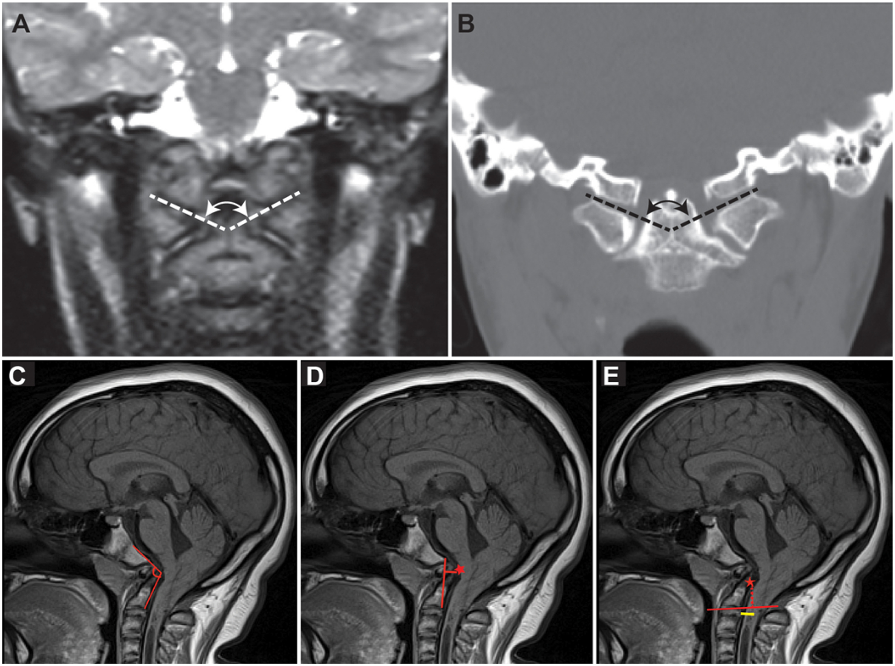
Methods: The authors analyzed the prevalence of condylar hypoplasia and atlas arch anomalies for patients in the Park-Reeves Syringomyelia Research Consortium database who underwent PFD+OCF. Condylar hypoplasia was defined by an atlanto-occipital joint axis angle (AOJAA) ≥ 130°. Atlas assimilation and arch anomalies were identified on presurgical radiographic imaging. This PFD+OCF cohort was compared with a control cohort of patients who underwent PFD alone. The control group was matched to the PFD+OCF cohort according to age, sex, and duration of symptoms at a 2:1 ratio.
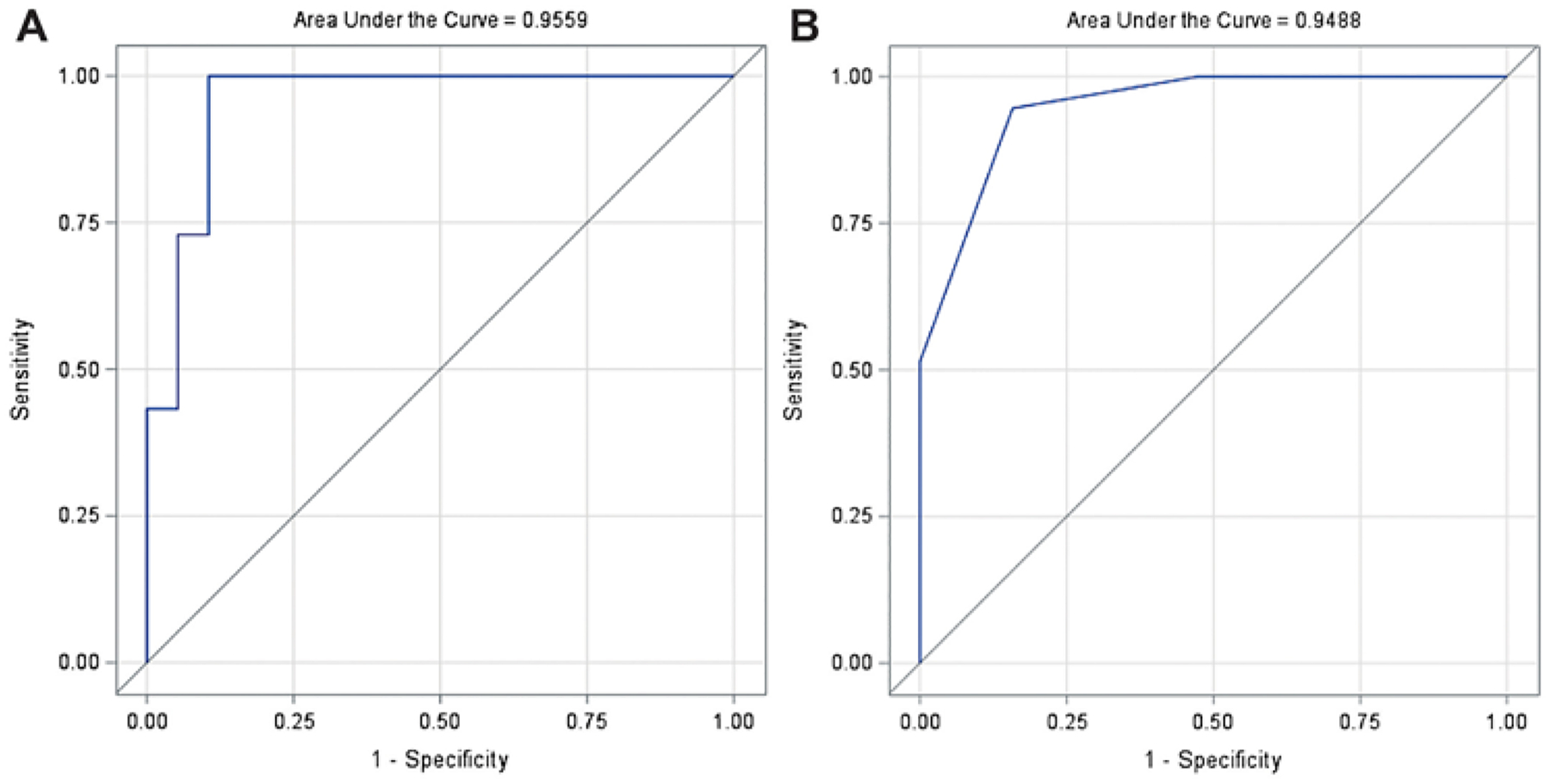
Results: Clinical features and radiographic atlanto-occipital joint parameters were compared between 19 patients in the PFD+OCF cohort and 38 patients in the PFD-only cohort. Demographic data were not significantly different between cohorts (p > 0.05). The mean AOJAA was significantly higher in the PFD+OCF group than in the PFD group (144° ± 12° vs 127° ± 6°, p < 0.0001). In the PFD+OCF group, atlas assimilation and atlas arch anomalies were identified in 10 (53%) and 5 (26%) patients, respectively. These anomalies were absent (n = 0) in the PFD group (p < 0.001). Multivariate regression analysis identified the following 3 CVJ radiographic variables that were predictive of OCF occurrence after PFD: AOJAA ≥ 130° (p = 0.01), clivoaxial angle < 125° (p = 0.02), and occipital condyle-C2 sagittal vertical alignment (C-C2SVA) ≥ 5 mm (p = 0.01). A predictive model based on these 3 factors accurately predicted OCF following PFD (C-statistic 0.95).
Conclusions: The authors' results indicate that the occipital condyle-atlas joint complex might affect the biomechanical integrity of the CVJ in children with CM-I and syringomyelia. They describe the role of the AOJAA metric as an independent predictive factor for occurrence of OCF following PFD. Preoperative identification of these skeletal abnormalities may be used to guide surgical planning and treatment of patients with complex CM-I and coexistent osseous pathology.
Keywords: Chiari malformation; atlas assimilation; condylar hypoplasia; congenital; occipitocervical fusion; syringomyelia.
Conflict of interest statement
Disclosures
The authors report no conflict of interest concerning the materials or methods used in this study or the findings specified in this paper.
Figures
References
-
- Menezes AH. Craniovertebral junction database analysis: incidence, classification, presentation, and treatment algorithms. Childs Nerv Syst. 2008; 24(10): 1101–1108. - PubMed
-
- Menezes AH, Fenoy KA. Remnants of occipital vertebrae: proatlas segmentation abnormalities. Neurosurgery. 2009; 64(5): 945–954. - PubMed
-
- Strahle JM, Taiwo R, Averill C, et al. Radiological and clinical predictors of scoliosis in patients with Chiari malformation type I and spinal cord syrinx from the Park-Reeves Syringomyelia Research Consortium. J Neurosurg Pediatr. 2019; 24(5): 520–527. - PubMed
-
- Akbari SHA, Rizvi AA, CreveCoeur TS, et al. Socioeconomic and demographic factors in the diagnosis and treatment of Chiari malformation type I and syringomyelia. J Neurosurg Pediatr. 2021; 29(3): 288–297. - PubMed
-
- Hadley MN, Zabramski JM, Browner CM, Rekate H, Sonntag VK. Pediatric spinal trauma. Review of 122 cases of spinal cord and vertebral column injuries. J Neurosurg. 1988; 68(1): 18–24. - PubMed
MeSH terms
Supplementary concepts
Grants and funding
LinkOut - more resources
Full Text Sources
Medical
Miscellaneous
