. 2024 Jun 6;32(6):706-714.e3.
doi: 10.1016/j.str.2024.03.005. Epub 2024 Apr 4.

MLL4 binds TET3

Affiliations

MLL4 binds TET3

Dustin C Becht et al. Structure. .

Abstract

Human mixed lineage leukemia 4 (MLL4), also known as KMT2D, regulates cell type specific transcriptional programs through enhancer activation. Along with the catalytic methyltransferase domain, MLL4 contains seven less characterized plant homeodomain (PHD) fingers. Here, we report that the sixth PHD finger of MLL4 (MLL4PHD6) binds to the hydrophobic motif of ten-eleven translocation 3 (TET3), a dioxygenase that converts methylated cytosine into oxidized derivatives. The solution NMR structure of the TET3-MLL4PHD6 complex and binding assays show that, like histone H4 tail, TET3 occupies the hydrophobic site of MLL4PHD6, and that this interaction is conserved in the seventh PHD finger of homologous MLL3 (MLL3PHD7). Analysis of genomic localization of endogenous MLL4 and ectopically expressed TET3 in mouse embryonic stem cells reveals a high degree overlap on active enhancers and suggests a potential functional relationship of MLL4 and TET3.

Keywords: MLL4; PHD finger; TET3; interaction; structure.


Conflict of interest statement

Declaration of interests The authors declare no competing interests.

Figures

Figure 1.
Figure 1.. MLL4PHD456 associates with TET3.
(a) MLL4 domain architecture. The triple PHD finger cassette MLL4PHD456, consisting of PHD4, PHD5 and PHD6, is denoted. (b, c) Schematic of the human proteome peptide-phage display library (b) used to identify the TET3 sequence (c) as a binding partner of MLL4PHD456. Intrinsically disordered regions are indicated by rectangles. (d) TET3 domain architecture. The DNA-binding CxxC domain, the Cys-rich domain (CD), the catalytic DSBH (double-stranded beta helix) domain and LCI (low complexity insert) are shown. (e) Binding affinity of MLL4PHD456 to TET3-FAM peptide as measured by MST. Kd represents the average of four independent measurements ± SEM. Point errors represent SEM. (f, g) Superimposed 1H,15N HSQC spectra of MLL4PHD456 collected upon titration with TET31451–1471 (f) and H4K16ac (g) peptides. Spectra are color coded according to the protein:peptide molar ratio. Data in (g) are taken from .
Figure 2.
Figure 2.. TET3 is a target of MLL4PHD6.
(a) Overlay of 1H,15N HSQC spectra of MLL4PHD6 in the presence of increasing amounts of TET31451–1471 peptide. Spectra are color coded according to the protein:peptide molar ratio. (b) Binding affinity of MLL4PHD6 to TET3-FAM peptide was measured by MST. Kd represents the average of three independent measurements ± SEM. Point errors represent SEM. (c) Superimposed 1H,15N HSQC spectra of MLL3PHD7 collected upon titration with the TET31451–1471 peptide. Spectra are color coded according to the protein:peptide molar ratio. (d) Binding affinity of MLL3PHD7 to TET3-FAM peptide was measured by MST. Kd represents the average of three independent measurements ± SEM. Point errors represent SEM. (e) Superimposed 1H,15N HSQC spectra of MLL3PHD4 collected upon titration with the TET31451–1471 peptide. Spectra are color coded according to the protein:peptide molar ratio.
Figure 3.
Figure 3.. Molecular mechanism for the recognition of TET3 by MLL4PHD6.
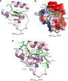
(a) A ribbon diagram of the solution NMR structure of MLL4PHD6 (pink) in complex with TET3 (green). Grey circles are zinc ions. (b) Electrostatic surface potential of MLL4PHD6 is shown with blue and red colors representing surface positive and negative charges, respectively. TET3 is depicted as green sticks. TET3 residues and residues in the negatively charged patches of MLL4PHD6 are labeled. (c) The structure of the MLL4PHD6-TET3 complex. The TET3 residues are shown as green sticks and labeled. The residues of MLL4PHD6 involved in the interaction are shown as pink sticks and labeled. Dashed lines indicate short distances.
Figure 4.
Figure 4.. Contribution of the TET3 residues.
(a) Superimposed 1H,15N HSQC spectra of wild type MLL4PHD6 collected upon titration with the indicated mutated TET3 peptides. Spectra are color coded according to the protein:peptide molar ratio. (b) Amino acid sequence of TET31451–1471 with residues mutated in various cancers highlighted in dark orange and labeled. (c) Structural overlay of MLL4PHD6 (pink) in complex with TET3 (green), MLL4PHD6 (wheat) in complex with H4K16ac peptide (orange) (PDB ID 6O7G) and MLL3PHD7 (grey) in complex with unmodified H4 peptide (yellow) (PDB ID 6MLC). (d) Overlay of the AlphaFold2 predicted model of the MLL4PHD456 structure (PHD4, PHD5, zinc knuckle and PHD6 are colored wheat, cyan, light blue and gray, respectively) with the structure of MLL4PHD6 (pink) in complex with TET3 (green) and with the structure of MLL4PHD6 in complex with H4K16ac peptide (orange) (PDB 6O7G). Only H4K16ac peptide from the structure of the MLL4PHD6-H4K16ac complex is shown for clarity.
Figure 5.
Figure 5.. Ectopic TET3 and endogenous MLL4 colocalize on active enhancers in mouse embryonic stem cells.
(a) Expression of TET3 during neuronal differentiation of ESCs. RNA-Seq data were sourced from GSE154475 . FPKM indicates gene expression levels. (b-d) Genomic colocalization of ectopically expressed TET3 and endogenous MLL4 in mouse embryonic stem cells (ESCs). TET3 ChIP-seq data was sourced from GSE94688 and MLL4, H3K4me1, and H3K27ac ChIP-seq data, as well as ATAC-seq data, were sourced from GSE154475 . (b) Proportional representation of the total genomic regions bound by TET3 and MLL4. (c) Genomic distribution of TET3 binding sites. Promoter was defined as the transcription start site ± 1kb. (d) Heat maps illustrating genomic binding patterns of TET3 and MLL4, accompanied by H3K4me1 enrichment profiles centered on TET3 binding regions. Enhancers were designated as promoter-distal regions with H3K4me1 marks, while promoter-distal regions lacking H3K4me1 were classified as other regions. (e-g) Colocalization of TET3 and MLL4 on ESC enhancers. (e) Relative representation of TET3 and MLL4 binding on enhancers. (f) Motif analysis performed on enhancers bound by MLL4+ or MLL4 TET3. (g) Gene ontology (GO) analysis of enhancers bound by MLL4+ or MLL4 TET3. (h-k) Colocalization of TET3 and MLL4 on active enhancers in ESCs. (h) Proportional depiction of TET3 and MLL4 binding on active enhancers, characterized by H3K27ac marks. (i) Heat maps displaying genomic profiles around the center of TET3-bound active enhancers. (j) Box plots illustrating normalized read counts on MLL4+ or MLL4− TET3-bound active enhancers, excluding outliers. Statistical significance was assessed using the Wilcoxon rank sum test. (k) ChIP-seq profiles of TET3, MLL4, H3K4me1, and H3K27ac are shown on the Nanog and Oct4 loci. Active enhancers colocalized by TET3 and MLL4 are highlighted in shaded regions.

References

    1. Schindler U, Beckmann H, Cashmore AR, and Schindler TF (1993). HAT3.1, a novel Arabidopsis homeodomain protein containing a conserved cysteine-rich region. Plant Journal 4, 137–150. - PubMed
    1. Li H, Ilin S, Wang W, Duncan EM, Wysocka J, Allis CD, and Patel DJ (2006). Molecular basis for site-specific read-out of histone H3K4me3 by the BPTF PHD finger of NURF. Nature 442, 91–95. - PMC - PubMed
    1. Peña PV, Davrazou F, Shi X, Walter KL, Verkhusha VV, Gozani O, Zhao R, and Kutateladze TG (2006). Molecular mechanism of histone H3K4me3 recognition by plant homeodomain of ING2. Nature 442, 100–103. - PMC - PubMed
    1. Shi X, Hong T, Walter KL, Ewalt M, Michishita E, Hung T, Carney D, Pena P, Lan F, Kaadige MR, et al. (2006). ING2 PHD domain links histone H3 lysine 4 methylation to active gene repression. Nature 442, 96–99. - PMC - PubMed
    1. Wysocka J, Swigut T, Xiao H, Milne TA, Kwon SY, Landry J, Kauer M, Tackett AJ, Chait BT, Badenhorst P, et al. (2006). A PHD finger of NURF couples histone H3 lysine 4 trimethylation with chromatin remodelling. Nature 442, 86–90. - PubMed

Publication types

MeSH terms

LinkOut - more resources