. 2024 May;23(5):100760.
doi: 10.1016/j.mcpro.2024.100760. Epub 2024 Apr 3.

The One Hour Human Proteome

Affiliations

The One Hour Human Proteome

Lia R Serrano et al. Mol Cell Proteomics. 2024 May.

Abstract

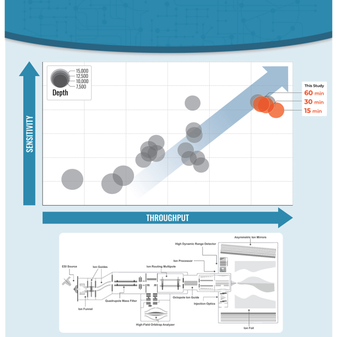
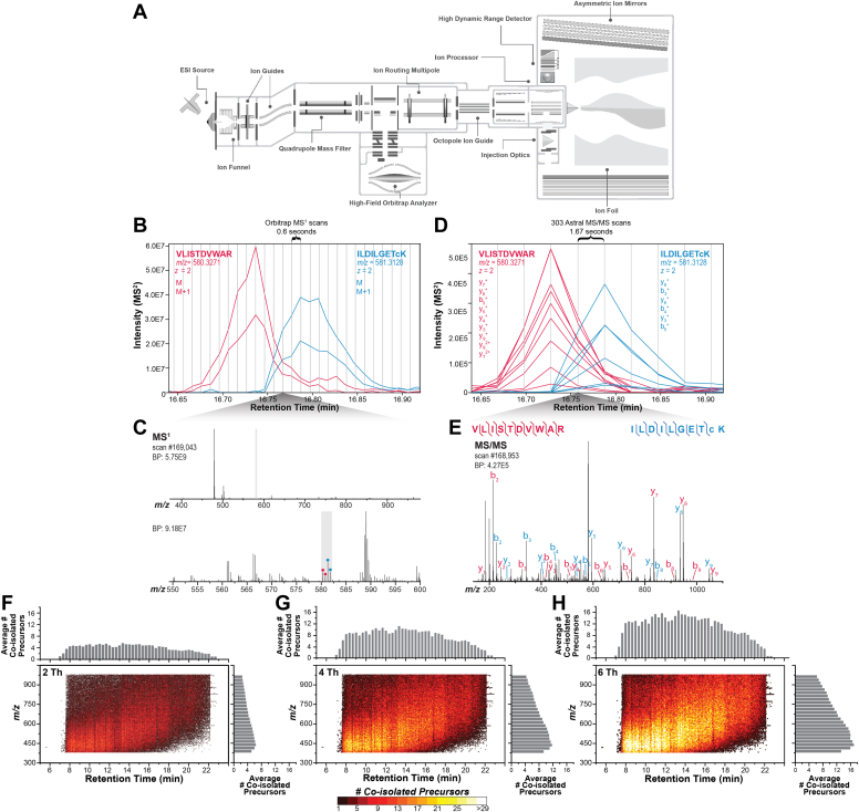
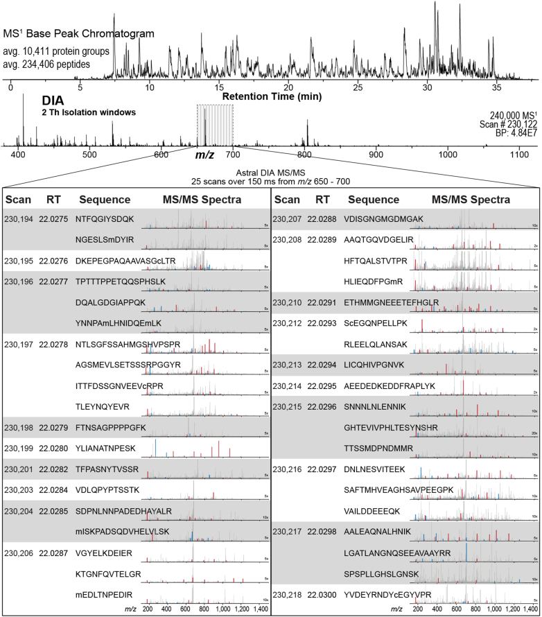
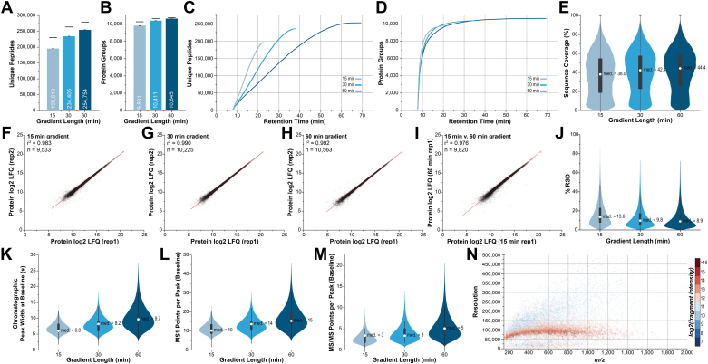
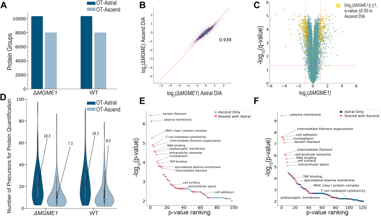
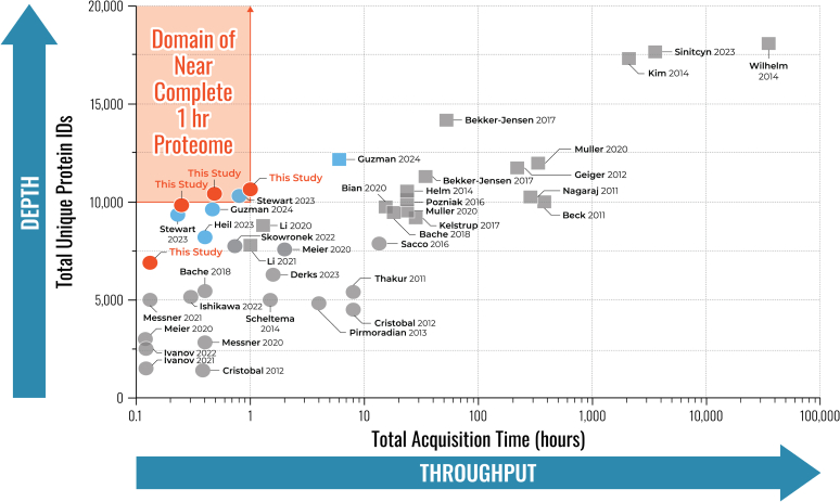
We describe deep analysis of the human proteome in less than 1 h. We achieve this expedited proteome characterization by leveraging state-of-the-art sample preparation, chromatographic separations, and data analysis tools, and by using the new Orbitrap Astral mass spectrometer equipped with a quadrupole mass filter, a high-field Orbitrap mass analyzer, and an asymmetric track lossless (Astral) mass analyzer. The system offers high tandem mass spectrometry acquisition speed of 200 Hz and detects hundreds of peptide sequences per second within data-independent acquisition or data-dependent acquisition modes of operation. The fast-switching capabilities of the new quadrupole complement the sensitivity and fast ion scanning of the Astral analyzer to enable narrow-bin data-independent analysis methods. Over a 30-min active chromatographic method consuming a total analysis time of 56 min, the Q-Orbitrap-Astral hybrid MS collects an average of 4319 MS1 scans and 438,062 tandem mass spectrometry scans per run, producing 235,916 peptide sequences (1% false discovery rate). On average, each 30-min analysis achieved detection of 10,411 protein groups (1% false discovery rate). We conclude, with these results and alongside other recent reports, that the 1-h human proteome is within reach.

Keywords: CRISPR; data independent acquisition; high throughput; instrumentation; mass spectrometry; proteome; proteomics; single shot; technology development.


Conflict of interest statement

Conflict of interest The authors declare the following competing financial interest(s): J. J. C. is a consultant for Thermo Fisher Scientific, Seer, and 908 Devices. T. N. A., E. D., A. P., M. Z., D. H., H. S., C. H., A. M., and V. Z. are employees of Thermo Fisher Scientific.

Figures

None
Graphical abstract
Fig. 1
Fig. 1
Instrument schematic and explanation of narrow-bin DIA methods.A, Orbitrap Astral instrument schematic. B, MS1 extracted ion chromatograms for two peptides of similar m/z which coelute and are coisolated, even with narrow DIA bins during the 15-min method. The monoisotopic and 13C isotope extracted ion chromatograms are shown for both peptides. C, MS1 spectrum at 16.76 min with inset showing two peptides coisolated within 2 Th gray box. D, MS/MS extracted ion chromatograms of product ions of the two coisolated peptides. E, MS/MS spectrum at 16.75 min with annotation of product ions for two identified peptide sequences. Vertical bars indicate frequency of Orbitrap MS1 and MS/MS scans in B and D, respectively. FH, the complexity of DIA MS/MS spectra is greatly reduced using narrow quadrupole isolation. Each dot represents a unique MS/MS scan and color corresponds to the number of PSMs in each scan, as identified by Spectronaut; 4 Th and 6 Th bins are theoretical, generated via in silico widening of bins of the 2 Th data processing. DIA, data-independent acquisition; MS, mass spectrometry; MS/MS, tandem mass spectrometry; PSM, peptide-spectrum match.
Fig. 2
Fig. 2
Example of Q-Orbitrap-Astral data-independent acquisition. Data-independent acquisition with high-resolution Orbitrap MS1 scans collected in parallel with narrow-bin, high-resolution Astral MS/MS scans. At a retention time of 22.02 min, scan #230,122, an Orbitrap full scan, was acquired while hundreds of DIA MS/MS were acquired in the Astral mass analyzer. For the m/z range from 650 to 700, 25 DIA scans were acquired over 150 ms resulting in 38 unique peptide identifications. The b- and y-type fragment ions are denoted in blue and red, respectively. Spectra are shown zoomed relative to the base beak due to limited fragmentation of coisolated, singly charged ions. DIA, data-independent acquisition; MS, mass spectrometry; MS/MS, tandem mass spectrometry.
Fig. 3
Fig. 3
Quantitative reproducibility and protein identification metrics for LC-MS gradients. Average (A) unique peptides and (B) protein groups for each experiment. Black bars denote total unique peptides or protein groups across all three replicates. Cumulative (C) peptides and (D) protein groups and (E) protein sequence coverage distributions for each LC-MS/MS method. Duplicate Q-Orbitrap-Astral data-independent acquisition analyses of HAP1 lysate with (F) 15-min, (G) 30-min, and (H) 60-min gradients and (I) comparison of protein label-free quantitation (LFQ) between 15- and 60-min gradients. J, relative standard deviation (RSD) of LC-MS/MS experiments performed in triplicate. K, distribution of LC peak width, (L) MS1 data points per peak, and (M) MS/MS data points per peak at baseline resolution. N, peak resolution relative to m/z of all b- and y-type product ions detected by the Astral analyzer, within 5-ppm of expected m/z, during the 15-min gradient method. LC-MS/MS, liquid chromatography with tandem mass spectrometry.
Fig. 4
Fig. 4
Comparison of differential expression analyses upon CRISPR-Cas9 MGME1 KO between 30-min Orbitrap Astral and 61-min Orbitrap Ascend DIA methods.A, unique WT and ΔMGME1 KO HAP1 protein groups identified across methods. B, correlation of log2-transformed fold changes measured by OT-Ascend and that measured by OT-Astral have a Pearson correlation coefficient of 0.939 (n = 7304). C, two-sided t tests for each quantified protein present in three KO and three WT replicates (n = 9462) were performed assuming equal variance. Negative log10-transformed q-values were plotted against log2-transformed fold changes between KO and WT for the OT-Astral-quantified proteins (green and yellow). OT-Astral delivered 449 significantly upregulated or downregulated proteins. Subsequently, 234 significantly changed proteins from the OT-Astral dataset were also determined to be significantly upregulated or downregulated by the OT-Ascend method (yellow). D, subset of proteins that pass the significance threshold within OT-Ascend dataset but not the OT-Astral dataset (143 of 380) have a higher median number of precursors that were used for protein quantitation in the Astral dataset. E-F, GO-term enrichment was performed on proteins quantified in all three KO and WT replicates that were determined to have a fold-change of at least two and a q-value <0.05. There were 40 shared significantly enriched (p-value <0.01) GO-terms (red), and the top 11 enriched in each dataset were corroborated in the comparison dataset. DIA, data-independent acquisition; GO, gene ontology.
Fig. 5
Fig. 5
Throughput and depth are shown for several LC-MS/MS-based human proteomics investigations spanning the past decade. The number of unique human proteins identified are plotted against the total LC-MS/MS acquisition time. Squares denote fractionated experiments, and circles denote single-shot experiments. Note, these data report protein groups identified, not necessarily quantified. LC-MS/MS, liquid chromatography with tandem mass spectrometry.

Similar articles

Cited by

References

    1. Liu Y., Beyer A., Aebersold R. On the dependency of cellular protein levels on mRNA abundance. Cell. 2016;165:535–550. - PubMed
    1. Aebersold R., Mann M. Mass spectrometry-based proteomics. Nature. 2003;422:198–207. - PubMed
    1. Han X., Aslanian A., Yates J.R. Mass spectrometry for proteomics. Curr. Opin. Chem. Biol. 2008;12:483–490. - PMC - PubMed
    1. Yates J.R., Ruse C.I., Nakorchevsky A. Proteomics by mass spectrometry: approaches, advances, and applications. Annu. Rev. Biomed. Eng. 2009;11:49–79. - PubMed
    1. Omenn G.S., Lane L., Overall C.M., Pineau C., Packer N.H., Cristea I.M., et al. The 2022 report on the human proteome from the HUPO human proteome project. J. Proteome Res. 2022;22:1024–1042. - PMC - PubMed