Review
. 2024 Apr 25;67(8):5999-6026.
doi: 10.1021/acs.jmedchem.3c02112. Epub 2024 Apr 5.

Channeling Nicotinamide Phosphoribosyltransferase (NAMPT) to Address Life and Death

Affiliations
Review

Channeling Nicotinamide Phosphoribosyltransferase (NAMPT) to Address Life and Death

Ganga Reddy Velma et al. J Med Chem. .

Abstract

Nicotinamide phosphoribosyltransferase (NAMPT) catalyzes the rate-limiting step in NAD+ biosynthesis via salvage of NAM formed from catabolism of NAD+ by proteins with NADase activity (e.g., PARPs, SIRTs, CD38). Depletion of NAD+ in aging, neurodegeneration, and metabolic disorders is addressed by NAD+ supplementation. Conversely, NAMPT inhibitors have been developed for cancer therapy: many discovered by phenotypic screening for cancer cell death have low nanomolar potency in cellular models. No NAMPT inhibitor is yet FDA-approved. The ability of inhibitors to act as NAMPT substrates may be associated with efficacy and toxicity. Some 3-pyridyl inhibitors become 4-pyridyl activators or "NAD+ boosters". NAMPT positive allosteric modulators (N-PAMs) and boosters may increase enzyme activity by relieving substrate/product inhibition. Binding to a "rear channel" extending from the NAMPT active site is key for inhibitors, boosters, and N-PAMs. A deeper understanding may fulfill the potential of NAMPT ligands to regulate cellular life and death.


Conflict of interest statement

The authors declare the following competing financial interest(s): G.R.J.T. is an inventor on patents assigned to the University of Illinois.

Figures

Figure 1
Figure 1
NAD+ biosynthesis, catabolism, and NAMPT enzyme mechanism. (A) The three biosynthetic pathways (kynurenine, Preiss–Handler, salvage) are shown with key enzymes and metabolites. Enzymes responsible for NAD+ catabolism are depicted in blue. See Figure S1 for full chemical structures of NAD+ biosynthesis intermediates and products. (B) Chemical reaction catalyzed by NAMPT converting NAM to NMN. (C) NAMPT inhibitor A-1326133 is a substrate for NAMPT and is converted to a PR adduct, which is an NMN analogue. (D) Structure of the archetypal NAMPT inhibitor FK866. (E) FK866 (magenta) and NAM (orange) bound to NAMPT showing the zones comprising the active site and rear channel (PDB 2GVJ, 8DSI).
Figure 2
Figure 2
Cell killing by FK866 in NAMPT-addicted cancer cells. (A) Correlation of cell death from siNAMPT versus FK866 treatment. (B) Correlation of cell death from siNAPRT versus FK866 treatment. (C) Correlation of NAMPT amplification (copy number NAMPTc#) or expression (NAMPTex) versus cell death showing dependence of FK866 sensitivity on NAMPT copy number (c#). (D) Cell death from FK866 versus siNAMPT (black, vehicle; orange, 10 nM FK866; pink, 50 nM; red, 100 nM; R2 = 0.9). Cell death measured by propidium iodide staining (Z score) data are from Chowdhry et al. and replotted. Each symbol represents a data point for an individual cell line.
Figure 3
Figure 3
NAMPT structural biology. (A) Comparison of the structures of NAPRT (PDB 4YUB) and NAMPT with FK866 (PDB 2GVJ) showing the homodimeric structures with active sites at the interface. The active sites are highlighted by showing active site residues in enhanced colors and docking NAM at the NAPRT active site. Both the depth of the rear channel and ready access to the active site are shown in the NAMPT structure in contrast to NAPRT. (B) H-bonding network (yellow dashes) at the doorsill between the nucleobase pocket and rear channel of NAMPT showing bound NAM (blue) and a network of three to four water molecules (in addition to three conserved water molecules, a fourth is often observed in crystal structures, which does not H-bond with NAM or NAMPT residues). (C) GMX1778 bound to NAMPT showing replacement of the water molecules by direct H-bonding of the ligand with residues in the doorsill (PDB 4O12). (D) Crystal structure of NAMPT active site and rear channel with four different cocrystallized inhibitors superposed to demonstrate the variety of warheads accommodated and the larger variety of cap groups accommodated in the rear channel (PDB 2GVJ, 6AZJ, 4LVA, 4KFN).
Scheme 1
Scheme 1
Scheme 2
Scheme 2
Scheme 3
Scheme 3
Figure 4
Figure 4
NAMPT substrate and substrate/inhibitors. (A) The enzymic conversion of NAM via NMN to NAD+. (B) PR-adduct formation for N-heterocyclic NAMPT inhibitors exemplified by as GNE-617. (C) Cartoon representation of the structural elements of the NAMPT homodimer. (D) The ATPase reaction phosphorylates His-247, leading to PRPP binding and an active site primed for phosphoribosyl transfer. (E) NAM binding leads to product formation. (F) Inhibitor cocrystal structures are of the apoenzyme. (G) Inhibitor binding to the primed active site can lead to phosphoribosylation to give a PR adduct. (H) Several crystal structures of PR adducts bound to the apoenzyme have been reported. (I–N) Clamps extracted from crystal structures, with substrate/inhibitor truncated to the N-heterocycle for clarity: (I) NAM (PDB 8DSD); (J) NMN (PDB 2H3D); (K) PR–GNE-617 (PDB 4L4L); (L) adenine in alternate clamp (PDB 8DSH); PR–SAR154782 (PDB 5LX5) in π-clamp (M) and alternate clamp configurations (N).
Figure 5
Figure 5
Vacor and other pyridyl compounds hijack enzymes binding nicotinamides. (A) Vacor is converted to analogues of NMN, NR, and NAD+ (VMN, VR, and VAD+, respectively), which are biologically active. VMN is claimed to dominate the biological effects of Vacor. (B) VMN is argued to activate SARM1 to catabolize NAD+ to yield cADPR, ADPR, and NAM, leading to axonal degeneration in neurons. (C) Enzymes with NADase or ART activity, such as SARM1 and CD38, are proposed to be hijacked by N-heterocyclic drugs (including NAMPT inhibitors) leading to ADPR adducts that mediate their biological activity. (D). The CD38 inhibitor, 78c, is also a substrate for CD38, which forms an ADPR adduct.
Figure 6
Figure 6
From NAMPT inhibitors to activators. (A) Structures of pyridyl and other N-heterocyclic NAMPT inhibitors and activators showing the evolution from 3-pyridyl inhibitors to non-pyridyl activators. (B) Phenolic activators include validated HTS hits and optimized compounds such as NAT5r with crystallographic evidence for binding to the rear channel (Table S3). (C) Optimized N-PAMs from two different chemical series shown by crystal structures to bind to the rear channel (Table S3).
Figure 7
Figure 7
(A) Superposition of crystal structures showing NAMPT active site with His-BeF3 mimicking phospho-H247, substrates (NAM, PRPP), or products (NMN, PPi) (PDB 3DKL, 3DHF). (B) The high energy phosphoenzyme intermediate breaks down via capture of PO3 by water, PPi, or ATP. (C) After autophosphorylation by ATP to give the phosphoenzyme (E*), the NAMPT enzyme mechanism partitions toward either (green) productive NAM binding and turnover or (red) nonproductive NAM binding. (D) Superposition of NAMPT crystal structures shows the nicotinamide ring H-bonding network with three water molecules in the doorsill (NP-A1 shown in gold; PDB 3DKL, 3DHF, 8DSC, 8DSD, 8DSE). (E) Crystal structure of quercitrin occupying the rear channel in the presence of an ADP analogue bound (PDB 8DSH). (F) The dependence of enzyme activity on [NAM] leads to a bell-shaped profile (the curve depicted is a fit of data at cellular concentrations of ATP and PRPP). The simulation shown, of a 6-fold right shift, leads to increased enzyme activity. (G) NAM, NP-A1, and three waters bound to NAMPT (PDB 8DSC). (H) Superposition of N-PAMs and NAMPT activators in the rear channel (PDB 8F7L, 8DTJ, 8DSC, 8DSD).
Figure 8
Figure 8
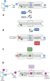
Mechanisms of orthosteric inhibition and allosteric activation. Schematic representation of small molecule regulation of NAMPT activity via binding to the rear channel. (A) High affinity NAM binding occurs after ATP autophosphorylation and PRPP binding leading to productive turnover to NMN. (B) Low affinity NAM binding leads to nonproductive ATP consumption and breakdown of the phosphoenzyme, which is inhibited by N-PAMs. (C) NAMPT inhibitors occupy the rear channel and nucleobase pocket preventing productive NAM binding. (D) NAMPT inhibitors that act as substrates are converted to PR adducts (NMN analogues) that may be further transformed to NAD+ analogues.

References

    1. Luongo T. S.; Eller J. M.; Lu M. J.; Niere M.; Raith F.; Perry C.; Bornstein M. R.; Oliphint P.; Wang L.; McReynolds M. R.; Migaud M. E.; Rabinowitz J. D.; Johnson F. B.; Johnsson K.; Ziegler M.; Cambronne X. A.; Baur J. A. SLC25A51 is a mammalian mitochondrial NAD(+) transporter. Nature 2020, 588, 174–179. 10.1038/s41586-020-2741-7. - DOI - PMC - PubMed
    1. Kory N.; Uit de Bos J.; van der Rijt S.; Jankovic N.; Gura M.; Arp N.; Pena I. A.; Prakash G.; Chan S. H.; Kunchok T.; Lewis C. A.; Sabatini D. M. MCART1/SLC25A51 is required for mitochondrial NAD transport. Sci. Adv. 2020, 6, eabe5310.10.1126/sciadv.abe5310. - DOI - PMC - PubMed
    1. Girardi E.; Agrimi G.; Goldmann U.; Fiume G.; Lindinger S.; Sedlyarov V.; Srndic I.; Gurtl B.; Agerer B.; Kartnig F.; Scarcia P.; Di Noia M. A.; Lineiro E.; Rebsamen M.; Wiedmer T.; Bergthaler A.; Palmieri L.; Superti-Furga G. Epistasis-driven identification of SLC25A51 as a regulator of human mitochondrial NAD import. Nat. Commun. 2020, 11, 6145.10.1038/s41467-020-19871-x. - DOI - PMC - PubMed
    1. Imai S.; Guarente L. NAD+ and sirtuins in aging and disease. Trends Cell Biol. 2014, 24, 464–471. 10.1016/j.tcb.2014.04.002. - DOI - PMC - PubMed
    1. Yoshino J.; Baur J. A.; Imai S. I. NAD(+) intermediates: the biology and therapeutic potential of NMN and NR. Cell Metab. 2018, 27, 513–528. 10.1016/j.cmet.2017.11.002. - DOI - PMC - PubMed

Publication types

MeSH terms