. 2024 Apr 5;12(4):e008174.
doi: 10.1136/jitc-2023-008174.

Combining CRISPR-Cas9 and TCR exchange to generate a safe and efficient cord blood-derived T cell product for pediatric relapsed AML

Affiliations

Combining CRISPR-Cas9 and TCR exchange to generate a safe and efficient cord blood-derived T cell product for pediatric relapsed AML

Vania Lo Presti et al. J Immunother Cancer. .

Abstract

Background: Hematopoietic cell transplantation (HCT) is an effective treatment for pediatric patients with high-risk, refractory, or relapsed acute myeloid leukemia (AML). However, a large proportion of transplanted patients eventually die due to relapse. To improve overall survival, we propose a combined strategy based on cord blood (CB)-HCT with the application of AML-specific T cell receptor (TCR)-engineered T cell therapy derived from the same CB graft.

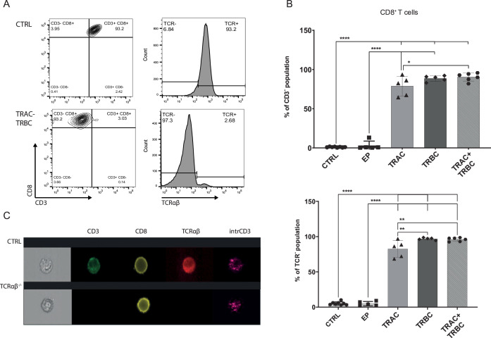
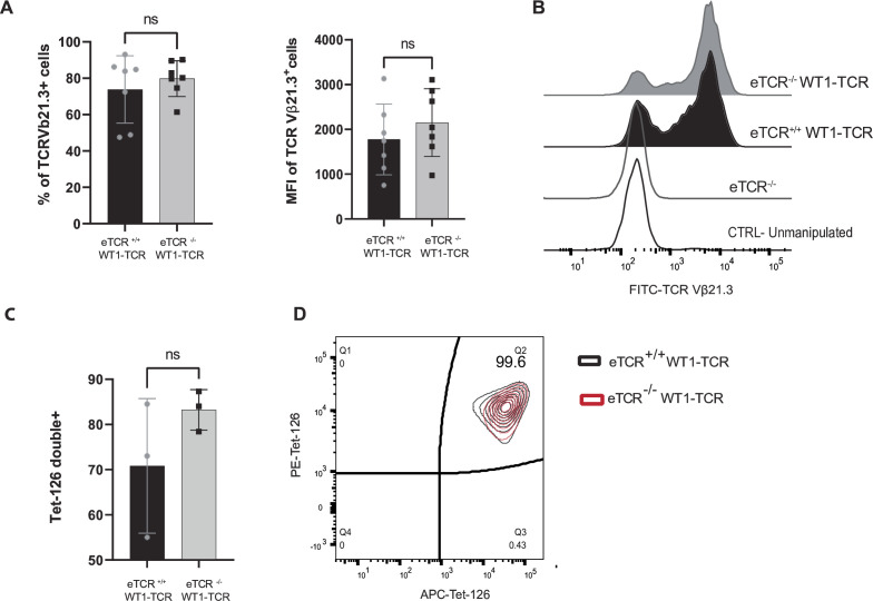
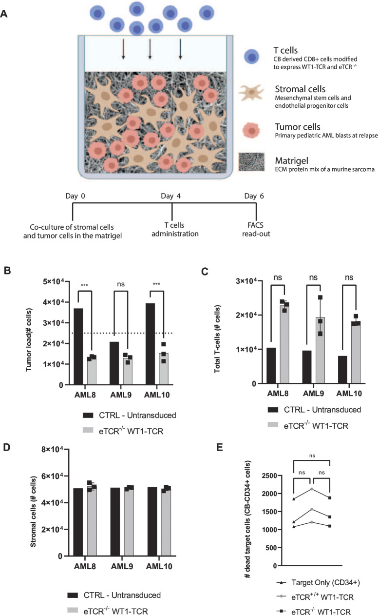
Methods: We produced CB-CD8+ T cells expressing a recombinant TCR (rTCR) against Wilms tumor 1 (WT1) while lacking endogenous TCR (eTCR) expression to avoid mispairing and competition. CRISPR-Cas9 multiplexing was used to target the constant region of the endogenous TCRα (TRAC) and TCRβ (TRBC) chains. Next, an optimized method for lentiviral transduction was used to introduce recombinant WT1-TCR. The cytotoxic and migration capacity of the product was evaluated in coculture assays for both cell lines and primary pediatric AML blasts.

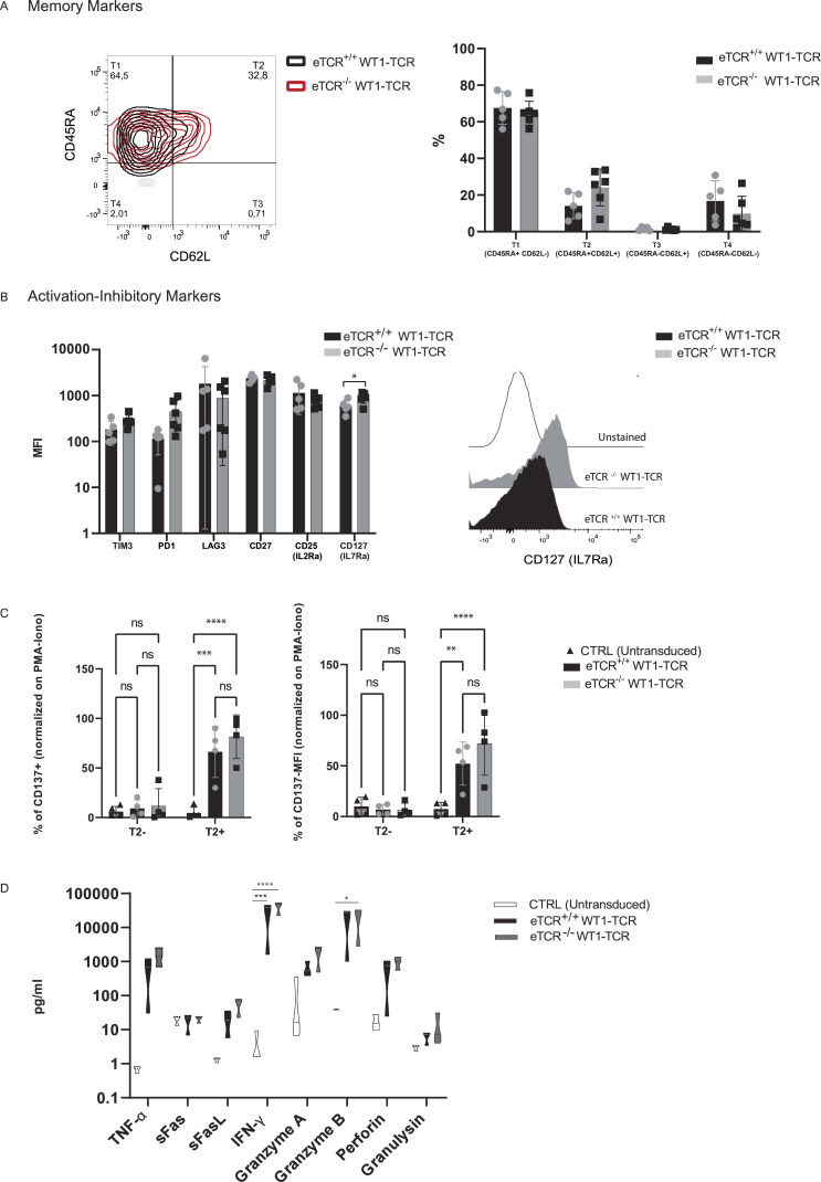
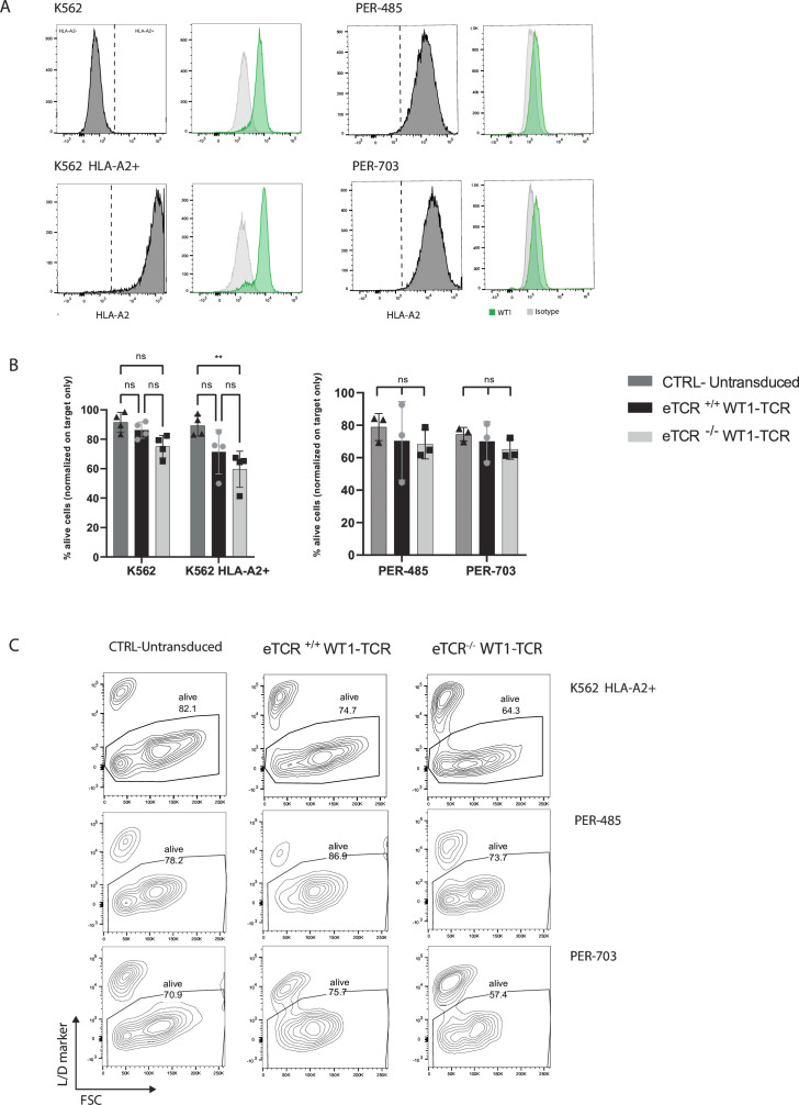
Results: The gene editing and transduction procedures achieved high efficiency, with up to 95% of cells lacking eTCR and over 70% of T cells expressing rWT1-TCR. WT1-TCR-engineered T cells lacking the expression of their eTCR (eTCR-/- WT1-TCR) showed increased cell surface expression of the rTCR and production of cytotoxic cytokines, such as granzyme A and B, perforin, interferon-γ (IFNγ), and tumor necrosis factor-α (TNFα), on antigen recognition when compared with WT1-TCR-engineered T cells still expressing their eTCR (eTCR+/+ WT1-TCR). CRISPR-Cas9 editing did not affect immunophenotypic characteristics or T cell activation and did not induce increased expression of inhibitory molecules. eTCR-/- WT1-TCR CD8+ CB-T cells showed effective migratory and killing capacity in cocultures with neoplastic cell lines and primary AML blasts, but did not show toxicity toward healthy cells.

Conclusions: In summary, we show the feasibility of developing a potent CB-derived CD8+ T cell product targeting WT1, providing an option for post-transplant allogeneic immune cell therapy or as an off-the-shelf product, to prevent relapse and improve the clinical outcome of children with AML.

Keywords: CD8-Positive T-Lymphocytes; Cell Engineering; Hematologic Neoplasms; Pediatrics; T cell Receptor - TCR.


Conflict of interest statement

Competing interests: JK reports grants from Gadeta, Novartis, and Miltenyi Biotec and is the inventor of patents dealing with γδT cell-related aspects, as well as the cofounder and shareholder of Gadeta. ZS is an inventor of patents dealing with γδT cell-related aspects.

Figures

Figure 1
Figure 1
CRISPR-Cas9-mediated TCRαβ gene editing targeting the alpha (TRAC) and beta (TRBC) chains in CB-CD8+ T cells. (A) Representative flow cytometry plot of TCRαβ membrane complexes in expanded and unedited CB-CD8+ T cells (CTRL) and CB-CD8+ transfected with ribonucleoprotein complex targeting the TRAC and TRBC locus separately or simultaneously. Flow cytometry analysis was performed 4 days after electroporation. (B)Percentage of CD3-negative population (top panel) and TCRαβ-negative population (bottom panel) comparing single targeting of TRAC (n=5), TRBC (n=5), and simultaneous TRAC+TRBC (n=6) with CTRL cells. (C) Representative images of single cells using ImageStream. CTRL is shown in the top panel and gene-edited CB-CD8+ T cells with both TRAC+TRBC gRNAs in the bottom panel. Data points refer to biological replicates and independent CB samples. Data are shown as mean±SD; *p≤0.05, ***p≤0.001, ****p≤0.0001. CB, cord blood; TCR, T cell receptor; CTRL, unedited CB-CD8+ T cells; EP, sham electroporeted .
Figure 2
Figure 2
Transduction efficiency and expression of recombinant TCR after lentiviral transduction in eTCR+/+ and eTCR−/− CB-CD8+ T cells. (A) Percentage and median fluorescence intensity of TCRVb21.3+ cells determined by flow cytometry 6 days after lentiviral transduction of eTCR+/+ (black) and eTCR−/− (gray) CB-CD8+ T cells. (B) Representative comparative flow cytometry plot of TCRVb21.3 (WT1-TCR) expression in transduced eTCR+/+ (black; n=7) and eTCR−/− (gray; n=7) CB-CD8+ T cells compared with untransduced eTCR+/+ and eTCR−/− CB-CD8+ T cells (white lines). (C) Tetramer reactivity of eTCR+/+ WT1-TCR (black; n=3) and eTCR−/− WT1-TCR (gray; n=3) CB-CD8+ T cells. (D) Representative flow cytometry plot of tetramer binding in eTCR+/+ WT1-TCR (black; n=3) and eTCR−/− WT1-TCR (red; n=3) CB-CD8+ T cells. Data points refer to biological replicates and independent CB samples. Data are shown as mean±SD. CB, cord blood; CTRL, control, eTCR, endogenous T cell receptor; FITC, fluorescein isothiocyanate, MFI, mean fluorescent intensity; TCR, T cell receptor; WT1, Wilms tumor 1.
Figure 3
Figure 3
Immune phenotype and activation capacity of eTCR+/+ WT1-TCR and eTCR−/− WT1-TCR. (A) The left panel shows a representative flow cytometry plot of CD45RA and CD62L expression in eTCR+/+ WT1-TCR (black) and eTCR−/− WT1-TCR (red); unstained cells are depicted in light gray. The right panel shows the percentage of cells characterized by several maturation markers in eTCR+/+ WT1-TCR (black; n=5) and eTCR−/− WT1-TCR (gray; n=5) CB-CD8+ T cells. Subpopulations were defined as T1 (CD45RA+, CD62L), T2 (CD45+, CD62L+), T3 (CD45RA, CD62L+), and T4 (CD45RA, CD62L). (B) The left panel shows the expression of multiple receptors on the surface of eTCR+/+ WT1-TCR (black) and eTCR−/− WT1-TCR (gray) calculated as MFI. The right panel shows a representative flow cytometry plot of CD127(IL7RA) expression in eTCR+/+ WT1-TCR (black) and eTCR−/− WT1-TCR (grey). Data points refer to biological replicates; >5 independent CB samples (n >5) . Data are shown as mean±SD. (C) Percentage (left panel) and MFI (right panel) of CD137+ T cells in untransduced CTRL (black triangle), eTCR+/+ WT1-TCR (black), and eTCR−/− WT1-TCR (gray) after 24 hours of coculture with T2 cells (T2−) or T2 cells loaded with the specific WT1 peptide (T2+). (D) Cytokine production after 24 hours of coculture with T2+ cells of CTRL-untransduced (white), eTCR+/+ WT1-TCR (black), and eTCR−/− WT1-TCR (gray) CB-CD8+ T cells. Data points refer to biological replicates; four independent CB samples. Data are shown as mean±SD; *p≤0.05, **p≤0.01, ***p≤0.001, ****p≤0.0001. CB, cord blood; CTRL, control, eTCR, endogenous T cell receptor; IL7RA, interleukin-7 receptor alpha, MFI, mean fluorescent intensity; PMA, phorbol myristate acetate; sFas, soluble Fas; sFasL, soluble Fas Ligand; TCR, T cell receptor; TNF⍺, tumor necrosis factor-⍺; IFNɣ, interferon-ɣ; WT1, Wilms tumor 1.
Figure 4
Figure 4
Cytotoxic capacity of eTCR+/+ WT1-TCR and eTCR−/− WT1-TCR. (A) HLA-A2 expression (gray) and WT1 intracellular expression (green) in target tumor cell lines (K562, K562 HLA-A2+, PER-485, PER-703). (B) Percentage of viable target cells after 16 hours of coculture with CTRL -untransduced (dark gray), eTCR+/+ WT1-TCR (black), and eTCR−/− WT1-TCR (gray). The percentage of viable target cells in coculture was normalized to the percentage of viable target cells in standard culture without the presence of CB-CD8+ T cells. (C) Representative flow cytometry plot depicts target cell viability after 16 hours of coculture with CB-CD8+ T cells. Data points refer to biological replicates; four independent CB samples for K562 and K562 HLA-A2+, and three independent CB samples for PER-485 and PER-703. Data are shown as mean±SD; **p≤0.01. CB, cord blood; CTRL, control; eTCR, endogenous T cell receptor; TCR, T cell receptor; WT1, Wilms tumor 1.
Figure 5
Figure 5
Cytotoxic capacity of eTCR−/− WT1-TCR in a 3D bone marrow niche model of pediatric AML. (A) Representative illustration of the main components of the 3D model and timeline of cell administration. (B) Cytotoxic capacity of eTCR−/− WT1-TCR (gray) and CTRL-untransduced (black) CB-CD8+ T cells after 4 days of coculture in the 3D model, based on the number of primary AML cells left in the model. Cell number was measured via flow cytometry and compared with the initial number of tumor cells used on day 0 (dotted line). (C) Proliferation capacity on antigen recognition of eTCR−/− WT1-TCR (gray) and untransduced CTRL (black) CB-CD8+ T cells after 4 days of coculture in the 3D model. (D) Viability of stromal cells after 4 days of coculture in the 3D model in the presence of eTCR−/− WT1-TCR (gray) and CTRL untransduced (black) CB-CD8+ T cells. Cell number is measured via flow cytometry. No statistically significant difference was detected. Data are shown as mean±SD from three biological replicates for eTCR−/− WT1-TCR and one for untransduced CTRL cells; ***p≤0.001. (E) Safety profile of eTCR−/− WT1-TCR (black square) and eTCR+/+ WT1-TCR (gray circle) in coculture (16 hours) with CB-derived CD34+ cells (black triangle). Data are shown from three CB-CD34+ donors in coculture with one CB donor for eTCR−/− WT1-TCR and eTCR+/+ WT1-TCR. 3D, three-dimensional; AML, acute myeloid leukemia; CB, cord blood; CTRL, control, ECM, extracellular matrix, eTCR, endogenous T cell receptor; TCR, T cell receptor; WT1, Wilms tumor 1.

Similar articles

Cited by

References

    1. Rasche M, Zimmermann M, Borschel L, et al. . Successes and challenges in the treatment of pediatric acute myeloid leukemia: a retrospective analysis of the AML-BFM trials from 1987 to 2012. Leukemia 2018;32:2167–77. 10.1038/s41375-018-0071-7 Available: 10.1038/s41375-018-0071-7 - DOI - DOI - PMC - PubMed
    1. Rosenfeld C, Cheever MA, Gaiger A. Wt1 in acute leukemia, chronic myelogenous leukemia and myelodysplastic syndrome: therapeutic potential of Wt1 targeted therapies. Leukemia 2003;17:1301–12. 10.1038/sj.leu.2402988 - DOI - PubMed
    1. Bergmann L, Miething C, Maurer U, et al. . High levels of Wilms' tumor gene (Wt1) mRNA in acute myeloid leukemias are associated with a worse long-term outcome. Blood 1997;90:1217–25. - PubMed
    1. Chapuis AG, Egan DN, Bar M, et al. . T cell receptor gene therapy targeting WT1 prevents acute myeloid leukemia relapse post-transplant. Nat Med 2019;25:1064–72. 10.1038/s41591-019-0472-9 Available: 10.1038/s41591-019-0472-9 - DOI - DOI - PMC - PubMed
    1. Rocha V, Labopin M, Sanz G, et al. . Transplants of umbilical-cord blood or bone marrow from unrelated donors in adults with acute leukemia. N Engl J Med 2004;351:2276–85. 10.1056/NEJMoa041469 - DOI - PubMed

Substances