. 2024 Apr 5;12(4):e008794.
doi: 10.1136/jitc-2024-008794.

Preclinical development of novel PD-L1 tracers and first-in-human study of [68Ga]Ga-NOTA-RW102 in patients with lung cancers

Affiliations

Preclinical development of novel PD-L1 tracers and first-in-human study of [68Ga]Ga-NOTA-RW102 in patients with lung cancers

You Zhang et al. J Immunother Cancer. .

Abstract

Background: The programmed cell death protein-1 (PD-1)/programmed death receptor ligand 1 (PD-L1) axis critically facilitates cancer cells' immune evasion. Antibody therapeutics targeting the PD-1/PD-L1 axis have shown remarkable efficacy in various tumors. Immuno-positron emission tomography (ImmunoPET) imaging of PD-L1 expression may help reshape solid tumors' immunotherapy landscape.

Methods: By immunizing an alpaca with recombinant human PD-L1, three clones of the variable domain of the heavy chain of heavy-chain only antibody (VHH) were screened, and RW102 with high binding affinity was selected for further studies. ABDRW102, a VHH derivative, was further engineered by fusing RW102 with the albumin binder ABD035. Based on the two targeting vectors, four PD-L1-specific tracers ([68Ga]Ga-NOTA-RW102, [68Ga]Ga-NOTA-ABDRW102, [64Cu]Cu-NOTA-ABDRW102, and [89Zr]Zr-DFO-ABDRW102) with different circulation times were developed. The diagnostic efficacies were thoroughly evaluated in preclinical solid tumor models, followed by a first-in-human translational investigation of [68Ga]Ga-NOTA-RW102 in patients with non-small cell lung cancer (NSCLC).

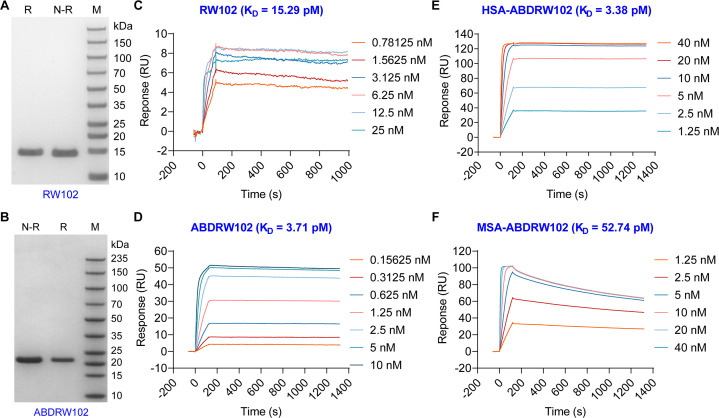
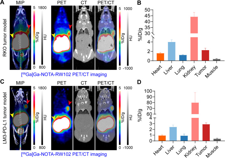
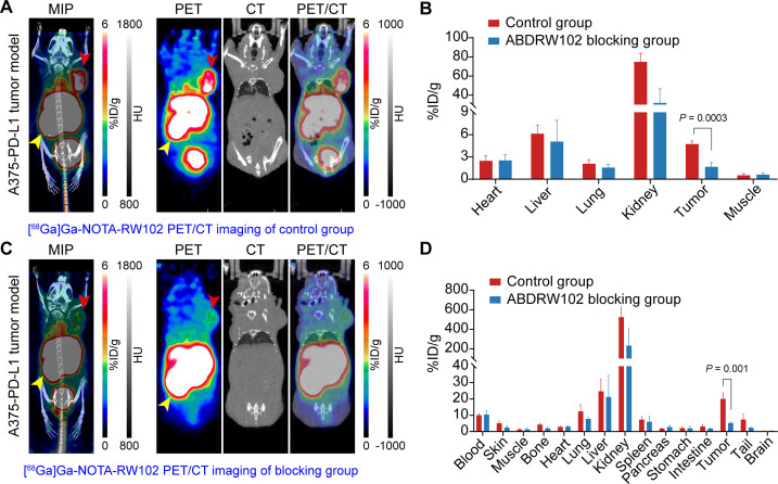
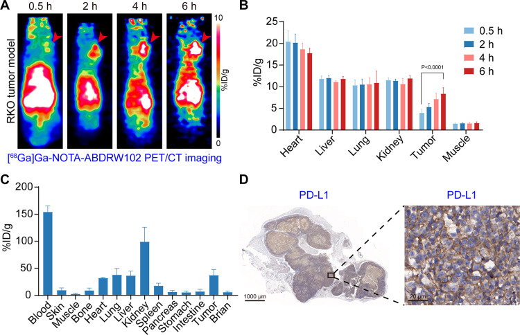
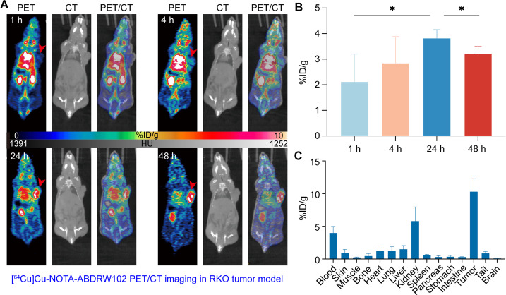
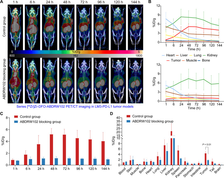
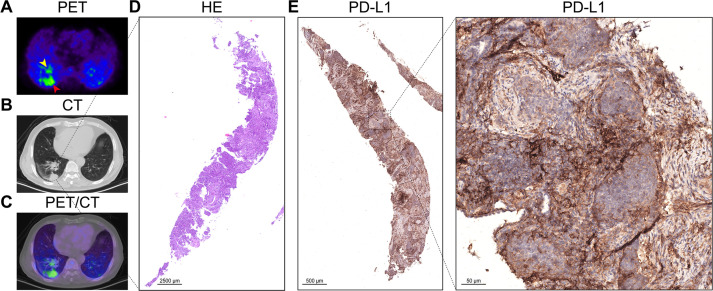
Results: While RW102 has a high binding affinity to PD-L1 with an excellent KD value of 15.29 pM, ABDRW102 simultaneously binds to human PD-L1 and human serum albumin with an excellent KD value of 3.71 pM and 3.38 pM, respectively. Radiotracers derived from RW102 and ABDRW102 have different in vivo circulation times. In preclinical studies, [68Ga]Ga-NOTA-RW102 immunoPET imaging allowed same-day annotation of differential PD-L1 expression with specificity, while [64Cu]Cu-NOTA-ABDRW102 and [89Zr]Zr-DFO-ABDRW102 enabled longitudinal visualization of PD-L1. More importantly, a pilot clinical trial shows the safety and diagnostic value of [68Ga]Ga-NOTA-RW102 immunoPET imaging in patients with NSCLCs and its potential to predict immune-related adverse effects following PD-L1-targeted immunotherapies.

Conclusions: We developed and validated a series of PD-L1-targeted tracers. Initial preclinical and clinical evidence indicates that immunoPET imaging with [68Ga]Ga-NOTA-RW102 holds promise in visualizing differential PD-L1 expression, selecting patients for PD-L1-targeted immunotherapies, and monitoring immune-related adverse effects in patients receiving PD-L1-targeted treatments.

Trial registration number: NCT06165874.

Keywords: Biomarker; Lung Cancer; Nuclear medicine; fMRI / PET.


Conflict of interest statement

Competing interests: WW, YZ and JL are inventors of a pending patent describing the reported PD-L1 tracers. WW is a consultant of Alpha Nuclide (Ningbo) Medical Technology Co., Ltd. WC declares conflict of interest with the following corporations: Actithera, Inc., Portrai, Inc., rTR Technovation Corporation, Four Health Global Pharmaceuticals Inc., and POP Biotechnologies, Inc.

Figures

Figure 1
Figure 1
Characterization of RW102 and ABDRW102. The purity of RW102 (A) and ABDRW102 (B) examined by sulfate-polyacrylamide gel electrophoresis was >95%. (C and D) The surface plasmon resonance studies showed the association and dissociation kinetics of RW102 (C) and ABDRW102 (D) interacting with recombinant human programmed death receptor ligand 1 protein. (E and F) ABDRW102 also showed high binding affinities to human serum albumin (HSA, E) and murine serum albumin (MSA, F). M, marker; N-R, non-reducing condition; R, reducing condition.
Figure 2
Figure 2
[68Ga]Ga-NOTA-RW102 immunoPET imaging in subcutaneous tumor models. (A and C) [68Ga]Ga-NOTA-RW102 immunoPET imaging in subcutaneous RKO (A, 7.64±1.42 MBq, n=3) and LM3-PD-L1 (C, 4.35±0.47 MBq, n=4) models 0.5 hours post-injection of the tracer. Maximum intensity projection (MIP) and coronal images showed clear delineation of the tumors (red arrowheads) and kidneys (yellow arrowheads), from which the tracer was excreted. (B and D) Quantitative data showed the tracer uptake and distribution patterns in RKO (B) and LM3-PD-L1 (D) models. immunoPET, immuno-positron emission tomography; PD-L1, programmed death receptor ligand 1.
Figure 3
Figure 3
[68Ga]Ga-NOTA-RW102 immunoPET imaging in A375-PD-L1 tumor models without or with ABDRW102 blocking. (A and C) [68Ga]Ga-NOTA-RW102 immunoPET imaging in the control group (A) and ABDRW102 blocking group (C). (B) Quantitative regions of interest analysis results in the control group and ABDRW102 blocking group. (D) Ex vivo biodistribution data showed the detailed distribution and uptake patterns of [68Ga]Ga-NOTA-RW102 in the control group and ABDRW102 blocking group. immunoPET, immuno-positron emission tomography; PD-L1, programmed death receptor ligand 1.
Figure 4
Figure 4
Serial [68Ga]Ga-NOTA-ABDRW102 immunoPET imaging in RKO cancer models (5.87±0.27 MBq, n=6). (A) Representative PET images of [68Ga]Ga-NOTA-ABDRW102 at multiple time points. The images showed clear delineation of the tumors (red arrowheads). (B) Regions of interest data of [68Ga]Ga-NOTA-ABDRW102 immunoPET imaging at different time points. (C) Ex vivo biodistribution data showing the detailed uptake and distribution of [68Ga]Ga-NOTA-ABDRW102 in the tumor, blood, major organs, and tissues. (D) Immunohistochemical verification of PD-L1 expression in tumors resected after immunoPET imaging. immunoPET, immuno-positron emission tomography; PD-L1, programmed death receptor ligand 1.
Figure 5
Figure 5
Serial [64Cu]Cu-NOTA-ABDRW102 immunoPET imaging in RKO models. (A) Representative PET images of [64Cu]Cu-NOTA-ABDRW102 at multiple time points. (B) Regions of interest data of [64Cu]Cu-NOTA-ABDRW102 immunoPET imaging in tumors at different time points. (C) Ex vivo biodistribution data showed the detailed uptake and distribution of [64Cu]Cu-NOTA-ABDRW102 in the tumor, blood, major organs, and tissues. immunoPET, immuno-positron emission tomography.
Figure 6
Figure 6
[89Zr]Zr-DFO-ABDRW102 immuno-positron emission tomography imaging in LM3-PD-L1 models (2.66±0.74 MBq, n=8). (A) Maximum intensity projection PET/CT images of the control group (up panel) and ABDRW102 blocking group (down panel) at multi-time points. (B) Regions of interest data showed the uptake kinetics of [89Zr]Zr-DFO-ABDRW102 in the control group and ABDRW102 blocking group at different time points. (C) Comparison of tumor uptake value between the control and ABDRW102 blocking groups at each time point. (D) Ex vivo biodistribution data showed the detailed uptake and distribution of [89Zr]Zr-DFO-ABDRW102 in the control and ABDRW102 blocking groups. PD-L1, programmed death receptor ligand 1; PET, positron emission tomography.
Figure 7
Figure 7
[68Ga]Ga-NOTA-RW102 immuno-positron emission tomography imaging of an adult patient with squamous lung cancer with 70% PD-L1 expression. (A–C) [68Ga]Ga-NOTA-RW102 PET/CT images of two tumor lesions in this patient (yellow and red arrowheads). (D) The H&E staining of the subpleural lesion in the lower lobe of the patient’s right lung. (E) The PD-L1 staining of the biopsied squamous lung cancer tissue showed 70% positivity. PD-L1, programmed death receptor ligand 1; PET, positron emission tomography.

References

    1. Wei W, Rosenkrans ZT, Liu J, et al. . Immunopet: concept, design, and applications. Chem Rev 2020;120:3787–851. 10.1021/acs.chemrev.9b00738 - DOI - PMC - PubMed
    1. Chakravarty R, Goel S, Cai W. Nanobody: the "magic bullet" for molecular imaging. Theranostics 2014;4:386–98. 10.7150/thno.8006 - DOI - PMC - PubMed
    1. Rashidian M, Ploegh H. Ploegh H: Nanobodies as noninvasive imaging tools. Immunooncol Technol 2020;7:2–14. 10.1016/j.iotech.2020.07.001 - DOI - PMC - PubMed
    1. Wei W, Younis MH, Lan X, et al. . Single-domain antibody theranostics on the horizon. J Nucl Med 2022;63:1475–9. 10.2967/jnumed.122.263907 - DOI - PMC - PubMed
    1. Keyaerts M, Xavier C, Heemskerk J, et al. . Phase I study of 68Ga-HER2-Nanobody for PET/CT assessment of HER2 expression in breast carcinoma. J Nucl Med 2016;57:27–33. 10.2967/jnumed.115.162024 - DOI - PubMed

Publication types

MeSH terms

Substances

Associated data