. 2024 Apr 5;12(4):e007716.
doi: 10.1136/jitc-2023-007716.

Targeting PARG induces tumor cell growth inhibition and antitumor immune response by reducing phosphorylated STAT3 in ovarian cancer

Affiliations

Targeting PARG induces tumor cell growth inhibition and antitumor immune response by reducing phosphorylated STAT3 in ovarian cancer

Antons Martincuks et al. J Immunother Cancer. .

Abstract

Background: Ovarian cancer is the most lethal gynecological malignancy, with limited treatment options after failure of standard therapies. Despite the potential of poly(ADP-ribose) polymerase inhibitors in treating DNA damage response (DDR)-deficient ovarian cancer, the development of resistance and immunosuppression limit their efficacy, necessitating alternative therapeutic strategies. Inhibitors of poly(ADP-ribose) glycohydrolase (PARG) represent a novel class of inhibitors that are currently being assessed in preclinical and clinical studies for cancer treatment.

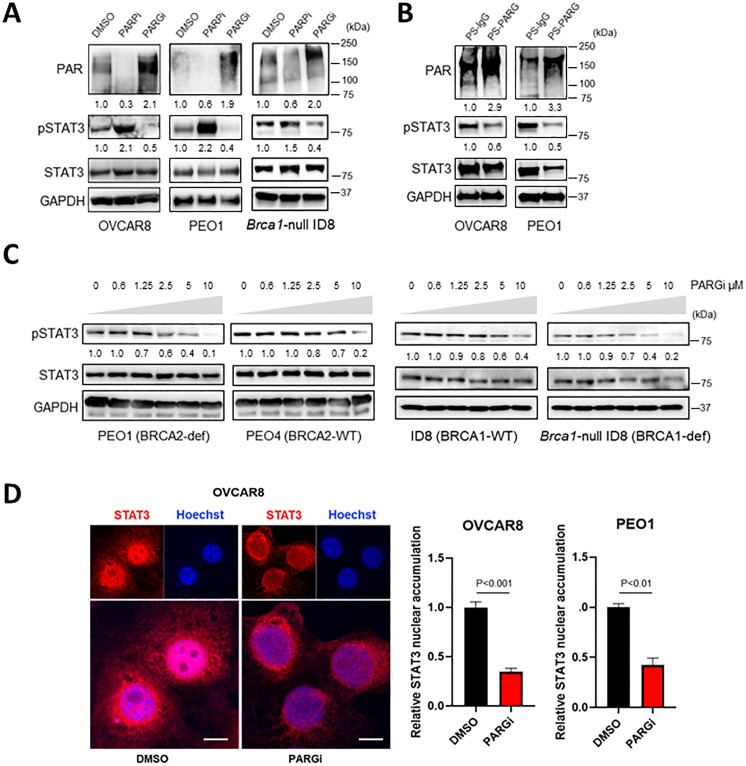
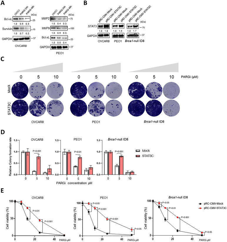
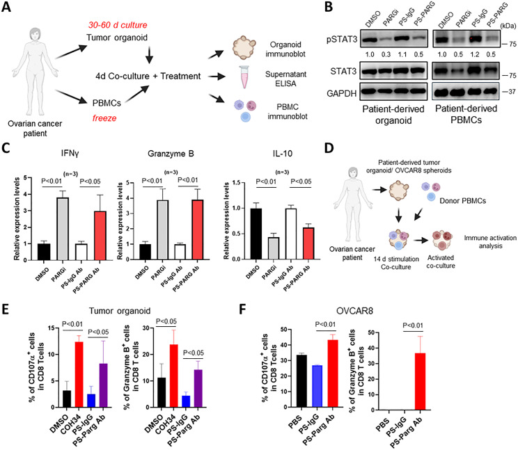
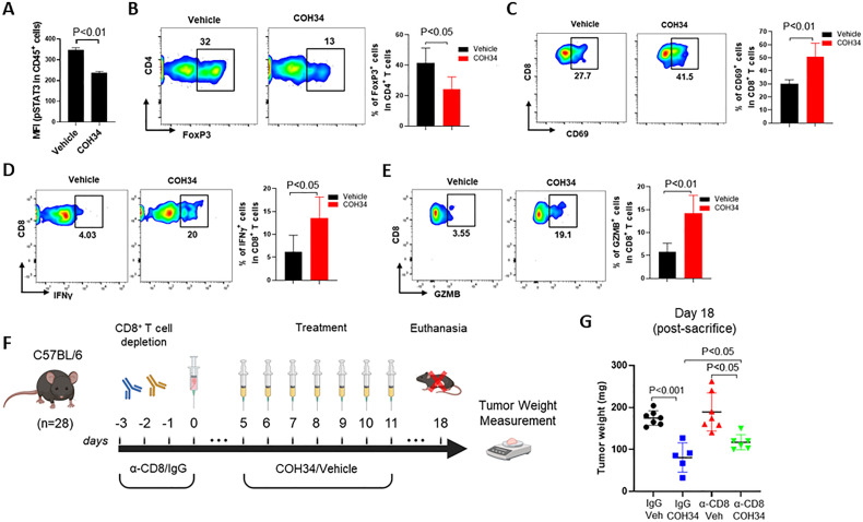
Methods: By using a PARG small-molecule inhibitor, COH34, and a cell-penetrating antibody targeting the PARG's catalytic domain, we investigated the effects of PARG inhibition on signal transducer and activator of transcription 3 (STAT3) in OVCAR8, PEO1, and Brca1-null ID8 ovarian cancer cell lines, as well as in immune cells. We examined PARG inhibition-induced effects on STAT3 phosphorylation, nuclear localization, target gene expression, and antitumor immune responses in vitro, in patient-derived tumor organoids, and in an immunocompetent Brca1-null ID8 ovarian mouse tumor model that mirrors DDR-deficient human high-grade serous ovarian cancer. We also tested the effects of overexpressing a constitutively activated STAT3 mutant on COH34-induced tumor cell growth inhibition.

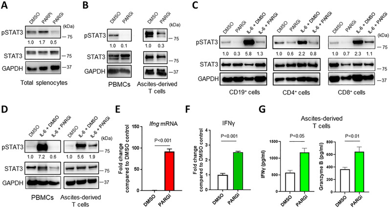
Results: Our findings show that PARG inhibition downregulates STAT3 activity through dephosphorylation in ovarian cancer cells. Importantly, overexpression of a constitutively activated STAT3 mutant in tumor cells attenuates PARG inhibitor-induced growth inhibition. Additionally, PARG inhibition reduces STAT3 phosphorylation in immune cells, leading to the activation of antitumor immune responses, shown in immune cells cocultured with ovarian cancer patient tumor-derived organoids and in immune-competent mice-bearing mouse ovarian tumors.

Conclusions: We have identified a novel antitumor mechanism underlying PARG inhibition beyond its primary antitumor effects through blocking DDR in ovarian cancer. Furthermore, targeting PARG activates antitumor immune responses, thereby potentially increasing response rates to immunotherapy in patients with ovarian cancer.

Keywords: Drug Evaluation, Preclinical; Lymphocyte Activation; Tumor Microenvironment.


Conflict of interest statement

Competing interests: None declared.

Figures

Figure 1
Figure 1
PARG inhibition decreases STAT3 Y705 phosphorylation and signaling. (A) Immunoblot analysis comparing total PARylation, phospho-STAT3 Tyr 705 (pSTAT3), total STAT3 (STAT3), and loading controls in BRCA-proficient OVCAR8, BRCA2-deficient PEO1, and Brca1-null ID8 cells after vehicle (DMSO), 10 µM olaparib (PARPi), or 10 µM COH34 (PARGi) overnight incubation. The image represents three independent experiments. (B) Immunoblotting of the indicated cell lines cultured with the PS-modified control (PS-IgG) or PARG (PS-PARG) antibody (20 µg/mL, overnight). The image represents two independent experiments. (C) Dose-dependent pSTAT3 inhibition by COH34 at indicated doses in BRCA-proficient and BRCA-deficient ovarian cancer cells analyzed by immunoblotting. The images represent two independent experiments. GAPDH was used as the loading control. (D) Immunofluorescent staining and confocal microscopic images (left) showing STAT3 (red) and nuclear staining (blue) of the indicated ovarian cancer cell lines treated with DMSO or 10 µM PARGi overnight. The scale bars represent 10 µm. Histograms (right) showing STAT3 nuclear accumulation. Data shown are mean±SD from three experiments. The protein levels of PAR or pSTAT3 shown in the immunoblotting images were quantified by band intensity using ImageJ software and normalized with GAPDH. DMSO, Dimethyl sulfoxide; GAPDH, Glyceraldehyde 3-phosphate dehydrogenase; PARG, poly(ADP-ribose) glycohydrolase; STAT3, signal transducer and activator of transcription 3.
Figure 2
Figure 2
PARGi-induced tumor cell growth inhibition is partially mediated by blocking STAT3. (A) A representative immunoblot of STAT3 downstream genes and loading controls in ovarian cancer cells treated with 10 µM COH34 at indicated time points. The experiment was performed twice. (B) Immunoblot comparing total STAT3 and loading controls in ovarian cancer cell lines transfected with either control vector (Mock) or constitutively active form of STAT3 (STAT3C). The image represents two independent experiments. (C) Representative colony formation assays of Mock transfected and STAT3C transfected cells from (B) in the presence of PARGi at indicated concentrations for 10 days. Images represent three independent experiments. (D) Relative colony-formation by the indicated Mock and STAT3C transfected ovarian cancer cell lines treated with increasing concentrations of COH34 (0 µM, 5 µM and 10 µM). (E) Cell viability analysis of the Mock transfected and STAT3C transfected ovarian cancer cell lines. Cells were treated with indicated concentrations of COH34 for 3 days (0, 6.25, 12.5, 25, and 50 µM). Error bars represent mean±SD of n=3 independent experiments, two-tailed, unpaired Student’s t-test. The protein levels of Bcl-xL, Survivin, MMP2, MMP9 or total STAT3 shown in the immunoblotting images were quantified by band intensity using ImageJ software and normalized with the levels of GAPDH. PARG, poly(ADP-ribose) glycohydrolase; STAT3, signal transducer and activator of transcription 3.
Figure 3
Figure 3
PARG inhibition (PARGi) decreases basal and IL-6-induced phosphorylated STAT3 (pSTAT3) in immune cells and stimulates immune activation in vitro. (A) Splenocytes from tumor-free mice were isolated and cultured in the presence of vehicle (DMSO), 10 µM olaparib (PARPi), or 10 µM COH34 (PARGi) overnight. The levels of pSTAT3 and total STAT3 were analyzed by immunoblotting. (B) Immunoblot comparing pSTAT3 in healthy donor-derived PBMCs and ovarian cancer patient ascites-derived CD3+ T cells after overnight incubation with either DMSO or 5 µM PARGi. (C) Immunoblotting of pSTAT3 in mouse naïve CD19+ B cells, CD4+ and CD8+ T cells pretreated with vehicle control or PARGi (5 µM, overnight) and stimulated with 20 ng/mL IL-6 for 30 min. (D) Analysis of IL-6-induced pSTAT3 similar to (C) in human immune cells from (B) after overnight pretreatment with either DMSO or 5 µM PARGi. (E) Real-time PCR of Ifng gene expression in mouse splenocytes cultured in the presence or absence of PARGi for 24 hours. Gene expression data are shown after normalization to Actb expression and are presented as the mean-fold induction (mean±SD) relative to unstimulated samples. (F) ELISA measuring IFN-γ levels in the supernatants from cocultures of mouse CD8+ T cells with murine ID8 tumor cells in the presence of vehicle or 10 µM PARGi for 24 hours. Data are presented as the mean-fold induction (mean±SD) relative to vehicle-treated samples. (G) Levels of IFN-γ and granzyme B in the supernatants from ovarian cancer patient ascites-derived CD3+ T cells after treatment with either 5 µM PARGi or DMSO for 24 hours as determined by ELISA. The protein levels of pSTAT3 shown in the immunoblotting images were quantified by band intensity using ImageJ software and normalized with the levels of GAPDH. DMSO, Dimethyl sulfoxide; GAPDH, Glyceraldehyde 3-phosphate dehydrogenase; PARG, poly(ADP-ribose) glycohydrolase; PBMCs, peripheral blood mononuclear cells; STAT3, signal transducer and activator of transcription 3.
Figure 4
Figure 4
PARG inhibition induces immune activation in ovarian tumor organoids. (A) Study design for (B,C) from ovarian cancer patient-derived tumor organoid/PBMC cocultures in the presence of 0.1% DMSO and 10 µM COH34 (96 hours), or 20 µg/mL PS-IgG and PS-PARG antibodies (48 hours). (B) Immunoblotting of phosphorylated STAT3 (pSTAT3) in ovarian cancer PDOs and patient-matched PBMCs after the PDO/PBMC cocultures were treated by DMSO, COH34, PS-IgG or PS-PARG antibodies. Data are representative of three independent experiments. (C) IFN-γ, granzyme B, and IL-10 levels in the PDO/PBMC coculture supernatants as measured by ELISA. Expression data are presented as mean-fold induction (mean±SD) relative to control samples from three different patients. (D) Study design for (E,F). PDOs or OVCAR8 spheroids and healthy donor PBMCs were prepared separately before coculturing. Tumor organoids (E) or OVCAR8 tumor cells (F) were cocultured with healthy donor PBMCs for 72 hours in the presence of 0.1% DMSO, 10 µM COH34, PS-IgG, or PS-PARG antibodies. Single-cell suspensions prepared from the cocultures of tumor organoids or OVCAR8 tumor cells and healthy donor PBMCs were analyzed by flow cytometry for activated T cells (GzmB+ or CD107a+) in CD8+ T cell populations. For tumor organoids, n=4; and for OVCAR8 tumor cells, n=3 (n represents the number of PBMC donors). The protein levels of pSTAT3 in the immunoblotting images were quantified by band intensity using ImageJ software and normalized with the levels of GAPDH. DMSO, Dimethyl sulfoxide; GAPDH, Glyceraldehyde 3-phosphate dehydrogenase; PARG, poly(ADP-ribose) glycohydrolase; PBMCs, peripheral blood mononuclear cells; PDO, patient-derived organoid; STAT3, signal transducer and activator of transcription 3.
Figure 5
Figure 5
In vivo PARG inhibition induces antitumor immune responses, and the antitumor effects are partially mediated by CD8+ T cells. 5×10⁶ Brca1-null ID8 tumor cells were injected subcutaneously into female C57BL/6 mice aged 7–10 weeks old. The tumor-bearing mice were treated with vehicle or COH34 (20 mg/kg) every day for 7 days. Single-cell suspensions prepared from the tumors were analyzed by flow cytometry to detect: (A) phosphorylated STAT3 (pSTAT3) in CD45+ immune cells. (B) FoxP3+ Tregs in CD4+ T cells and; (C, D, E) activated CD8+ T cells (CD69+ cells, IFN-γ+, and GZMB+). Data are shown as means±SD (n=4–5, each sample was pooled from 3 to 4 mice). (F) In vivo study design for (G). To deplete CD8+ T cells, C57BL/6 mice were injected intraperitoneally with rat anti-CD8 antibody or rat IgG2b (isotype control) on days −3 to –2, −1, and 0 relative to subcutaneous injection of Brca1-null ID8 tumor cells (day 0). When the tumors reached an average size of 100 mm³ on day 5, vehicle or COH34 (20 mg/kg) were administered by intraperitoneal injections every day for 7 days. (G) On day 18, mice were euthanized and tumor weight was measured. Data are shown as means±SD (n=5–7 mice per group). PARG, poly(ADP-ribose) glycohydrolase; STAT3, signal transducer and activator of transcription 3.

References

    1. Matulonis UA, Sood AK, Fallowfield L, et al. Ovarian cancer. Nat Rev Dis Primers 2016;2:16061. 10.1038/nrdp.2016.61 - DOI - PMC - PubMed
    1. Siegel RL, Miller KD, Wagle NS, et al. Cancer statistics, 2023. CA Cancer J Clin 2023;73:17–48. 10.3322/caac.21763 - DOI - PubMed
    1. Morand S, Devanaboyina M, Staats H, et al. Ovarian cancer Immunotherapy and personalized medicine. Int J Mol Sci 2021;22:6532. 10.3390/ijms22126532 - DOI - PMC - PubMed
    1. Drakes ML, Stiff PJ. Ovarian cancer: therapeutic strategies to overcome immune suppression. Adv Exp Med Biol 2021;1330:33–54. 10.1007/978-3-030-73359-9_3 - DOI - PubMed
    1. Fucikova J, Coosemans A, Orsulic S, et al. Immunological configuration of ovarian carcinoma: features and impact on disease outcome. J Immunother Cancer 2021;9:e002873. 10.1136/jitc-2021-002873 - DOI - PMC - PubMed

Publication types

MeSH terms

Substances