. 2024 Apr 5;23(1):122.
doi: 10.1186/s12933-024-02196-0.

Repressive H3K27me3 drives hyperglycemia-induced oxidative and inflammatory transcriptional programs in human endothelium

Affiliations

Repressive H3K27me3 drives hyperglycemia-induced oxidative and inflammatory transcriptional programs in human endothelium

Julia Sánchez-Ceinos et al. Cardiovasc Diabetol. .

Abstract

Background: Histone modifications play a critical role in chromatin remodelling and regulate gene expression in health and disease. Histone methyltransferases EZH1, EZH2, and demethylases UTX, JMJD3, and UTY catalyse trimethylation of lysine 27 on histone H3 (H3K27me3). This study was designed to investigate whether H3K27me3 triggers hyperglycemia-induced oxidative and inflammatory transcriptional programs in the endothelium.

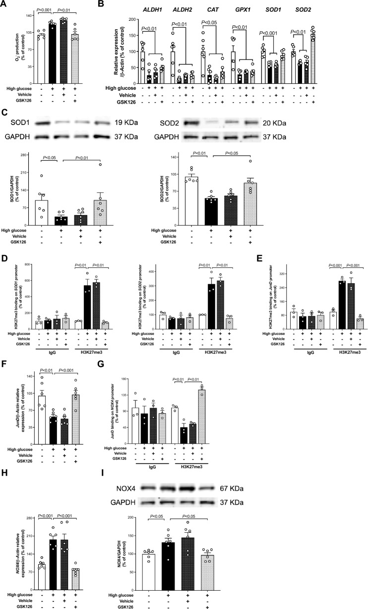
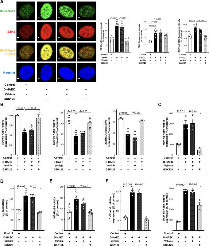
Methods: We studied human aortic endothelial cells exposed to high glucose (HAEC) or isolated from individuals with diabetes (D-HAEC). RT-qPCR, immunoblotting, chromatin immunoprecipitation (ChIP-qPCR), and confocal microscopy were performed to investigate the role of H3K27me3. We determined superoxide anion (O2-) production by ESR spectroscopy, NF-κB binding activity, and monocyte adhesion. Silencing/overexpression and pharmacological inhibition of chromatin modifying enzymes were used to modulate H3K27me3 levels. Furthermore, isometric tension studies and immunohistochemistry were performed in aorta from wild-type and db/db mice.

Results: Incubation of HAEC to high glucose showed that upregulation of EZH2 coupled to reduced demethylase UTX and JMJD3 was responsible for the increased H3K27me3. ChIP-qPCR revealed that repressive H3K27me3 binding to superoxide dismutase and transcription factor JunD promoters is involved in glucose-induced O2- generation. Indeed, loss of JunD transcriptional inhibition favours NOX4 expression. Furthermore, H3K27me3-driven oxidative stress increased NF-κB p65 activity and downstream inflammatory genes. Interestingly, EZH2 inhibitor GSK126 rescued these endothelial derangements by reducing H3K27me3. We also found that H3K27me3 epigenetic signature alters transcriptional programs in D-HAEC and aortas from db/db mice.

Conclusions: EZH2-mediated H3K27me3 represents a key epigenetic driver of hyperglycemia-induced endothelial dysfunction. Targeting EZH2 may attenuate oxidative stress and inflammation and, hence, prevent vascular disease in diabetes.

Keywords: Chromatin-modifying drugs; Diabetes; EZH2 inhibitor GSK126; Endothelial cells; Epigenetics; Inflammation; Oxidative stress.


Conflict of interest statement

The authors declare that no conflict of interest exists.

Figures

Fig. 1
Fig. 1
High glucose triggers repressive H3K27me3 via derangement of histone-modifying enzymes. A Representative western blot images and relative densitometric quantifications showing H3K27me3 expression in HAEC exposed to normal (5 mmol/l) and high (25 mmol/l) glucose (n = 6/group). B Histone methyltransferases (EZH1, EZH2) and demethylases (UTX, JMJD3, UTY) mRNAs in the two experimental groups (n = 6/group) assessed by RT-qPCR. C Representative western blot images and densitometric quantifications of EZH2, UTX, and JMJD3 expression in HAEC exposed to normal and high glucose (n = 6/group). D, E Representative western blot images and densitometric quantifications showing H3K27me3 expression after reprogramming of chromatin-modifying enzymes (n = 6/group). F H3K27me3 protein expression in the presence of EZH2 selective inhibitor GSK126 (5 µmol/l) or vehicle alone (n = 6/group). G EZH2 gene expression assessed by RT-qPCR (n = 6/group) in HAEC exposed to the same experimental conditions. H Confocal microscopy images of H3K27me3 (green), EZH2 (red), and EZH2/H3K27me3 colocalization (yellow), and relative quantification of fluorescence intensity. Cell nuclei are stained with Hoechst (blue). Scale bar = 2 μm. (n = 12/group)
Fig. 2
Fig. 2
EZH2-mediated H3K27me3 signature contributes to oxidative stress. A Electron spin resonance (ESR) spectroscopy analysis of O2 production, B expression of ROS scavenging enzymes ALDH1, ALDH2, CAT, GPX1, SOD1 and SOD2 genes and C representative Western blots images and densitometric quantifications of SOD1 and SOD2 proteins in HAEC exposed to normal (5 mmol/l) and high (25 mmol/l) glucose treated with EZH2 inhibitor GSK126 (5 µmol/l) or vehicle alone (n = 6/group). D ChIP-qPCR assay showing the binding of H3K27me3 to SOD1 and SOD2 promoters in high glucose-treated cells and the inhibitory effect exterted by GSK126 (5 µmol/l; n = 3/group). E The interaction of H3K27me3 with JunD promoter in HAEC exposed to high glucose was also abolished by EZH2 inhibitor GSK126 as shown by ChIP-qPCR assay (n = 3/group), F–I Downregulation of JunD mRNA (n = 6/group), G JunD binding on NOX4 promoter (n = 3/group), and subsequent upregulation of H NOX4 gene and I protein expression (n = 6/group) in HAEC exposed to high glucose were blunted by GSK126 (5 µmol/l) but not DMSO vehicle alone. IgG controls of ChIP-qPCR assay are also shown
Fig. 3
Fig. 3
EZH2-mediated H3K27me3 signature contributes to NF-κB p65-dependent inflammatory changes. A, C Effect of NADPH oxidase inhibitor apocynin (100 µmol/l) and GSK126 (5 µmol/l) on high glucose-induced increase of NF-κB p65 binding activity (n = 3–6/group). B, D RT-qPCR showing gene expression of inflammatory markers (n = 6/group) in HAECs exposed to high glucose alone in the presence of apocynin (100 µmol/l), GSK126 (5 µmol/l), or vehicle alone. E Representative images and relative quantification of monocyte adhesion to HAEC exposed to high glucose in the presence and in the absence of TNFa (5 mmol/l) and treated with GSK126 (5 µmol/l) or vehicle alone (n = 3/group). Scale bar = 100 μm. IL-6 interleukin-6, TNFα tumor necrosis factor α, MCP-1 monocyte chemoattractant factor-1, ICAM-1 intercellular adhesion molecule 1, VCAM-1 vascular cell adhesion molecule 1
Fig. 4
Fig. 4
GSK126 recovers high glucose-induced endothelial dysfunction. A Endothelium-dependent relaxations to acetylcholine (Ach) and endothelium-independent relaxations to sodium nitroprusside (SNP) in mouse aortic rings after exposure to normal (5 mmol/l) and high (25 mmol/l) glucose in the presence and in the absence of GSK126 (5 µmol/l; n = 4–6/group). B Representative confocal images and quantification of H3K27me3, EZH2, SOD1, and SOD2 protein expression in aortas from WT, db/db mice, and db/db mice exposed to vehicle or GSK126 (5 µmol/l; n = 6/group). Scale bar = 500 μm
Fig. 5
Fig. 5
Targeting EZH2-H3K27me3 epigenetic signature rescues abnormal phenotypes in endothelial cells isolated from patients with diabetes. A Representative confocal microscopy images of H3K27me3 (green), EZH2 (red), and EZH2/H3K27me3 colocalization (yellow) and densitometric quantification of fluorescence intensity. Cell nuclei are stained with Hoechst (blue). Scale bar = 2 μm. n = 6/group. B and C RT-qPCR arrays showing gene expression of SOD1, SOD2, JunD, and C NOX4, respectively (n = 3/group). D electron spin resonance (ESR) spectroscopy analysis of O2 production (n = 3/group). E NF-κB p65 binding activity (n = 6/group), and F gene expression of IL-6 and MCP-1 (n = 3/group). IL-6, interleukin-6; MCP-1, monocyte chemoattractant factor-1

Similar articles

Cited by

References

    1. Saeedi P, Petersohn I, Salpea P, Malanda B, Karuranga S, Unwin N, et al. Global and regional diabetes prevalence estimates for 2019 and projections for 2030 and 2045: results from the International Diabetes Federation Diabetes Atlas, 9th edition. Diabetes Res Clin Pract. 2019;157:107843. doi: 10.1016/j.diabres.2019.107843. - DOI - PubMed
    1. Einarson TR, Acs A, Ludwig C, Panton UH. Prevalence of cardiovascular disease in type 2 diabetes: a systematic literature review of scientific evidence from across the world in 2007–2017. Cardiovasc Diabetol. 2018;17:83. doi: 10.1186/s12933-018-0728-6. - DOI - PMC - PubMed
    1. Alexander Y, Osto E, Schmidt-Trucksäss A, Shechter M, Trifunovic D, Duncker DJ, et al. Endothelial function in cardiovascular medicine: a consensus paper of the European Society of Cardiology Working Groups on Atherosclerosis and Vascular Biology, Aorta and Peripheral Vascular Diseases, Coronary Pathophysiology and Microcirculation, and Thrombosis. Cardiovasc Res. 2021;117:29–42. doi: 10.1093/cvr/cvaa085. - DOI - PMC - PubMed
    1. Incalza MA, D’Oria R, Natalicchio A, Perrini S, Laviola L, Giorgino F. Oxidative stress and reactive oxygen species in endothelial dysfunction associated with cardiovascular and metabolic diseases. Vascul Pharmacol. 2018;100:1–19. doi: 10.1016/j.vph.2017.05.005. - DOI - PubMed
    1. Guzik TJ, Cosentino F. Epigenetics and immunometabolism in diabetes and aging. Antioxid Redox Signal. 2017;2018(29):257–274. - PMC - PubMed

Publication types