

CNS tumors with PLAGL1-fusion: beyond ZFTA and YAP1 in the genetic spectrum of supratentorial ependymomas
- PMID: 38581034
- PMCID: PMC10998316
- DOI: 10.1186/s40478-023-01695-7
CNS tumors with PLAGL1-fusion: beyond ZFTA and YAP1 in the genetic spectrum of supratentorial ependymomas
Abstract
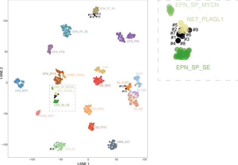
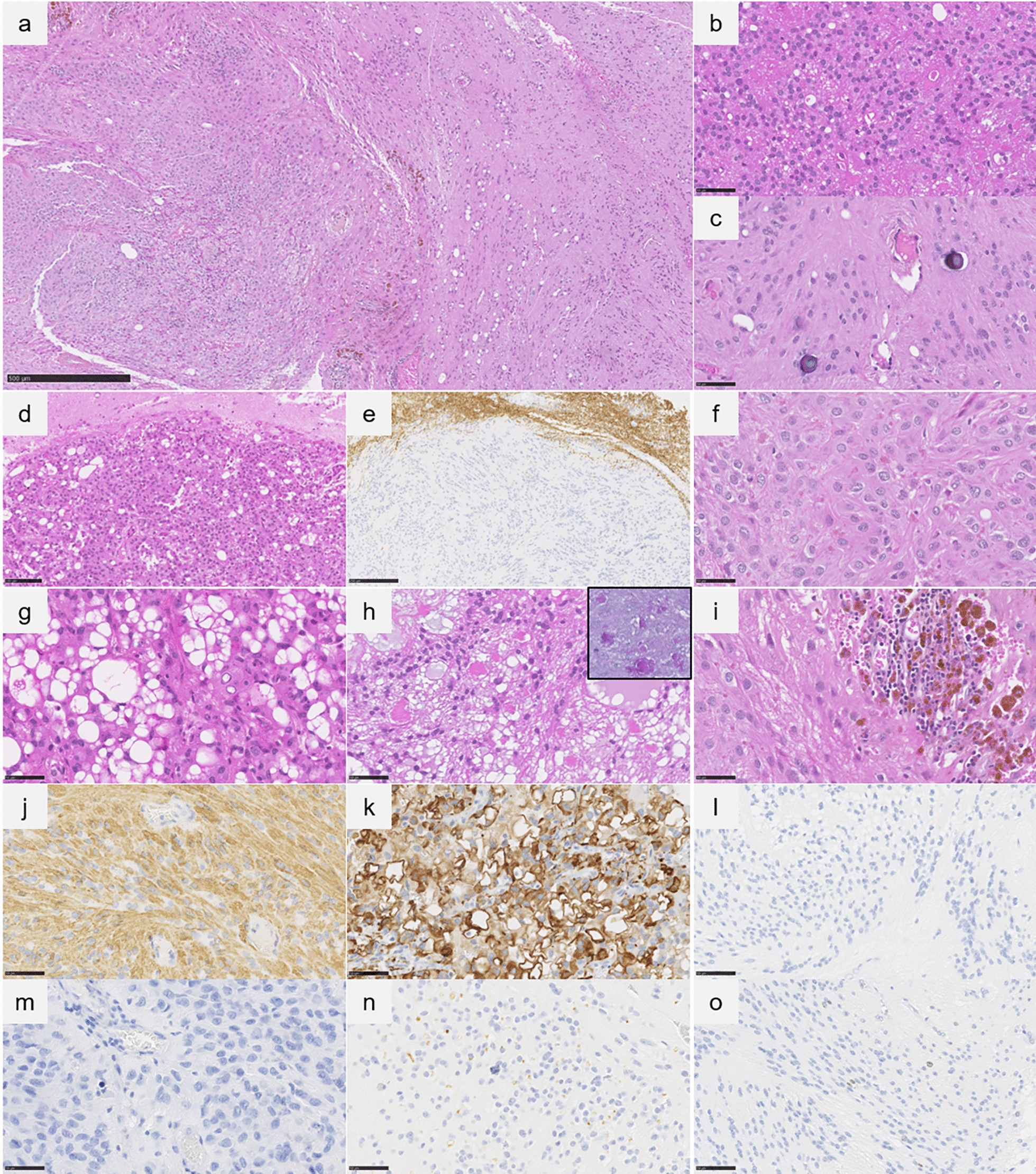
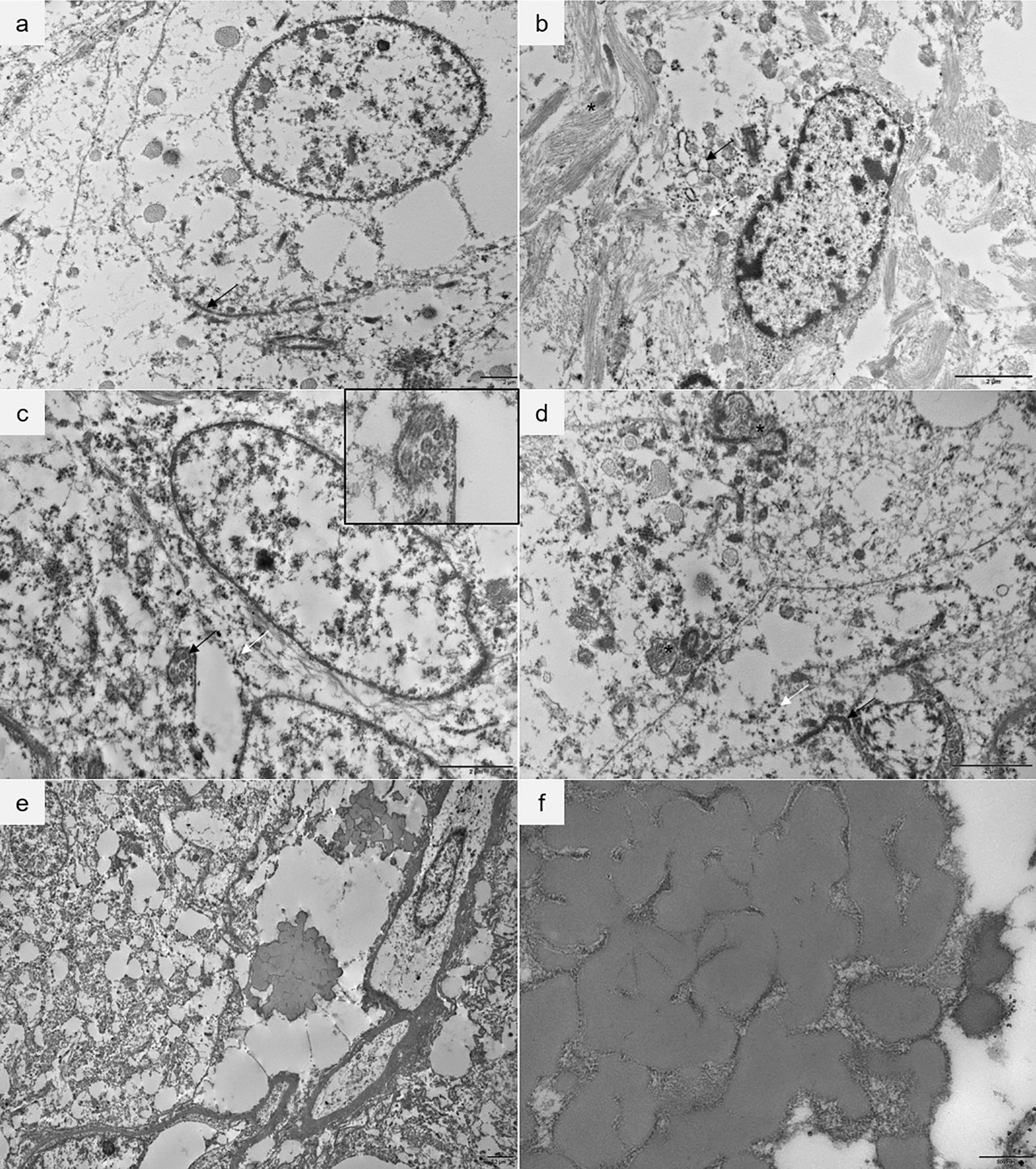
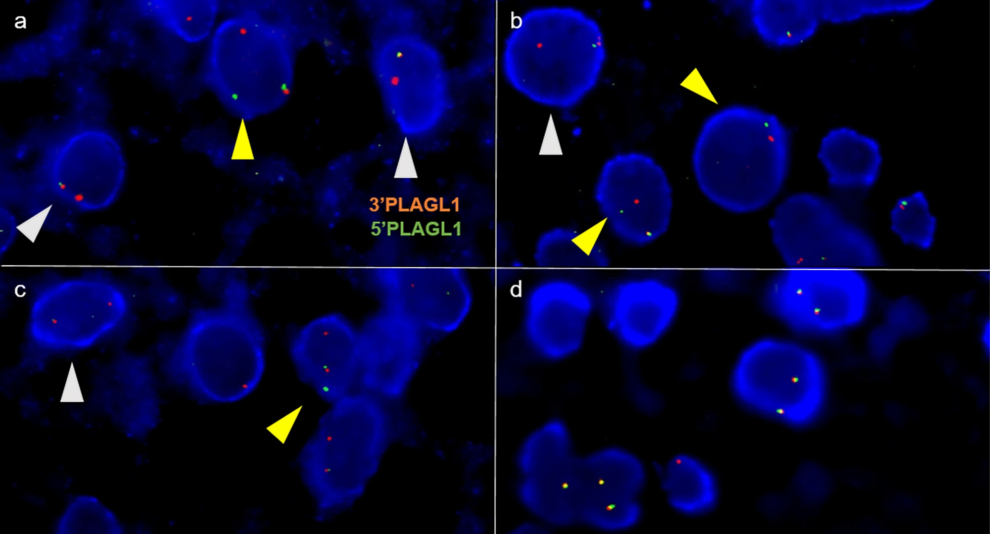
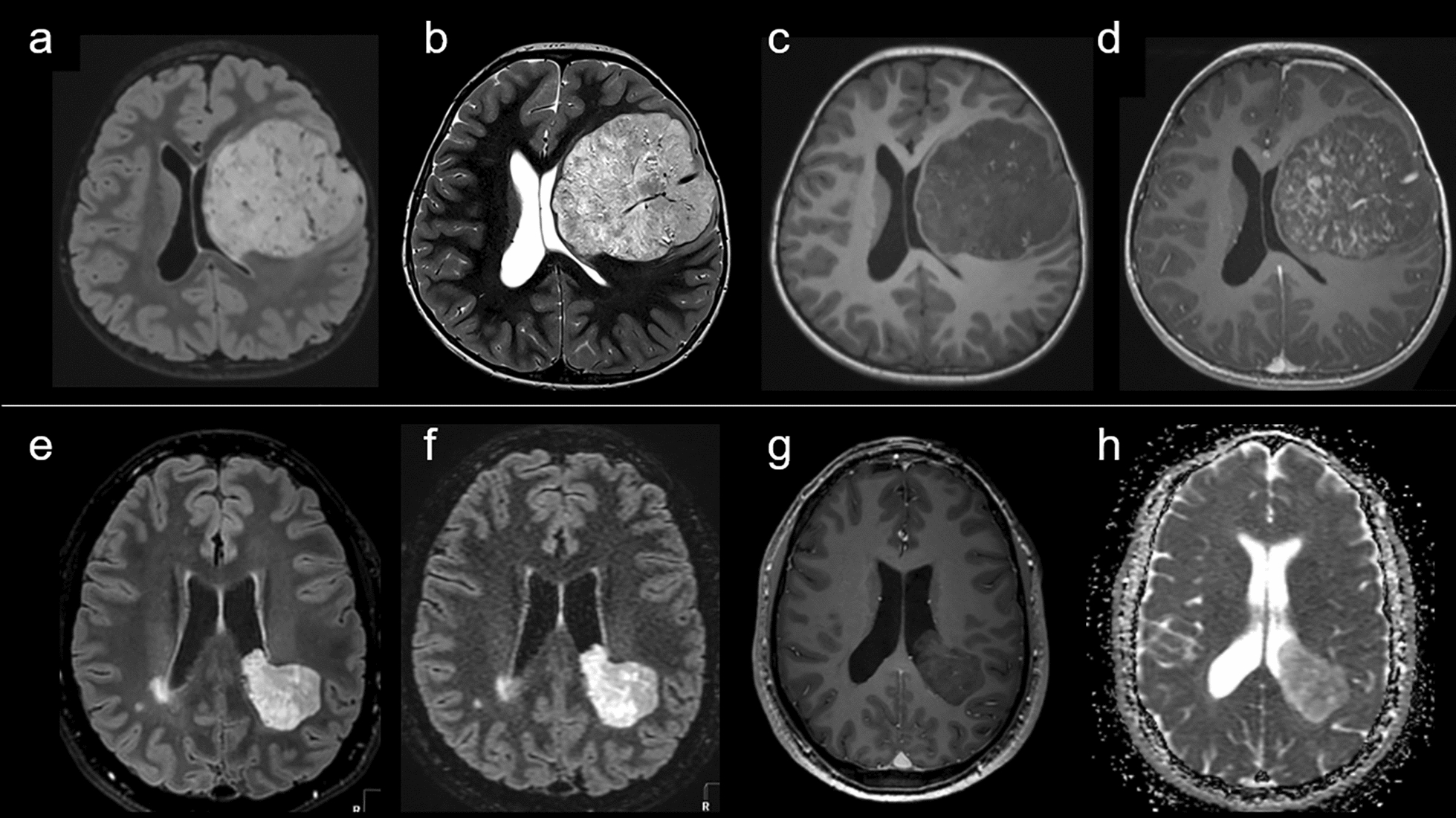
A novel methylation class, "neuroepithelial tumor, with PLAGL1 fusion" (NET-PLAGL1), has recently been described, based on epigenetic features, as a supratentorial pediatric brain tumor with recurrent histopathological features suggesting an ependymal differentiation. Because of the recent identification of this neoplastic entity, few histopathological, radiological and clinical data are available. Herein, we present a detailed series of nine cases of PLAGL1-fused supratentorial tumors, reclassified from a series of supratentorial ependymomas, non-ZFTA/non-YAP1 fusion-positive and subependymomas of the young. This study included extensive clinical, radiological, histopathological, ultrastructural, immunohistochemical, genetic and epigenetic (DNA methylation profiling) data for characterization. An important aim of this work was to evaluate the sensitivity and specificity of a novel fluorescent in situ hybridization (FISH) targeting the PLAGL1 gene. Using histopathology, immunohistochemistry and electron microscopy, we confirmed the ependymal differentiation of this new neoplastic entity. Indeed, the cases histopathologically presented as "mixed subependymomas-ependymomas" with well-circumscribed tumors exhibiting a diffuse immunoreactivity for GFAP, without expression of Olig2 or SOX10. Ultrastructurally, they also harbored features reminiscent of ependymal differentiation, such as cilia. Different gene partners were fused with PLAGL1: FOXO1, EWSR1 and for the first time MAML2. The PLAGL1 FISH presented a 100% sensitivity and specificity according to RNA sequencing and DNA methylation profiling results. This cohort of supratentorial PLAGL1-fused tumors highlights: 1/ the ependymal cell origin of this new neoplastic entity; 2/ benefit of looking for a PLAGL1 fusion in supratentorial cases of non-ZFTA/non-YAP1 ependymomas; and 3/ the usefulness of PLAGL1 FISH.
Keywords: DNA-methylation; Ependymoma; PLAGL1; Subependymoma.
© 2024. The Author(s).
Conflict of interest statement
The authors declare that they have no conflict of interest directly related to the topic of this article.
Figures
Similar articles
-
Concurrent ependymal and ganglionic differentiation in a subset of supratentorial neuroepithelial tumors with EWSR1-PLAGL1 rearrangement.Acta Neuropathol Commun. 2024 Sep 3;12(1):143. doi: 10.1186/s40478-024-01809-9. Acta Neuropathol Commun. 2024. PMID: 39228008 Free PMC article.
-
Ependymal Tumors: Overview of the Recent World Health Organization Histopathologic and Genetic Updates with an Imaging Characteristic.AJNR Am J Neuroradiol. 2024 Nov 7;45(11):1624-1634. doi: 10.3174/ajnr.A8237. AJNR Am J Neuroradiol. 2024. PMID: 38844368 Review.
-
Supratentorial non-RELA, ZFTA-fused ependymomas: a comprehensive phenotype genotype correlation highlighting the number of zinc fingers in ZFTA-NCOA1/2 fusions.Acta Neuropathol Commun. 2021 Aug 13;9(1):135. doi: 10.1186/s40478-021-01238-y. Acta Neuropathol Commun. 2021. PMID: 34389065 Free PMC article.
-
Glioneuronal tumors PATZ1-fused: clinico-molecular and DNA methylation signatures for a variety of morphological and radiological profiles.Acta Neuropathol Commun. 2025 May 24;13(1):114. doi: 10.1186/s40478-025-02037-5. Acta Neuropathol Commun. 2025. PMID: 40413488 Free PMC article.
-
Signs and symptoms to determine if a patient presenting in primary care or hospital outpatient settings has COVID-19.Cochrane Database Syst Rev. 2022 May 20;5(5):CD013665. doi: 10.1002/14651858.CD013665.pub3. Cochrane Database Syst Rev. 2022. PMID: 35593186 Free PMC article.
Cited by
-
A novel TEAD1::NCOA2 fusion that potentially expands the concept of supratentorial ependymoma, YAP1 fusion-positive.Acta Neuropathol. 2025 Feb 10;149(1):14. doi: 10.1007/s00401-025-02852-z. Acta Neuropathol. 2025. PMID: 39928140 Free PMC article. No abstract available.
-
PLAG-Family Amplified CNS Embryonal Tumour With PLAG1 Immunohistochemical Expression: Expanding the Spectrum of Diagnostic Tools.Neuropathol Appl Neurobiol. 2025 Apr;51(2):e70017. doi: 10.1111/nan.70017. Neuropathol Appl Neurobiol. 2025. PMID: 40207790 Free PMC article. No abstract available.
-
Advances in molecular prognostication and treatments in ependymoma.J Neurooncol. 2025 Apr;172(2):317-326. doi: 10.1007/s11060-024-04923-9. Epub 2025 Jan 6. J Neurooncol. 2025. PMID: 39757304 Review.
References
-
- Andreiuolo F, Varlet P, Tauziède-Espariat A, Jünger ST, Dörner E, Dreschmann V, Kuchelmeister K, Waha A, Haberler C, Slavc I, Corbacioglu S, Riemenschneider MJ, Leipold A, Rüdiger T, Körholz D, Acker T, Russo A, Faber J, Sommer C, Armbrust S, Rose M, Erdlenbruch B, Hans VH, Bernbeck B, Schneider D, Lorenzen J, Ebinger M, Handgretinger R, Neumann M, van Buiren M, Prinz M, Roganovic J, Jakovcevic A, Park S-H, Grill J, Puget S, Messing-Jünger M, Reinhard H, Bergmann M, Hattingen E, Pietsch T. Childhood supratentorial ependymomas with YAP1-MAMLD1 fusion: an entity with characteristic clinical, radiological, cytogenetic and histopathological features. Brain Pathol Zurich Switz. 2019;29:205–216. doi: 10.1111/bpa.12659. - DOI - PMC - PubMed
Publication types
MeSH terms
Substances
LinkOut - more resources
Full Text Sources
Medical
Molecular Biology Databases
Research Materials
Miscellaneous
