. 2024 Apr 5;15(1):99.
doi: 10.1186/s13287-024-03696-2.

How to differentiate induced pluripotent stem cells into sensory neurons for disease modelling: a functional assessment

Affiliations

How to differentiate induced pluripotent stem cells into sensory neurons for disease modelling: a functional assessment

Anil Kumar Kalia et al. Stem Cell Res Ther. .

Abstract

Background: Human induced pluripotent stem cell (iPSC)-derived peripheral sensory neurons present a valuable tool to model human diseases and are a source for applications in drug discovery and regenerative medicine. Clinically, peripheral sensory neuropathies can result in maladies ranging from a complete loss of pain to severe painful neuropathic disorders. Sensory neurons are located in the dorsal root ganglion and are comprised of functionally diverse neuronal types. Low efficiency, reproducibility concerns, variations arising due to genetic factors and time needed to generate functionally mature neuronal populations from iPSCs remain key challenges to study human nociception in vitro. Here, we report a detailed functional characterization of iPSC-derived sensory neurons with an accelerated differentiation protocol ("Anatomic" protocol) compared to the most commonly used small molecule approach ("Chambers" protocol). Anatomic's commercially available RealDRG™ were further characterized for both functional and expression phenotyping of key nociceptor markers.

Methods: Multiple iPSC clones derived from different reprogramming methods, genetics, age, and somatic cell sources were used to generate sensory neurons. Manual patch clamp was used to functionally characterize both control and patient-derived neurons. High throughput techniques were further used to demonstrate that RealDRGs™ derived from the Anatomic protocol are amenable to high throughput technologies for disease modelling.

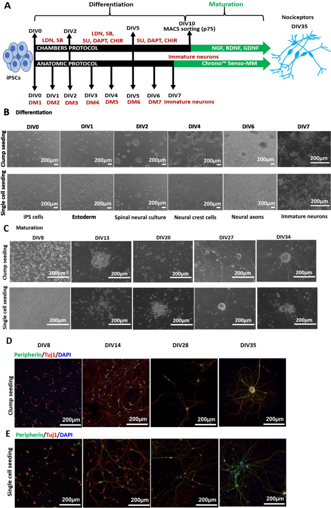
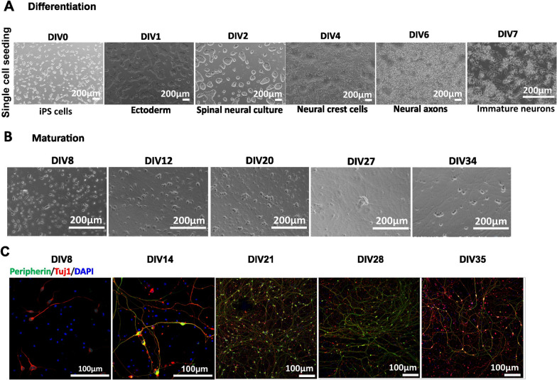
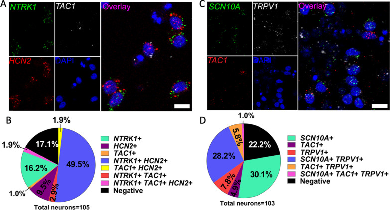
Results: The Anatomic protocol rendered a purer culture without the use of mitomycin C to suppress non-neuronal outgrowth, while Chambers differentiations yielded a mix of cell types. Chambers protocol results in predominantly tonic firing when compared to Anatomic protocol. Patient-derived nociceptors displayed higher frequency firing compared to control subject with both, Chambers and Anatomic differentiation approaches, underlining their potential use for clinical phenotyping as a disease-in-a-dish model. RealDRG™ sensory neurons show heterogeneity of nociceptive markers indicating that the cells may be useful as a humanized model system for translational studies.

Conclusions: We validated the efficiency of two differentiation protocols and their potential application for functional assessment and thus understanding the disease mechanisms from patients suffering from pain disorders. We propose that both differentiation methods can be further exploited for understanding mechanisms and development of novel treatments in pain disorders.

Keywords: Disease modelling; Human induced pluripotent stem cells; Pain; Sensory neurons; Sodium channel.


Conflict of interest statement

A.L. has a research agreement with Grunenthal. T.J.P has a research agreement with Grunenthal and Merck. A.L. and T.J.P. receive counselling fees from Grunenthal. P.W. and V.T. are shareholders and employees of Anatomic Incorporated. A.K.K. is currently employed at Grunenthal.

Figures

Fig. 1
Fig. 1
Anatomic protocol: differentiation and maturation (Ctrl1). A Schematic diagram outlining the steps for differentiation and maturation with the Anatomic compared to the Chambers protocol. Anatomic protocol requires 7 days of differentiation as compared to 10 days with Chambers protocol to achieve immature neurons. With Chambers protocol neurons were MACS sorted on DIV10 of differentiation. Neurons were then matured until DIV 35–40. DIV-Days in vitro, LDN-193189, SB431542 and SU5402, CHIR99021 and DAPT. DM-Differentiation medium, Chrono™ Senso-MM-Maturation medium. B Differentiation of Ctrl1 iPSCs with Anatomic protocol. Phase contrast images display single cell and clump seeding on DIV0 of differentiation. Differentiation involves formation of ectoderm within 24 h, spinal neural culture (DIV2), neural crest formation (DIV4) and generation of immature neurons by DIV7. Scale Bar—200 µm. C Maturation of Ctrl1 neurons with growth factors. Both seeding protocols resulted in formation of dense homogenous neuronal networks during the maturation period. No morphological differences could be observed during maturation from both protocols. Scale Bar—200 µm. D Immunostaining of neurons for Peripherin and Tuj1 on DIV8, 14, 28 and 35 with clump seeding. E Single cell seeded neurons staining for Peripherin and Tuj1 on DIV8, 14, 28 and 35. Scale Bar—200 µm. Peripherin-green, Tuj1-red and DAPI-blue fluorescence. DIV-Days in vitro
Fig. 2
Fig. 2
Electrophysiological characterization of Anatomic-derived neurons (Ctrl1 and Ctrl2). A Comparison of percentage mature neurons (first AP generated with a square pulse current injection with steps of 10pA) between clump and single cell seeding. i. Representative mature and immature AP traces ii. Percentage of mature neurons: Clump Ctrl1- DIV14: 60% (n = 3 out of 5), DIV28: 100% (n = 5 out of 5), DIV35: 83% (n = 5 out of 6) and Single cell Ctrl1- DIV14: 20% (n = 1 out of 5), DIV28: 100% (n = 5 out of 5), DIV35: 100% (n = 6 out of 6). Ctrl2—Percentage of mature neurons: DIV14: (67% (n = 4 out of 6), DIV21: 71% (n = 5 out of 7), DIV28: 100% (n = 7 out of 7) and DIV35: 100% (n = 5 out of 5). Number of mature cells out of total cells patched are denoted for each recording day above the bar. B RMP of Ctrl1 and Ctrl2 neurons. C Cell capacitance (pF) for Ctrl1 and Ctrl2 neurons. D TTXr currents recorded in the presence of 500 nM TTX. i. Voltage protocol, ii. Representative current traces, iii. Current density measured on DIV14, 28 and 35. E Voltage clamp recordings of inward sodium and outward potassium currents. i. Voltage protocol ii. Na+ and K+ current traces. iii. Sodium current density measured on DIV14 and 28 of maturation. iv. Potassium current density measured on DIV14 and 28 of maturation. F Tonic firing in response to ramp current stimuli i. Ramp current stimuli, ii. AP firing in response to ramp current stimuli 1nA/1 s. iii Percentage tonic neurons. Data shown as mean ± SEM. One-way Anova Bonferroni's multiple comparisons test
Fig. 3
Fig. 3
Anatomic protocol: Differentiation and Maturation (Ctrl2). A Differentiation: Phase contrast images display single cell seeding of iPSCs on DIV0 of differentiation and generation of immature neurons by DIV7. Scale bar—200 µm. B Maturation: Maturation of neurons DIV7-35. Differentiation resulted in a homogenous neuronal network during the maturation period. Scale Bar 200 µm. C Immunostaining of neurons for Peripherin and Tuj1 on DIV8, 14, 21, 28 and 35. Scale Bar—100 µm. DIV-Days in vitro. Peripherin-green, Tuj1-red and DAPI-blue fluorescence
Fig. 4
Fig. 4
Chambers protocol: Differentiation and Maturation (Ctrl1). A Phase contrast images during the differentiation and maturation of iPSC-derived from Ctrl1 subject. DIV0 iPSCs, DIV10 generation of immature neurons, DIV35 shows ganglion-like morphology (marked with blue arrows). DIV18 and 35 also shows the culture outgrown with other non-neuronal cell types (marked with red arrows). Scale bar—200 µm. B Neurons at DIV35 from Anatomic protocol results in homogenous neuronal network. Scale bar—200 µm. C Immunostaining confirms peripheral neuronal identity of neurons from DIV11, 14, 21, 28 and 35 of maturation. DIV-Days in vitro. Scale bar − 100 µm. Peripherin-green, Tuj1-red and DAPI-blue fluorescence
Fig. 5
Fig. 5
Comparison of electrophysiological characteristics of Anatomic and Chambers-derived neurons (Ctrl1). A Comparison of percentage of mature APs (First AP generated with a square pulse current injection) Percentage Mature neurons- DIV14: 43% (n = 2 out of 7), DIV28: 100% (n = 5 out of 5) and DIV35: 100% (n = 5 out of 5) for neurons generated with Chambers protocol. Number of cells having APs with overshoot above 0 mV out of total cells patched are denoted for each recording day above the bar. B RMP measured from both the protocols. One way Anova Bonferroni's multiple comparisons test. C Cell capacitance (pF) measured from both the protocols. One way Anova Bonferroni's multiple comparisons test. D TTXr currents recorded in the presence of 500 nM TTX. Mann–Whitney test. E, F Voltage clamp recordings of inward Na+ and K+ current density measured on DIV14 of maturation. Mann–Whitney test G Tonic firing neurons in response to ramp current stimuli. Chambers protocol- DIV28: n = 4 (4/4 Tonic, 1 cell no AP), DIV35: n = 5 (5/5 Tonic). H The average number of APs generated in response to ramp current stimuli. n = 5 for both protocols. Multiple t test. DIV-Days in vitro. Data are shown as mean ± SEM
Fig. 6
Fig. 6
Disease modelling of IEM and SFN pain disorders using the Anatomic protocol. A, B Cell size and RMP did not show significant differences observed for both IEM and SFN groups as compared to control. C Rheobase indicate significant lesser current injection needed to generate an AP for both SFN and IEM group. D AP threshold shows significant shift to depolarized potentials for IEM and SFN-derived neurons as compared to control. E Significant shift of afterhyperpolarization (AHP) to more depolarized potentials for SFN patient as compared to control. F Ramp current stimuli 500 pA/500 ms resulted in a significant increase in no. of APs firing from SFN-derived neurons as compared to control group. G Time needed to generate 1st AP is significantly reduced for both IEM and SFN-derived neurons as compared to control. H In response to ramp current injections, there was a significant increase in number of APs fired from both patient-derived neurons (Ctrl n = 10, IEM n = 16, SFN n = 22). Two way ANOVA. AG One way ANOVA with Tukey's multiple comparison test. Data are shown as mean ± SEM
Fig. 7
Fig. 7
Gene expression in Anatomic RealDRG™ neurons. A Expression of mRNA for NTRK1 (TRKA, green), TAC1 (preprotachykinin-1; white) and HCN2 (red) seen in Anatomic protocol cells at DIV14 co-labeled with DNA marker DAPI (blue). B Pie chart for staining in A showing distributions of cells expressing indicated markers. C Expression of mRNA for SCN10A (NaV1.8, green), TRPV1 (white), TAC1 (red) seen in Anatomic protocol cells at DIV16 co-labeled with DNA marker DAPI (blue). D Pie chart for staining in C showing distributions of cells expressing indicated markers. Images are cropped from 40X images. Scale bar—10 µm
Fig. 8
Fig. 8
Anatomic RealDRG™ can be adapted for FLIPRPenta high-throughput Ca2+ assays. A Ca2+ responses of sensory neurons at DIV15 plated with varying cell densities (1000–10,000 cells/well) on 384-well plates. B KCl-induced Ca2+ responses at DIV15, DIV21 and DIV35 C sample trace from DIV21. D Veratridine-induced Ca2+ responses at DIV15, 21 and 35 E sample trace showing complete inhibition of responses by TTX (1 µM). F Capsaicin-induced Ca2+ responses emerge at DIV35, G capsaicin sample trace. H 24 h treatment with IL6/soluble IL6-R does not affect the response properties of sensory neurons at DIV35. Data are presented as mean ± S.D. from n > 3 replicates
Fig. 9
Fig. 9
Voltage- and current-clamp recordings of Anatomic RealDRG™ on Qube384. A Representative current traces with holding voltage at − 90 mV. B Current-clamp recording: holding voltage at − 90 mV, ramp current clamp was elicited by injecting current from − 100 pA to 100 pA with 500 or 1000 ms duration. C, D Qube384 experiment success rates and current expression levels during culture period of 28 days. C Success rate indicates all the cells passed the criteria of Rmem > 200 MΩ, the rates were decreased with longer culture period, which are 54 ± 5% (n = 3), 52 ± 5% (n = 3), 41 ± 5% (n = 4), and 43 ± 7% (n = 4), respectively, for DIV16, 21, 28, and 35 days. For the cells passed membrane resistance and cell size filters, average expression levels of Kv, Nav channels, and AP firings are 90–97%, 60–77%, and 60–67%, respectively. D Current densities were measured at depolarizations to − 10 mV, 20 ms for Nav currents, and + 60 mV, 500 ms for Kv currents. E Current traces with internal solution of KF (black) and after exchanging to CsF based internal solution (red). Currents were elicited by 10 mV stepwise voltage increasing from − 90 to + 60 mV for 300 ms. F Current–voltage relationship curve were all normalized to the current amplitudes at + 60 mV before IC exchange. G Family of current traces in control and 0.5 µM TTX groups by using the same voltage protocol in Fig. 1A. H Current–voltage relationship curves, all the current amplitudes were normalized to the control currents at voltage of − 10 mV (Data showed DIV21 cells). I Current traces at voltage − 10 mV for control (red), 0.5 µM TTX (blue), and 10 µM A-803467 in 0.5 µM TTX (Magenta). J Current densities of 0.5 µM TTX and 10 µM A-803467 in 0.5 µM TTX for DIV21, 28, and 35
Fig. 10
Fig. 10
MEA recordings of Anatomic RealDRG™ sensory neurons over the maturation period. A Cells were plated over the electrode array region within a typical well of the Axion 48 well cytoview MEA plate shown here at DIV16 (16 electrodes/well). Scale bar—100 µm. B Data visualization from RealDRGs cultured on MEAs. Heat plot across the 48 well MEA indicating spike activity. Each colored circle is an electrode with detected APs. C Representative traces showing a single active electrode before and during a 42 °C temperature ramp at DIV36. D Representative raster plots tracking the per-electrode responsiveness to 42 °C heat ramp from a single timepoint each during week 2, 3, 4, and 5. E The percent active electrode yield (AEY) from 384 electrodes in a 48-well plate for three recording sessions each during week 2, 3, 4, and 5 after plating of cells. Blue trace describes the AEY during 42 °C trials, the 37 °C electrode data (prior to heating) is shown in black. F The average Mean Firing Rate (MFR, Hz, mean ± SEM) for 384 electrodes in a 48-well plate for three recording sessions each during week 2, 3, 4, and 5 after plating of cells. Blue bars describe the MFR during 42 °C trials at each timepoint, the 37 °C trials MFR is shown in black. Asterisks denote significance with Sidak’s multiple comparisons (**p < 0.01, ***p < 0.001, ****p < 0.0001). G Pie charts describing the evolution of responsiveness to 42 °C from the 174 active electrodes identified at week 5 from their response in the previous weeks. The four types of responses during week 5 were: consistent (positive response from 3 of 3 42 °C trials/wk), responders (1 ≤ positive trials < 3 trials/wk), negative (spontaneously active with negative responses to 42 °C), and inactive electrodes (< 1 spk/min)

Update of

References

    1. Gorecka J, Kostiuk V, Fereydooni A, Gonzalez L, Luo J, Dash B, Isaji T, Ono S, Liu S, Lee SR, Xu J, Liu J, Taniguchi R, Yastula B, Hsia HC, Qyang Y, Dardik A. The potential and limitations of induced pluripotent stem cells to achieve wound healing. Stem Cell Res Ther. 2019;10(1):87. doi: 10.1186/s13287-019-1185-1. - DOI - PMC - PubMed
    1. Méndez-Maldonado K, Vega-López GA, Aybar MJ, Velasco I. Neurogenesis from neural crest cells: molecular mechanisms in the formation of cranial nerves and ganglia. Front Cell Dev Biol. 2020;8:635. doi: 10.3389/fcell.2020.00635. - DOI - PMC - PubMed
    1. Cox JJ, Woods CG, Kurth I. Peripheral sensory neuropathies—pain loss vs. pain gain. Med Gen. 2020;32(3):233–241. doi: 10.1515/medgen-2020-2039. - DOI
    1. Alsaloum M, Waxman SG. iPSCs and DRGs: Stepping stones to new pain therapies. Trends Mol Med. 2022;28(2):110–122. doi: 10.1016/j.molmed.2021.11.005. - DOI - PMC - PubMed
    1. Renthal W, Chamessian A, Curatolo M, Davidson S, Burton M, Dib-Hajj S, Dougherty PM, Ebert AD, Gereau RW, 4th, Ghetti A, Gold MS, Hoben G, Menichella DM, Mercier P, Ray WZ, Salvemini D, Seal RP, Waxman S, Woolf CJ, Stucky CL, Price TJ. Human cells and networks of pain: Transforming pain target identification and therapeutic development. Neuron. 2021;109(9):1426–1429. doi: 10.1016/j.neuron.2021.04.005. - DOI - PMC - PubMed

MeSH terms

LinkOut - more resources