

Dynamic Changes of Capsular-Intraocular Lens Adhesion in Plate-Haptic Hydrophilic and Loop-Haptic Hydrophobic Eyes
- PMID: 38581607
- PMCID: PMC11109074
- DOI: 10.1007/s40123-024-00933-y
Dynamic Changes of Capsular-Intraocular Lens Adhesion in Plate-Haptic Hydrophilic and Loop-Haptic Hydrophobic Eyes
Abstract
Introduction: The aim of this work is to investigate the dynamic changes of capsular-intraocular lens (IOL) adhesion in plate-haptic hydrophilic and loop-haptic hydrophobic eyes.
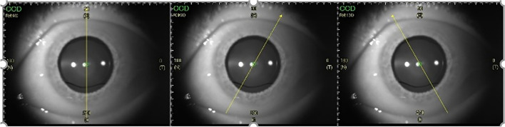
Methods: Cataract eyes that met the inclusion criteria were randomly assigned to receive implantation of a plate-haptic hydrophilic or loop-haptic IOL. The anterior capsular adhesion, posterior capsular adhesion, and the configurations of the capsular bend were evaluated using swept-source optical coherence tomography at 1 day, 1 week, 1 month, and 3 months postoperatively.
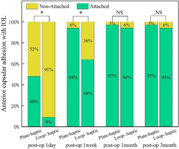
Results: In total, 66 eyes of 66 patients were eligible for the analysis: 33 in the plate-haptic group and 33 in the loop-haptic group. The contact between the anterior capsule and IOL in the plate-haptic group was earlier than that in the loop-haptic group upon comparing the measurements taken at 1 day and 1 week (p = 0.001, p = 0.003, respectively). The complete attachment of the posterior capsule and IOL in the plate-haptic group was significantly greater at 1 week, 1 month, and 3 months (p = 0.001, p = 0.000, p = 0.001, respectively). The capsular bend index of the plate-haptic group was significantly greater than that of the loop-haptic group at each time points except at 1 day (p = 0.007, p = 0.049, p = 0.005, respectively). Furthermore, a new type of capsular bend, "cocked adhesion," was observed in the plate-haptic eyes.
Conclusions: The plate-haptic IOL demonstrated excellent capsular adhesion compared to the loop-haptic IOL, which was probably attributed to haptic compressibility. A special cocked configuration of the capsular bend in plate-haptic IOL was observed for the first time. Further studies are warranted to confirm the effect of the new type of capsular bend.
Keywords: Capsular adhesion; Intraocular lens; Loop-haptic; Plate-haptic.
© 2024. The Author(s).
Conflict of interest statement
All named authors confirm that they have no competing interests to declare.
Figures


Similar articles
-
Influence of a Capsular Tension Ring on Capsular Bag Behavior of a Plate Haptic Intraocular Lens: An Intraindividual Randomized Trial.Ophthalmology. 2024 Apr;131(4):445-457. doi: 10.1016/j.ophtha.2023.10.031. Epub 2023 Oct 31. Ophthalmology. 2024. PMID: 37914042 Clinical Trial.
-
Capsular bag stability and posterior capsule opacification of a plate-haptic design microincision cataract surgery intraocular lens: 3-year results of a randomised trial.Br J Ophthalmol. 2013 Dec;97(12):1565-8. doi: 10.1136/bjophthalmol-2013-303710. Epub 2013 Sep 24. Br J Ophthalmol. 2013. PMID: 24064942 Clinical Trial.
-
Relationship of Posterior Capsular Opacification and Capsular Bend Type Investigation Based on Swept-source Optical Coherence Tomography.Curr Eye Res. 2020 Jan;45(1):17-23. doi: 10.1080/02713683.2019.1645183. Epub 2019 Jul 29. Curr Eye Res. 2020. PMID: 31348676
-
Posterior capsulorhexis combined with optic buttonholing: an alternative to standard in-the-bag implantation of sharp-edged intraocular lenses? A critical analysis of 1000 consecutive cases.Graefes Arch Clin Exp Ophthalmol. 2008 Jun;246(6):787-801. doi: 10.1007/s00417-008-0779-6. Epub 2008 Apr 19. Graefes Arch Clin Exp Ophthalmol. 2008. PMID: 18425525 Free PMC article. Review.
-
Secondary IOL Implantation Without Capsular Support.Am J Ophthalmol. 2025 Aug 17:S0002-9394(25)00426-X. doi: 10.1016/j.ajo.2025.08.023. Online ahead of print. Am J Ophthalmol. 2025. PMID: 40829699 Review. No abstract available.
Cited by
-
How do intraocular lens materials influence the outcome of cataract surgery?Curr Opin Ophthalmol. 2025 Jan 1;36(1):18-24. doi: 10.1097/ICU.0000000000001095. Epub 2024 Oct 23. Curr Opin Ophthalmol. 2025. PMID: 39446645 Free PMC article. Review.
References
Grants and funding
LinkOut - more resources
Full Text Sources
Miscellaneous
