. 2024 May 15;436(10):168557.
doi: 10.1016/j.jmb.2024.168557. Epub 2024 Apr 4.

HIV-1 Integrase Assembles Multiple Species of Stable Synaptic Complex Intasomes That Are Active for Concerted DNA Integration In vitro

Affiliations

HIV-1 Integrase Assembles Multiple Species of Stable Synaptic Complex Intasomes That Are Active for Concerted DNA Integration In vitro

Min Li et al. J Mol Biol. .

Abstract

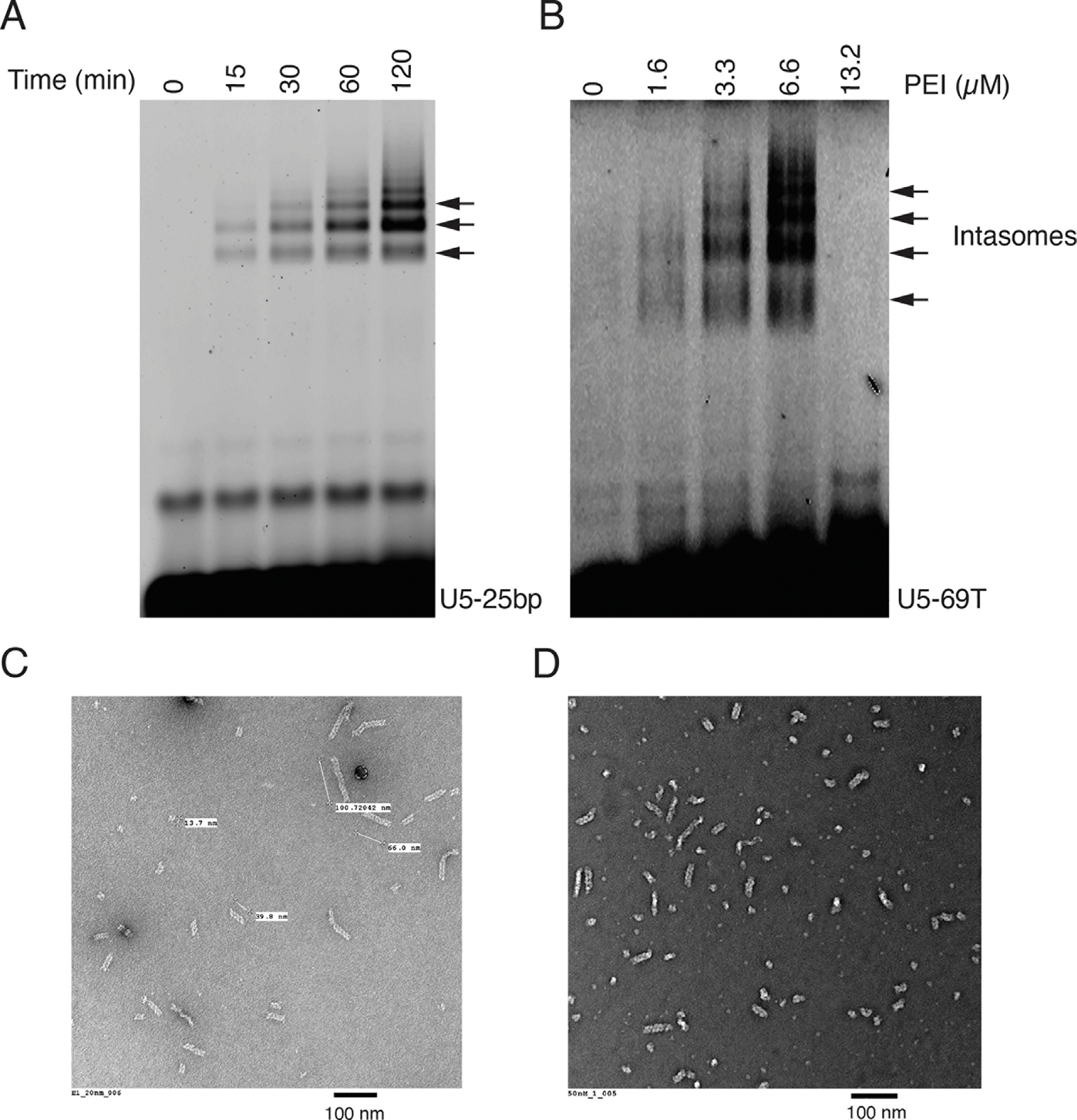
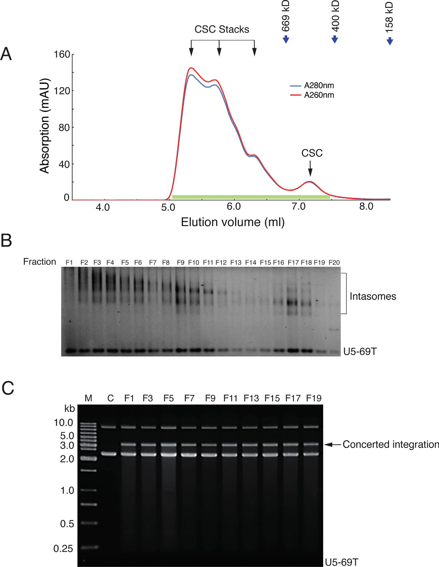
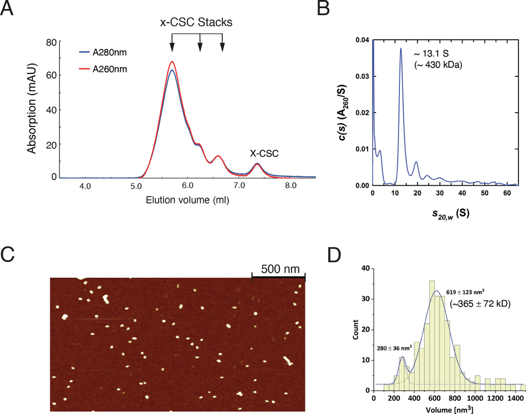
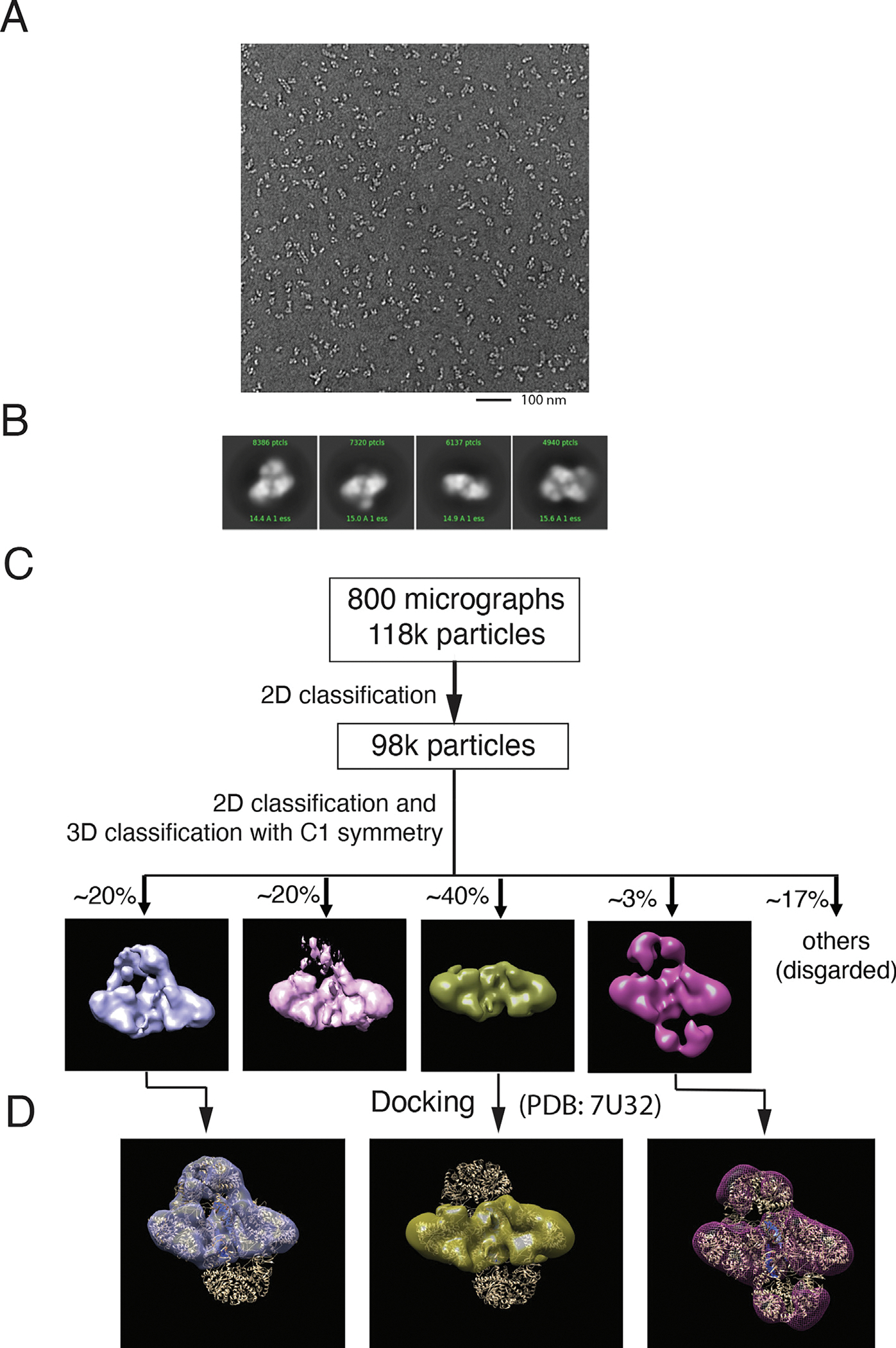
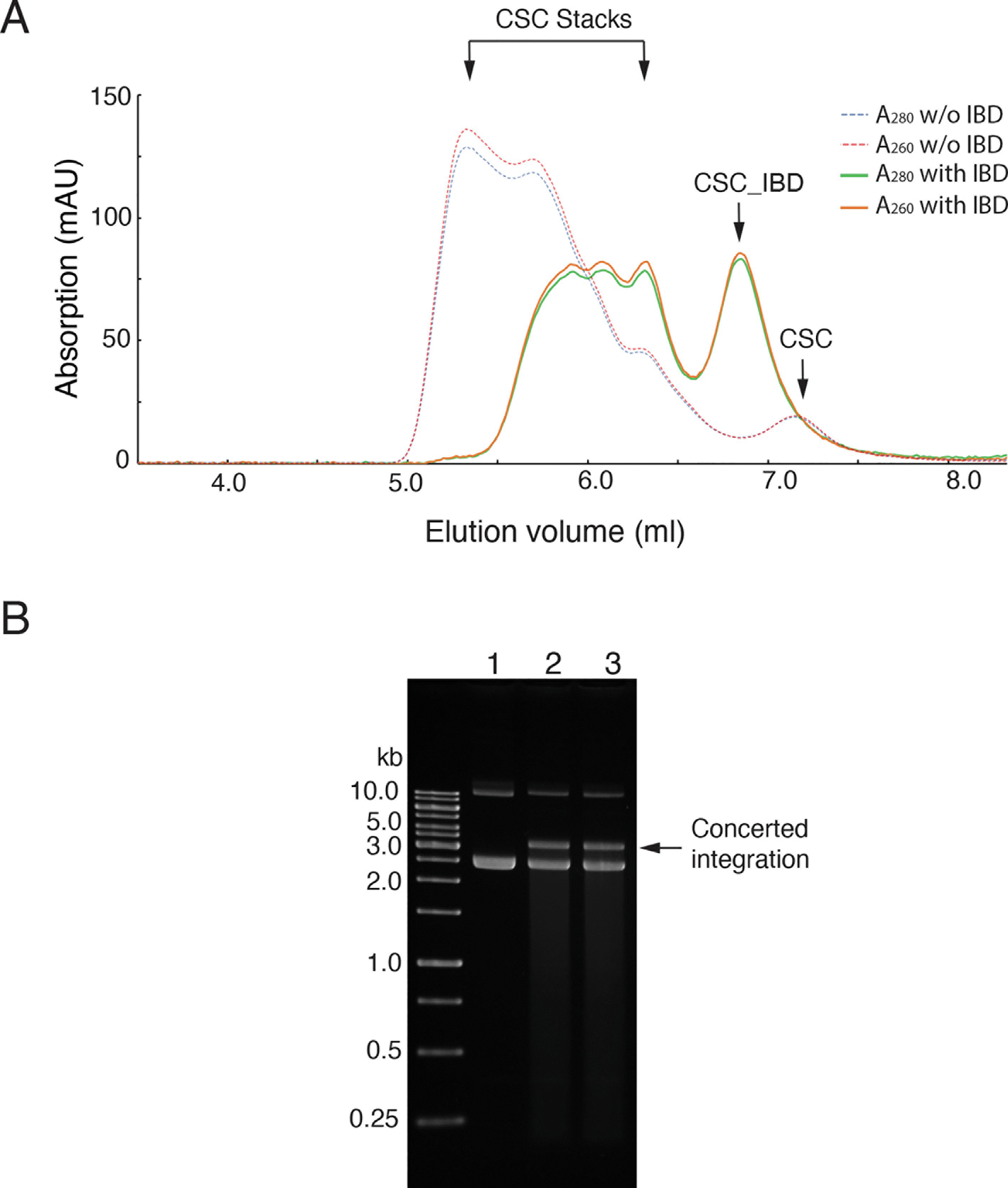
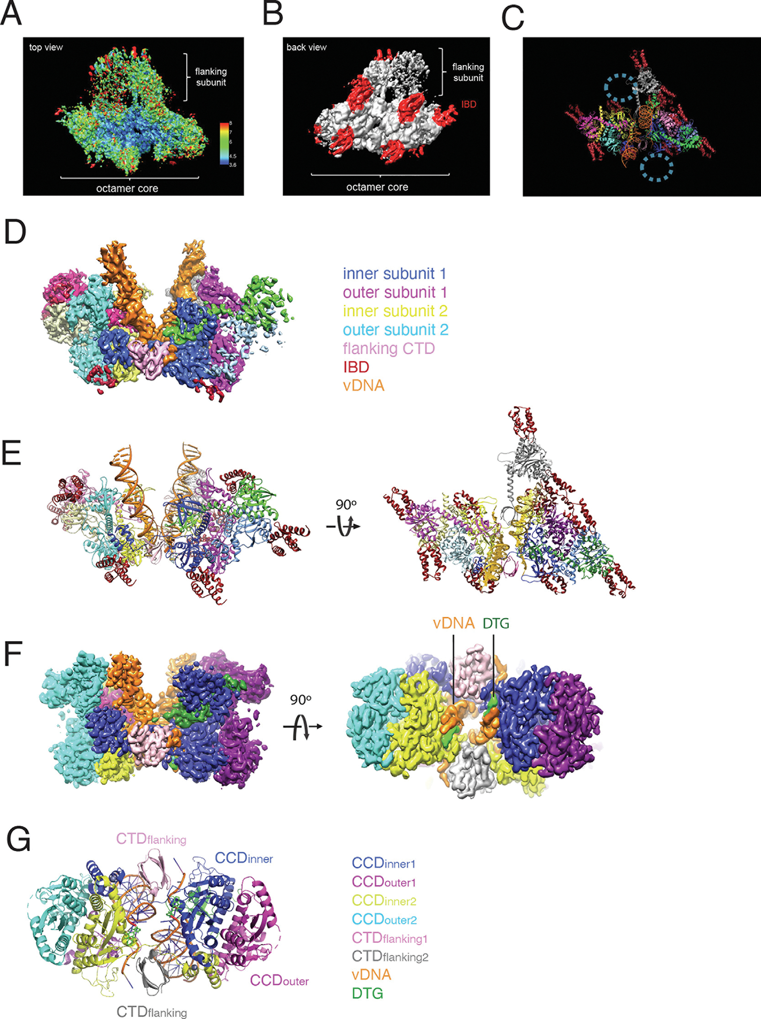
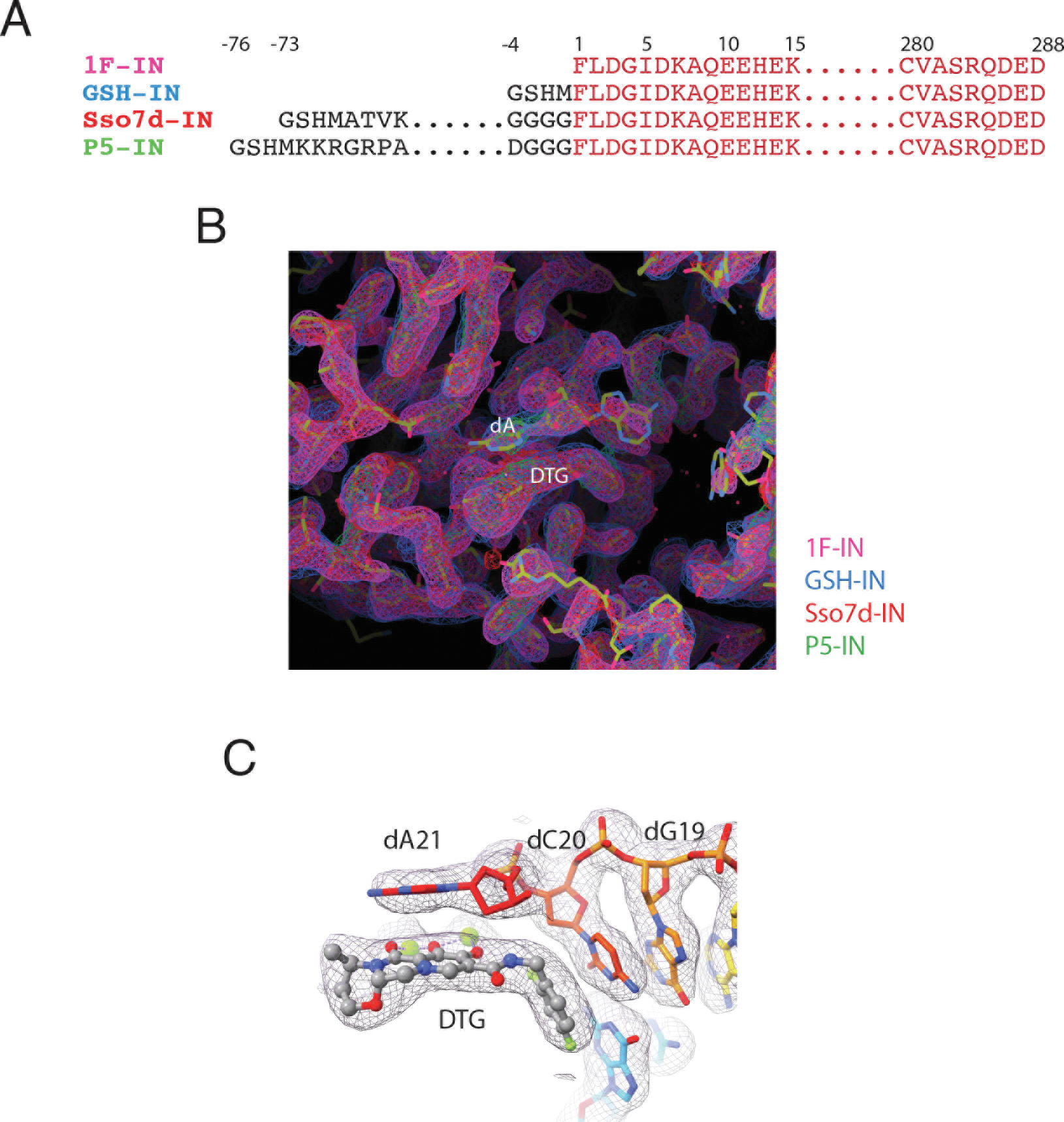
Retroviral DNA integration is mediated by nucleoprotein complexes (intasomes) in which a pair of viral DNA ends are bridged by a multimer of integrase (IN). Most of the high-resolution structures of HIV-1 intasomes are based on an HIV-1 IN with an Sso7d protein domain fused to the N-terminus. Sso7d-IN aggregates much less than wild-type IN and has been critical for structural studies of HIV-1 intasomes. Unexpectedly, these structures revealed that the common core architecture that mediates catalysis could be assembled in various ways, giving rise to both tetrameric and dodecameric intasomes, together with other less well-characterized species. This differs from related retroviruses that assemble unique multimeric intasomes, although the number of protomers in the intasome varies between viruses. The question of whether the additional Sso7d domain contributes to the heterogeneity of HIV-1 intasomes is therefore raised. We have addressed this by biochemical and structural studies of intasomes assembled with wild-type HIV-1 IN. Negative stain and cryo-EM reveal a similar range of multimeric intasome species as with Sso7d-IN with the same common core architecture. Stacks of intasomes resulting from domain swapping are also seen with both wild-type and Sso7d-IN intasomes. The propensity to assemble multimeric intasome species is, therefore, an intrinsic property of HIV-1 IN and is not conferred by the presence of the Sso7d domain. The recently solved intasome structures of different retroviral species, which have been reported to be tetrameric, octameric, dodecameric, and hexadecameric, highlight how a common intasome core architecture can be assembled in different ways for catalysis.

Keywords: HIV; integrase; integration; nucleoprotein complex; retrovirus.


Conflict of interest statement

Declaration of competing interest The authors declare that they have no known competing financial interests or personal relationships that could have appeared to influence the work reported in this paper.

Figures

Figure 1.
Figure 1.
Both Sso7d-IN and wild-type IN assemble heterogenous intasome species. A. Electrophoretic mobility shift assays (EMSA) of intasomes assembled with Sso7d-IN. B. EMSA of intasomes assembled with wild-type IN. Heterogeneity of Sso7d intasomes (C) and wild-type HIV-1 intasomes (D) visualized by negative stain electron microscopy (FEI Morgagni).
Figure 2.
Figure 2.
Size-exclusion chromatography and integration activity of intasomes assembled with wild-type IN. A. Elution profile of wild-type intasomes on TSKgel UltraSW HPLC (Tosho Bioscience) column. Larger species (CSC stacks) that are proto-intasome oligomeric stacks and the discrete cleaved synaptic complex (CSC) intasome species are indicated with arrows. The elution volumes of protein standards are shown. B. Fractions 1 to 20 (lane 1–20), corresponding to 5.1 min to 7.5 min elution volume were analyzed by native 3% agarose gel electrophoresis, and detected by a fluorescence scanner. Multiple species of intasomes are indicated; it is noted that the larger intasome species can partially dissociate during the gel electrophoresis. C. Every other fraction from F1 to F19 (5.1 ml to 7.5 ml elution volume) were normalized by A260nm and tested for strand transfer activity in the presence of 5 mM Mg2+ and 300 ng of supercoiled plasmid DNA. Integration products were separated on a 1.5% TBE agarose gel and visualized by ethidium bromide staining. Lane 2, Control with intasomes omitted from the reaction; lane 3–12, 2.5 nM intasomes from fraction F1-F19 was added into the reaction mixture. Concerted integration products are indicated by an arrow.
Figure 3.
Figure 3.
Analytical Ultracentrifuge and Atomic Force Microscopy (AFM) analysis of wild-type intasomes. A. Wild-type intasomes were crosslinked with 2.0 mM BS3 before TSKgel UltraSW gel filtration analysis. B. Fractions eluting between 7.1 ml to 7.5 ml (xCSC) were analyzed by sedimentation velocity at 35,000 rpm and 20 °C using the absorbance. A major species at 13.1 S has an estimated molar mass of 430 kDa. C. Representative AFM image of intasomes. xCSC was diluted in 20 mM Hepes pH 8.0, 0.75 M NaCl buffer to 1 nM and visualized by AFM. Scale bar: 500 nm. D. Histogram of the volume distribution of the intasomes. A fit to two Gaussian components is superimposed on the histogram. The volume of 619 ± 123 nm3 corresponds to a molecular mass of between 323 ± 65 and 365 ± 72 kDa depending on the hydration ratio of the intasome. The broad volume distribution is mainly the result of the tip-sample convolution due to the finite tip size. The variability of the molecular mass mean and variance stems from uncertainty of the hydration ratio of the intasomes which, for most proteins, ranges between 0.3 and 0.4.
Figure 4.
Figure 4.
Negative staining Electron Microscopy analysis of wild-type CSC intasomes. A. Representative negative staining-EM micrograph of wild-type HIV-1 IN CSC (scale bar, 100 nm). B. Representative 4 major 2D classes, only top views of the 2D classes are presented. C. Flow chart for negative staining EM data processing. Multiple rounds of 2D and 3D classification were used for removing obvious junk and damaged particles. C1 symmetry was applied for the 3D classification. Note that all classes contain “stable octamer core”, the major difference among the classes is the heterogeneity of flanking subunits region. Class 4 is consistent with hexadecamer intasomes and only represents about 3% of the total picked particles. D. MVV hexadecameric intasome atomic model (PDB: 7U32) was fitted in the three classes maps to demonstrate the missing flanking subunit in the heterogeneous HIV intasome species.
Figure 5.
Figure 5.
LEDGF-IBD can reduce the heterogeneity of intasomes and has no effect on the integration activity of intasomes. A. Wild-type intasomes were incubated at RT for 2 hrs with or without IBD and analyzed by TSKgel UltraSW gel filtration. Mono-dispersed and stacks of CSCs are indicated with arrows. The total amount of larger species of intasome are reduced by incubation with IBD. B, Equal amount of CSC and CSC_IBD was incubated with pGEM-9zf in the presence of 5 mM MgCl2. Integration products were analyzed on a 1.5% TBE agarose gel and visualized by ethidium bromide staining. Lane 1, intasomes were omitted from the reaction; lane 2, 2.5 nM wild-type intasomes were added to the reaction mixture; lane 3, 2.5 nM CSC_IBD was added in the reaction mixture; concerted integration products are indicated by an arrow.
Figure 6.
Figure 6.
Cryo-EM structure determination of wild-type CSC_IBD intasomes. A. 3D reconstruction density map of CSC_IBD refined with C1 imposed. Colors are according to the local resolution estimated by ResMap, and the color scale bar is shown on its right. B. Back view of the 3D reconstruction map. IBD densities are highlighted with red color. C. Atomic model of CSC_IBD. Regions with circles likely represent additional flexible IN protomers; due to the heterogeneity they could not be reliably interpreted. D. Segmentation of wild-type CSC_IBD cryo-EM map. Integrase protomers and vDNAs are color coded. E. Atomic model of the wild-type CSC_IBD. F. 3D reconstruction density map of CSC_IBD refined with CIC mask and C2 symmetry imposed. DTG molecules are indicated with green color. G. Atomic model of wild-type CIC.
Figure 7.
Figure 7.
The conserved intasome cores of different wild-type integrase variants are identical. A. Sequence comparison of the integrase constructs tested in this study. Construct 1F-IN (in red) has a native N-terminus. GSH-IN contains 4 extra amino acids (G-S-H-M) at the N terminus upon thrombin cleavage of affinity tag. P5-IN and Sso7d-IN are hyperactive fusion integrase proteins with an AT-hook peptide and Sso7d protein respectively, at the N terminus. B. Comparison of cryo-EM maps of different constructs. The maps are colored coded according to labels on the right, all the maps are overlapped to each other. Only atomic model 1F-IN is shown. C. DTG binding region of 1F-IN map superimposed with the final model.

Similar articles

Cited by

References

    1. Lesbats P, Engelman AN, Cherepanov P, (2016). Retroviral DNA integration. Chem. Rev. 116, 12730–12757. - PMC - PubMed
    1. Craigie R (2018). Nucleoprotein intermediates in HIV-1 DNA integration: structure and function of HIV-1 intasomes. In Virus Protein and Nucleoprotein Complexes (Harris JR & Bhella D, eds), pp. 189–210. - PMC - PubMed
    1. Engelman A, Mizuuchi K, Craigie R, (1991). HIV-1 DNA integration: mechanism of viral DNA cleavage and DNA strand transfer. Cell 67, 1211–1221. - PubMed
    1. Passos DO, Li M, Yang RB, Rebensburg SV, Ghirlando R, Jeon Y, et al., (2017). Cryo-EM structures and atomic model of the HIV-1 strand transfer complex intasome. Science 355, 89–92. - PMC - PubMed
    1. Passos DO, Li M, Jozwik IK, Zhao XZ, Santos-Martins D, Yang R, et al., (2020). Structural basis for strand-transfer inhibitor binding to HIV intasomes. Science 367, 810. - PMC - PubMed

Publication types

LinkOut - more resources