. 2024 Apr 6;8(1):86.
doi: 10.1038/s41698-024-00556-3.

Enhanced plant-derived vesicles for nucleotide delivery for cancer therapy

Affiliations

Enhanced plant-derived vesicles for nucleotide delivery for cancer therapy

Sara Corvigno et al. NPJ Precis Oncol. .

Abstract

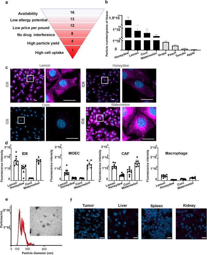
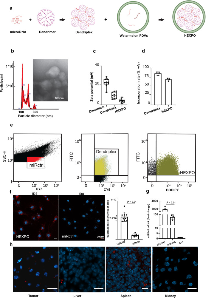
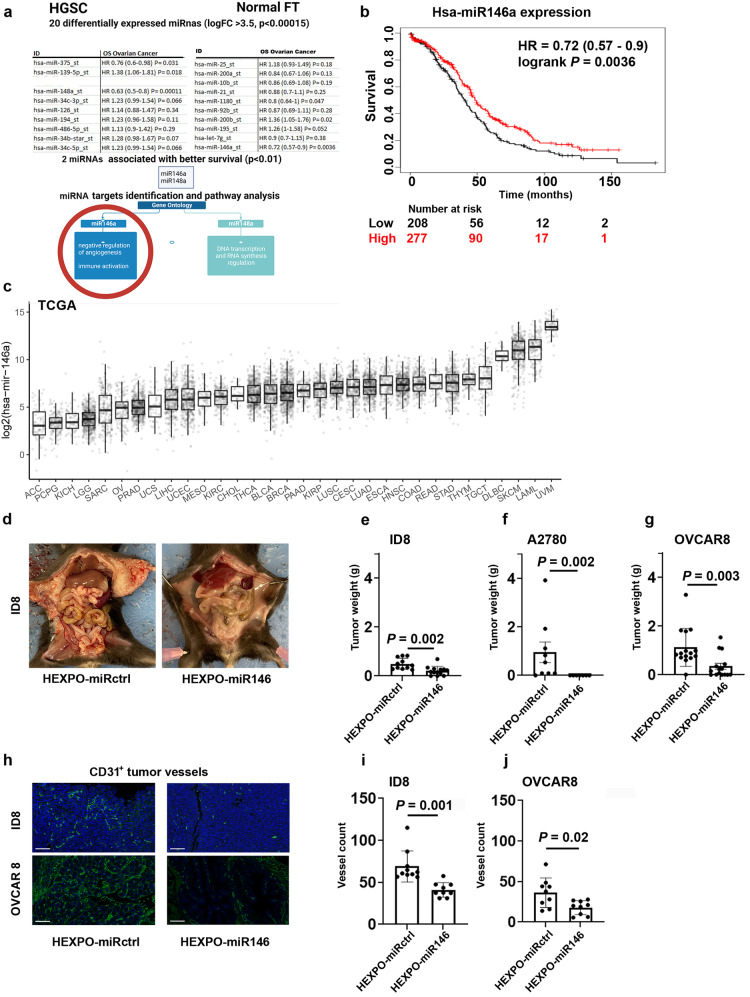
Small RNAs (microRNAs [miRNAs] or small interfering RNAs [siRNAs]) are effective tools for cancer therapy, but many of the existing carriers for their delivery are limited by low bioavailability, insufficient loading, impaired transport across biological barriers, and low delivery into the tumor microenvironment. Extracellular vesicle (EV)-based communication in mammalian and plant systems is important for many physiological and pathological processes, and EVs show promise as carriers for RNA interference molecules. However, some fundamental issues limit their use, such as insufficient cargo loading and low potential for scaling production. Plant-derived vesicles (PDVs) are membrane-coated vesicles released in the apoplastic fluid of plants that contain biomolecules that play a role in several biological mechanisms. Here, we developed an alternative approach to deliver miRNA for cancer therapy using PDVs. We isolated vesicles from watermelon and formulated a hybrid, exosomal, polymeric system in which PDVs were combined with a dendrimer bound to miRNA146 mimic. Third generation PAMAM was chosen due to its high branching structure and versatility for loading molecules of interest. We performed several in vivo experiments to demonstrate the therapeutic efficacy of our compound and explored in vitro biological mechanisms underlying the anti-tumor effects of miRNA146, which are mostly related to its anti-angiogenic activity.


Conflict of interest statement

S.C., Y.L., E.B., E.S., N.N.B., A.L.A., S.N., C.R.A., H.C., Y.W., L.Z., S.L., and G.L.B. declare that they have no competing interests. These authors declare the following competing interests: H.L. is a shareholder and scientific advisor of Precision Scientific Ltd. A.K.S. declares the following competing financial or non-financial interests: he is a shareholder of BioPath and a consultant for Merck, AstraZeneca, Onxeo, ImmunoGen, Ivlon, GSK, and Kiyatec.

Figures

Fig. 1
Fig. 1. Selection and characterization of plant-derived vesicles (PDVs).
a Scheme of the systematic approach adopted to select watermelon PDVs. b Particle yield from selected species. c Cell uptake of PDVs from selected species. The left panels show 20× magnification of random fields, and the right panels show 63× magnification of the same areas. Scale bar = 20 µm. d Quantification of cell uptake. Bars represent the means and standard deviations of the ratios of positive cells to fluorescent control microRNA (miRNA) on total cells counted on the basis of the 4′,6-diamidino-2-phenylindole (DAPI) signal. e Characterization of PDVs according to Nanotracking analysis and transmission electron microscopy (TEM) imaging results (Scale bar = 100 nm). f Fluorescence-based imaging of the tumor, liver, spleen, and kidney from an ID8 model after intraperitoneal administration of fluorescent watermelon PDVs. Scale bar = 10 µm. In the bar graphs, the whiskers represent the standard deviation from the mean. MOECs mouse ovarian endothelial cells, CAFs cancer-associated fibroblasts, mL milliliter, nm nanometer, µm micrometer.
Fig. 2
Fig. 2. Hybrid exosomal polymeric (HEXPO) nanoparticle formulation and characterization.
a Schematic representation of HEXPO formulation. b Characterization of HEXPO with nanotracking analysis and transmission electron microscopy (TEM) imaging. c Measurement of HEXPO charge. d Quantification of dendriplex incorporation into HEXPO, measured through spectrophotometry. e Quantification of the loading efficiency of control microRNA (miRctrl) plus dendrimers into HEXPO, measured via small-particle flow cytometry. Gating for the population of interest was based on negative controls (unstained compensation beads, unstained PDVs, and 1:1000 diluted BODIPY), single color controls (BODIPY-stained PDVs, Cy5-labeled polystyrene beads, and FITC-labeled compensation beads bound to PAMAM dendrimer), and fluorescence minus one (FMO) controls (cy5-labeled polystyrene beads loaded in BODIPY-stained PDVs, FITC-conjugated PAMAM dendrimers loaded in BODIPY-stained PDVs, and cy5-labeled-miRNA control loaded in FITC-conjugated PAMAM dendrimers bound to compensation beads). f Uptake of fluorescent miRctrl after HEXPO treatment of ID8 cells (left) and free miRctrl treatment (right), with quantification. Scale bar = 20 µm. g Quantitative polymerase chain reaction of miR146a after HEXPO and miR146 treatment (versus untreated control) of ID8 cells. h Fluorescence-based imaging of the tumor, liver, spleen, and kidneys from an ID8 model after intraperitoneal administration of HEXPO with fluorescent miRctrl. In the histograms, the whiskers represent standard deviation from the mean. Scale bar = 10 µm. CY5 cyanine 5, Bodipy TR Bodipy tracker, Fc fold change, mV millivolt, w/v weight per volume, nm nanometer, miRctrl miRNA-negative control, n of cells number of cells, miR146 miRNA 146a, Ctrl control cells, µm micrometer, P p value.
Fig. 3
Fig. 3. Selection of miR146a.
a Scheme of the systematic approach adopted to select miR146a. b Kaplan–Meier curve representing the survival of patients with ovarian cancer with high or low expression of miR146a using data from a publicly available database (kmplot.com). c MiR146a expression from The Cancer Genome Atlas (TCGA) RNA sequencing data of different tumors (https://ropensci.org/blog/2021/11/16/how-to-cite-r-and-r-packages/). d ID8 in vivo model treated with hybrid exosomal polymeric control microRNA (HEXPO-miRctrl) and hybrid exosomal polymeric miR146a (HEXPO-miR146). Quantification of tumor weight in e ID8, f A2780, and g OVCAR8 models (two treatment groups per model). h Representative images of CD31 vessel-specific staining of formalin-fixed, paraffin-embedded (FFPE) sections from ID8 and OVCAR8 models. Scale bar = 100 µm. Quantification of vessel numbers in sections from FFPE tumor samples from the i ID8 model and j OVCAR8 model. In the bar graphs, dots represent each sample, bars represent means, and whiskers represent the standard deviations from the mean (standard error of the mean for panel f). FT fallopian tube, OS overall survival, HR hazard ratio, P p-value, logFC logarithmic fold change of expression, TCGA The Cancer Genome Atlas Program, miRctrl miRNA negative control, g grams.
Fig. 4
Fig. 4. Dual anti-angiogenic effect of miR146a.
a Representative 5X images of wells from the tube-formation assay of control miRNA (miRctrl)–transfected RF24 cells treated with conditioned medium from miRctrl- and miR146a-transfected OVCAR8 and A2780 cells and miR146a-transfected RF24 cells. bd Quantification of tube counts from the tube formation assays in a. e Representative images from the Matrigel plug assay performed in nude mice, with quantification in f. g Angiogenesis array performed on conditioned media from miRctrl- and miR146a-transfected OVCAR8 and A2780 cells (in red SERPINE1 dots), with quantification in h. Heatmaps of the gene expression profiles of miRctrl- and miR146a-transfected i A2780 and OVCAR8 cells and j RF24 cells. k Representative image of Gene Ontology pathways enriched in upregulated and downregulated genes in miR146a-transfected RF24, A2780, and OVCAR8 cells. In the histograms, dots represent each sample, bars represent the mean, and whiskers represent the standard deviations from the mean. CM conditioned medium, M medium, p p value, miRctrl miRNA-negative control, PBS phosphate-buffered saline, rhVEGF recombinant human vascular endothelial growth factor, OD optical densities, ICAM2 intercellular adhesion molecule 2, PODXL podocalyxin-like, SYTL1 synaptotagmin-like protein 1, PTPRH protein tyrosine phosphatase receptor type H, HYAL2 hyaluronidase 2, THBD thrombomodulin, NEU1 neuraminidase 1, PQLC2 PQ loop repeat‐containing, BRI3 brain protein I3, TMBIM1 transmembrane BAX inhibitor motif containing 1, OSTM1 osteopetrosis-associated transmembrane protein 1, RAB7A member RAS oncogene family, TMEM150C transmembrane protein 150C, TRPM2 transient receptor potential melastatin 2, TM6SF1 transmembrane 6 superfamily member 1, MCOLN1 mucolipin transient receptor potential cation channel 1, THBS1 thrombospondin 1, CARD10 caspase recruitment domain family member 10, EGR3 early growth response protein 3, PTGS2 prostaglandin-endoperoxide synthase 2, SERPINE1 serine proteinase inhibitor, Clade E1, APOLD1 apolipoprotein L domain containing 1, TNFRSF12A tumor necrosis factor receptor superfamily, member 12a, CYR61 cysteine-rich angiogenic inducer 61, BCL2L11 Bcl-2-like protein 11, CASP9 caspase 9, ABCB1 adenosine triphosphate (ATP)-binding cassette transporter 1, BCL6 B-cell lymphoma 6, CXCL8 C-X-C motif chemokine ligand 8, p value.

References

    1. Carthew RW, Sontheimer EJ. Origins and Mechanisms of miRNAs and siRNAs. Cell. 2009;136:642–655. doi: 10.1016/j.cell.2009.01.035. - DOI - PMC - PubMed
    1. Mainini F, Eccles MR. Lipid and Polymer-Based Nanoparticle siRNA Delivery Systems for Cancer Therapy. Molecules. 2020;25:2692. doi: 10.3390/molecules25112692. - DOI - PMC - PubMed
    1. Yadav S, Shekhawat M, Jahagirdar D, Kumar Sharma N. Natural and artificial small RNAs: a promising avenue of nucleic acid therapeutics for cancer. Cancer Biol. Med. 2017;14:242–253. doi: 10.20892/j.issn.2095-3941.2017.0038. - DOI - PMC - PubMed
    1. Zuckerman JE, Davis ME. Clinical experiences with systemically administered siRNA-based therapeutics in cancer. Nat. Rev. Drug Discov. 2015;14:843–856. doi: 10.1038/nrd4685. - DOI - PubMed
    1. Blanco E, Shen H, Ferrari M. Principles of nanoparticle design for overcoming biological barriers to drug delivery. Nat. Biotechnol. 2015;33:941–951. doi: 10.1038/nbt.3330. - DOI - PMC - PubMed