. 2024 Jul:308:122562.
doi: 10.1016/j.biomaterials.2024.122562. Epub 2024 Apr 1.

Engineered extracellular vesicle-based gene therapy for the treatment of discogenic back pain

Affiliations

Engineered extracellular vesicle-based gene therapy for the treatment of discogenic back pain

Shirley N Tang et al. Biomaterials. 2024 Jul.

Abstract

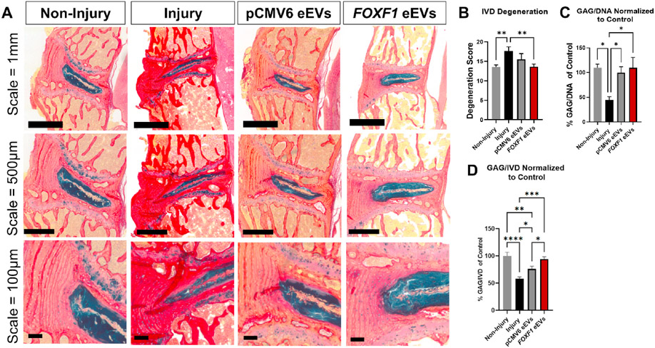
Painful musculoskeletal disorders such as intervertebral disc (IVD) degeneration associated with chronic low back pain (termed "Discogenic back pain", DBP), are a significant socio-economic burden worldwide and contribute to the growing opioid crisis. Yet there are very few if any successful interventions that can restore the tissue's structure and function while also addressing the symptomatic pain. Here we have developed a novel non-viral gene therapy, using engineered extracellular vesicles (eEVs) to deliver the developmental transcription factor FOXF1 to the degenerated IVD in an in vivo model. Injured IVDs treated with eEVs loaded with FOXF1 demonstrated robust sex-specific reductions in pain behaviors compared to control groups. Furthermore, significant restoration of IVD structure and function in animals treated with FOXF1 eEVs were observed, with significant increases in disc height, tissue hydration, proteoglycan content, and mechanical properties. This is the first study to successfully restore tissue function while modulating pain behaviors in an animal model of DBP using eEV-based non-viral delivery of transcription factor genes. Such a strategy can be readily translated to other painful musculoskeletal disorders.

Keywords: Cell reprogramming; Engineered extracellular vesicles; Intervertebral disc; Low back pain; Nanocarriers; Non-viral gene delivery.


Conflict of interest statement

Declaration of competing interest The authors declare that they have no known competing financial interests or personal relationships that could have appeared to influence the work reported in this paper.

Figures

Fig. 1.
Fig. 1.. Study Design: Overview of the study methods describing the surgery groups, eEV injections, and evaluated dependent variables.
(1) Primary mouse embryonic fibroblasts (PMEFs) were transfected via electroporation with pCMV6 or FOXF1 plasmids followed by engineered extracellular vesicle (eEV) isolation. (2) Surgery was performed on 15-week-old male and female wild type mice (N = 11–12 per experimental group, N = 5–6 per sex), where non-injury controls were animals that underwent surgical exposure of IVD with no puncture, injured animals sustained IVD puncture with saline injection, and pCMV6 eEV and FOXF1 eEVs treated animals received a single injection of eEVs, respectively in the L4/L5, L5/L6, and L6/S1 IVDs. Micrograph (right panel) highlighting IVDs (black arrows) within the surgical field. (3) Pain behavioral assessments were conducted throughout pre operation and 12 weeks post operation and treatment, with assessment of function and structure at 12 weeks by magnetic resonance imaging (MRI), micro computed tomography (μCT/microCT), mechanical tests, Alcian blue/picrosirius red staining (AB/PSR), Dimethyl Methylene Assay (DMMB), and immunohistochemistry (IHC).
Fig. 2.
Fig. 2.. Non-viral transfection of primary mouse fibroblasts with the developmental transcription factor FOXF1 leads to the generation of FOXF1-loaded engineered extracellular vesicles (eEVs).
(A) Schematic diagram illustrating how eEVs are derived from donor cells after transfection with a plasmid encoding for the transcription factor FOXF1. qRT-PCR analysis showing (B) robust upregulation of gene expression (mRNA transcripts) and (C) plasmid DNA copy numbers of FOXF1 in donor cells 24 h after transfection compared to pCMV6-transfected cells (control) (n = 3). (D) Nanosight analysis showing an average particle concentration in the order of ~1.3X10¹¹ particles per mL with an average size of ~280 nm for FOXF1 and pCMV6 eEVs (n = 3). (E) qRT-PCR showing that the level of FOXF1 mRNA packed in the eEVs was 4 orders of magnitude higher compared to pCMV6 eEVs (n = 3). (F) Absolute qRT-PCR analysis showing copy numbers of FOXF1 plasmid DNA packed in eEVs compared to pCMV6 eEVs (n = 3). (G) Western blot characterization confirmed the presence of the EV markers CD63 and TSG101, the cytoskeletal marker tubulin in the donor cells, FOXF1 eEVs and pCMV6 eEVs, and the expression of calnexin (endoplasmic reticulum protein) in the donor cells and the pCMV6 eEVs with a reduced expression in FOXF1 eEV formulation. All error bars are shown as standard error of the mean (SEM). *p < 0.05 and **p < 0.001. Two-tail t-test.
Fig. 3.
Fig. 3.. FOXF1 eEV treatment decreases evoked and spontaneous pain behaviors in a mouse lumbar disc puncture model.
(A) Grip Test/Hanging Wire of (left) female and (right) male mice over 12 weeks for Non-Injured, Injured, pCMV6 eEV, and FOXF1 eEV treated animals respectively recorded as grip time before falling (N = 9–10/sex). (B) Tail suspension assessment of (left) female and (right) male mice over 12 weeks was recorded as time mobile (N = 6–7/sex). (C) Cold Plate assessment of (left) female and (right) Male mice over 12 weeks recorded as the time to paw withdrawal or licking and graphed as hypersensitivity (N = 9–10/sex). (D) Open field assessment of (left) female and (right) male mice over 12 weeks recorded as the distance traveled (N = 9–10/sex). All data is normalized to the pre-op recorded values of each individual mouse to account for pain behaviors pre-operation and shown as the difference at 4–12 weeks post-operation compared to pre-op measures (Δ Time or Δ Distance). Statistics = Two Way ANOVA with multiple comparisons. Letters on each graph indicate significant differences (p < 0.05) between respective groups where Non-Injury = C, Injury = I, pCMV6 −eEV treated animals = S, and FOXF1 −eEV treated animals = F. All error bars are shown as standard error of the mean (SEM). *p < 0.01, **p < 0.001, ***p < 0.0001. Data for open field resting and rearing behaviors can be found in Supplemental Fig. 3. Additional parameters of this respective 2 Way ANOVA can be found in Supplemental Table 2.
Fig. 4.
Fig. 4.. FOXF1 eEV treatment mitigates structural changes of the IVD by maintaining tissue hydration and disc height after injury.
(A) Representative T2 sagittal magnetic resonance imaging (MRI) of non-injured, injured, pCMV6 eEV treated animals, and FOXF1eEV treated animals post euthanasia, depicting the entire lumbar spine (L1-L6) with arrows indicating L4/L5, L5/L6, and L6/S1 IVDs (below dashed line). (B) Average T2 IVD Disc Intensity of injured discs quantified from MRI as relative IVD intensity normalized to the intensity of control groups using ImageJ. (N = 11–12, ** = p < 0.01). (C) Representative microCT images of Non-Injured, Injured, pCMV6 eEV treated animals, and FOXF1eEV treated animals post euthanasia depicting the entire lumbar spine (L1-L6) with arrows indicating L4/L5, L5/L6, and L6/S1 IVDs. (D) MicroCT images were reconstructed using CTVox and analyzed on ImageJ for the average disc height of each IVD (posterior, middle, anterior heights) and normalized as % disc height with respect to control. (N = 11–12, * = p < 0.05, ** = p < 0.01). All Error bars indicate standard error mean (SEM). Statistics = one-way ANOVA with Fishers LSD.
Fig. 5.
Fig. 5.. Therapeutic intervention using FOXF1 eEVs promotes restoration of IVD structure by increasing tissue stability.
(A) Image of custom-designed gripping apparatus for mouse motion segments mechanical analysis and image of mouse motion segment (scale bar = 1 mm). (B) Schematic demonstrating the mechanical parameters assessed from axial testing, including the Neutral Zone (NZ) Stiffness parameters using the Stiffness threshold method from load-displacement curves [32]. (C) Quantified NZ stiffness normalized to IVD height and area for each group (N = 11–12, * = p < 0.05). (D) Average creep curves calculated from mean parameter values of the five-parameter viscoelastic model [33] see Supplemental Fig. 5. The normalized creep displacement response of injured IVDs (gray line) was magnified compared to non-injured and eEV treated IVDs. (E) The total normalized creep displacement of the injured IVDs was significantly greater than FOXF1 eEV treated IVDs (N = 11–12, * = p < 0.05). (F) The normalized elastic stiffness (SE) of the injured IVDs was significantly lower than the pCMV6 eEV group (N = 11–12, * = p < 0.05). Collectively, results demonstrate that the punctured IVDs deformed more under the same applied axial or creep loads than FOXF1 eEV treated IVDs suggesting FOXF1 eEV treatment restored mechanical function in axial and creep loading. *p < 0.05 Supplemental Data on overall loading behaviors and creep behaviors can be found in Supplemental Fig. 4 & 5.
Fig. 6.
Fig. 6.. IVD structure and extracellular matrix content is promoted via FOXF1 eEV treatment by increasing GAG accumulation.
(A) Alcian blue Picrosirius Red (AB/PSR) histological stains of L5/L6 mouse intervertebral disc motion segments for Non-Injured, Injured, pCMV6 eEV treated animals, and FOXF1eEV treated animals at different magnifications (scale bars = 1 mm, 500 μm, and 100 μm). (B) Quantified degeneration scores of IVDs in each group where higher scores indicated increased degeneration. (N = 9–11, ** = p < 0.01). (C) Proteoglycan (sGAG) content of whole isolated IVDs, digested in papain, and normalized to respective digest DNA and % of non-injured control (N = 11–12, * = p < 0.05). (D) sGAG content of whole isolated IVDs normalized to IVD dry weight post lyophilization. All Error bars indicate standard error mean (SEM). Statistics = one-way ANOVA with Fishers LSD.
Fig. 7.
Fig. 7.. FOXF1 Immunohistochemistry (IHC) of mouse L5/L6 intervertebral disc (IVD) motion segments depicting IVDs sandwiched between respective L5 and L6 vertebrae.
From left to right as shown are representative images of Non-Injured, Injured, pCMV6 eEV treated animals, and FOXF1eEV treated animals along with a negative antibody control at different magnifications (scale bars = 1 mm, 500 μm, and 100 μm). Red arrows indicate positive staining. Although no statistical differences were found in computing % positive FOXF1 signal for each group (Supplemental Fig. 8), positive signal distribution of FOXF1 eEV treated animals showed narrower colocalization of the FOXF1 positive regions with the cell nuclei compared to injured animals.

References

    1. Schwarzer AC, Aprill CN, Derby R, Fortin J, Kine G, Bogduk N, The prevalence and clinical features of internal disc disruption in patients with chronic low back pain, Spine 20 (1995) 1878–1883. - PubMed
    1. Luoma K, Riihimäki H, Luukkonen R, Raininko R, Viikari-Juntura E, Lamminen A, Low back pain in relation to lumbar disc degeneration, Spine 25 (2000) 487–492. - PubMed
    1. Wiet MG, Piscioneri A, Khan SN, Ballinger MN, Hoyland JA, Purmessur D, Mast Cell-Intervertebral disc cell interactions regulate inflammation, catabolism and angiogenesis in Discogenic Back Pain, Sci. Rep 7 (2017) 12492. - PMC - PubMed
    1. Richards J, Tang S, Gunsch G, Sul P, Wiet M, Flanigan DC, Khan SN, Moore S, Walter B, Purmessur D, Mast cell/proteinase activated receptor 2 (PAR2) mediated interactions in the pathogenesis of discogenic back pain, Front. Cell. Neurosci 13 (2019). - PMC - PubMed
    1. Smith LJ, Nerurkar NL, Choi KS, Harfe BD, Elliott DM, Degeneration and regeneration of the intervertebral disc: lessons from development, Dis Model Mech 4 (2011) 31–41. - PMC - PubMed

Publication types

MeSH terms

Substances

LinkOut - more resources