

Effect of poly-L-lactic acid and polydioxanone biostimulators on type I and III collagen biosynthesis
- PMID: 38584576
- PMCID: PMC10999943
- DOI: 10.1111/srt.13681
Effect of poly-L-lactic acid and polydioxanone biostimulators on type I and III collagen biosynthesis
Abstract
Objective: Safe, effective, and biocompatible minimally invasive procedures with the potential to stimulate collagen production have been made to recover dermal thickness and skin quality. The main of this animal model experiment was to observe the effect of poly-L-lactic acid (PLLA) and polydioxanone (PDO) biostimulators in collagen I and III after hypodermal injection.
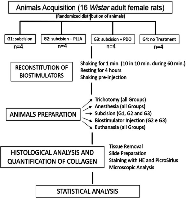
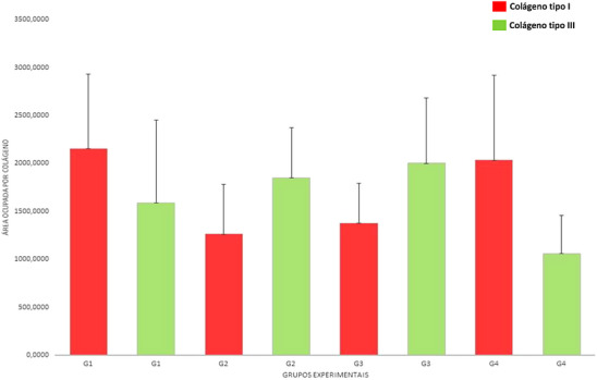
Methodology: Sixteen adult female rats (Wistar) were randomized into four groups and had dorsal treatment with: G1: hypodermic subcision (HS) only; G2: HS and PLLA hypodermic injection (HI), G3: HS and PDO HI; G4: Control, with no treatment.
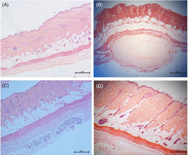
Results: In histochemical, it was observed hypodermal and dermal tissue in more organized thickness in G3 and in G4 when compared to G1 and G2. There was few difference in G1 compared to G4. The tissue of G2 showed irregularities in the arrangement of collagen fibers, less defined structure and lower distribution of type I collagen compared to the other groups. There is a greater tendency for the proportions of type III collagen among tissues treated with both biostimulators (G2 and G3). PLLA and PDO had relatively similar percentages of collagen when compared to G4. The amount of type I collagen was higher in tissues treated with subcision, while type III collagen was higher in tissues treated with both biostimulators.
Conclusion: G3 showed better performance in collagen production, although small, when compared with G2.
Keywords: biostimulators; collagen; polydioxanone; poly‐L‐Latic acid; rejuvenation; skin; subcision.
© 2024 The Authors. Skin Research and Technology published by John Wiley & Sons Ltd.
Figures

References
-
- Baumann L. Skin ageing and its treatment: review article. J Pathol. 2007;211:241‐251. - PubMed
-
- Zhang SY, Zhou XY, Zhou XL, et al. Subtype‐ specific inherited predisposition to pemphigus in the Chinese population. Br J Dermatol. 2019;180:828‐835. - PubMed
-
- El‐Domyati M, Attia S, Saleh F, et al. Intrinsic aging vs. photoaging: a comparative histopathological, immunohistochemical, and ultrastructural study of skin. Exp Dermatol. 2002;11:398‐405. - PubMed
MeSH terms
Substances
LinkOut - more resources
Full Text Sources
