Review
. 2024 Mar 22:15:1373507.
doi: 10.3389/fphar.2024.1373507. eCollection 2024.

Large conductance voltage-and calcium-activated K+ (BK) channel in health and disease

Affiliations
Review

Large conductance voltage-and calcium-activated K+ (BK) channel in health and disease

Felipe Echeverría et al. Front Pharmacol. .

Abstract

Large Conductance Voltage- and Calcium-activated K+ (BK) channels are transmembrane pore-forming proteins that regulate cell excitability and are also expressed in non-excitable cells. They play a role in regulating vascular tone, neuronal excitability, neurotransmitter release, and muscle contraction. Dysfunction of the BK channel can lead to arterial hypertension, hearing disorders, epilepsy, and ataxia. Here, we provide an overview of BK channel functioning and the implications of its abnormal functioning in various diseases. Understanding the function of BK channels is crucial for comprehending the mechanisms involved in regulating vital physiological processes, both in normal and pathological conditions, controlled by BK. This understanding may lead to the development of therapeutic interventions to address BK channelopathies.

Keywords: BK channel; KCNMA1; cellular excitability; channelopathies; molecular and cellular mechanisms; physiology.


Conflict of interest statement

The authors declare that the research was conducted in the absence of any commercial or financial relationships that could be construed as a potential conflict of interest. The handling editor DT declared a past co-authorship with the author RL. The author(s) declared that they were an editorial board member of Frontiers, at the time of submission. This had no impact on the peer review process and the final decision.

Figures

FIGURE 1
FIGURE 1
Model of structure and function of the BK channel. (A) Topology of monomeric BK channel α subunit showing the extracellular N-terminal region, the transmembrane domain (TMD) composed by the voltage sensor domain (VSD) where gating charges (R210 and R213 in the segment S4) reside (Carrasquel-Ursulaez et al., 2022) the pore domain (PD), and the intracellular C-terminal domain (CTD) where the RCK1 and RCK2 Ca2+ binding sites reside. (B) Schematic H-A allosteric gating model for BK channel activation by voltage and calcium (Horrigan and Aldrich, 2002). The PD can exist in close (C) or open (O) configurations defined by the equilibrium constant L. Each of the four VSD could be at rest (R) or activated (A) governed by the equilibrium constant J. The Ca2+ sensors may have calcium bound (X-Ca2+) or unbound (X) dominated by the equilibrium constant K. Each module is allosterically coupled to pore opening, governed by the indicated equilibrium constants The C for CTD-PD coupling, D for VSD-PD coupling, and E for VSD-CTD coupling. (C, D) Human BK channel cryo-EM Ca2+-bound structure (PDB:6V38) showing the non-domain swapped disposition of VSD and PD, whereas CTDs have a domain-swapped configuration forming the gating ring; the S6-RCK1 linker connects TMD and CTD.
FIGURE 2
FIGURE 2
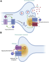
Role of BK channels in neuronal excitability and neurotransmitter release. BK channels act as feedback regulators to control neuronal excitability. Their activation caused by depolarization results in a massive efflux of K+ ions, hyperpolarizing the cell membrane. Through their voltage sensitivity and ability to detect changes in Ca2+ concentration, activation of BK channels modulates neuronal excitability and limits transmitter release (GABA). BK channels co-localize and form nanodomains with voltage-gated Ca2+ (CaV) channels in presynaptic compartments (A) and N-methyl-D-aspartate (NMDA) receptors in postsynaptic terminals (B). The co-localization of BK and CaV channels provides BK channels with an effective local Ca2+ concentration for their activation and function.
FIGURE 3
FIGURE 3
Effect of BK channel function on action potential (AP) shape, size, and firing rate. AP simulation using NEURON (Mahapatra et al., 2018). This model considers multiple ionic conductances, including calcium channels (L-type and T-type), voltage-gated potassium channels (Kv1, KCNQ), ATP-activated potassium channels, voltage- and calcium-activated potassium channels (BK, SK, IK), and a leak current. The plot shows three APs with different percentages of BK conductance (0%, 50%, 100%), exemplifying BK channel role in APs firing frequency. A reduction in BK channel conductance (GBK) increases smooth muscle cell excitability and may contribute to pathological conditions such as hypertension, erectile dysfunction (ED), and overactive bladder (OAB).
FIGURE 4
FIGURE 4
Role of the BK channel in regulating arterial tone and smooth muscle contraction. Depolarization of arterial smooth muscle cells by activation of G-protein coupled receptors (GPCRs) triggers the opening of Cav channels, leading to Ca2+ influx followed by sarcoplasmic reticulum Ca2+ release (Ca2+ wave) from the inositol triphosphate receptor (IP3R), which promotes cell contraction. Under conditions favoring vasodilation, Ca2+ release from the ryanodine receptor (RyR) is stimulated by luminal SR Ca2+ levels, resulting in a localized Ca2+ spark that activates the BK channel. This leads to membrane hyperpolarization and subsequent inhibition of Cav channel activity. The balanced interplay between BK, RyR, and Cav channels can promote relaxation or contraction depending on the regulation of their activity.

References

    1. Aaronson P. I., Sarwar U., Gin S., Rockenbauch U., Connolly M., Tillet A., et al. (2006). A role for voltage-gated, but not Ca 2+-activated, K + channels in regulating spontaneous contractile activity in myometrium from virgin and pregnant rats. Br. J. Pharmacol. 147 (7), 815–824. 10.1038/sj.bjp.0706644 - DOI - PMC - PubMed
    1. Alle H., Kubota H., Geiger J. R. P. (2011). Sparse but highly efficient Kv3 outpace BKca channels in action potential repolarization at hippocampal mossy fiber boutons. J. Neurosci. 31 (22), 8001–8012. 10.1523/JNEUROSCI.0972-11.2011 - DOI - PMC - PubMed
    1. Amberg G. C., Bonev A. D., Rossow C. F., Nelson M. T., Santana L. F. (2003). Modulation of the molecular composition of large conductance, Ca 2+ activated K+ channels in vascular smooth muscle during hypertension. J. Clin. Investigation 112 (5), 717–724. 10.1172/JCI18684 - DOI - PMC - PubMed
    1. Amberg G. C., Santana L. F. (2003). Downregulation of the BK channel beta1 subunit in genetic hypertension. Circulation Res. 93 (10), 965–971. 10.1161/01.RES.0000100068.43006.36 - DOI - PubMed
    1. Ancatén-González C., Segura I., Alvarado-Sánchez R., Chávez A. E., Latorre R. (2023). Ca2+- and voltage-activated K+ (BK) channels in the nervous system: one gene, a myriad of physiological functions. Int. J. Mol. Sci. 24 (4), 3407. 10.3390/ijms24043407 - DOI - PMC - PubMed

LinkOut - more resources