

A case of choroidal melanocytoma treated by transscleral resection: A clinicopathological study
- PMID: 38584718
- PMCID: PMC10997995
- DOI: 10.1016/j.ajoc.2024.102043
A case of choroidal melanocytoma treated by transscleral resection: A clinicopathological study
Abstract
Purpose: Choroidal melanocytoma is a rare benign melanocytic tumor. We report a case of choroidal melanocytoma that was definitively diagnosed by histopathological findings after local resection.
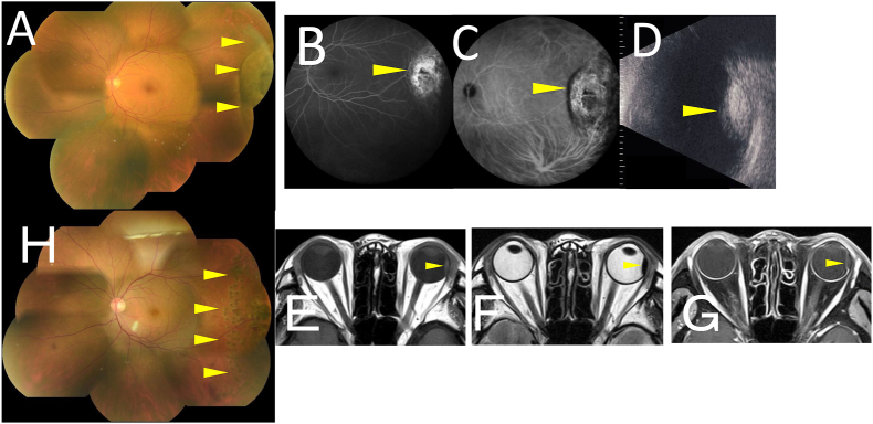
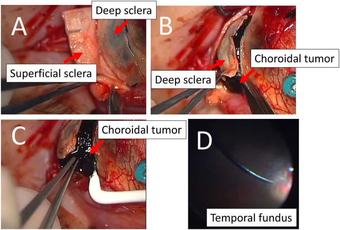
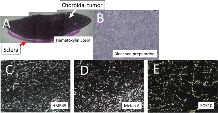
Observation: A 71-year-old female complained of blurred vision in her left eye. Her best-corrected visual acuity (BCVA) was 1.0. A dark-brown elevated lesion, measuring 5 papilla-diameter was found in the periphery of the fundus in her left eye. The mass showed hyperfluorescence on fluorescein angiography, early hypofluorescence and late hyperfluorescence on indocyanine green angiography. B-mode echography indicated the mass was originated from the choroid. Orbital magnetic resonance imaging showed isointense signal intensity on T1-weighted images (WI) and hypointense signal intensity on T2-WI, and poor Gadolinium enhancement on T1WI. The tumor was suspected to be melanocytoma, but it was difficult to differentiate from malignant melanoma. Transscleral tumor resection combined with 25-gauge vitrectomy was performed. Histopathological examinations led to the diagnosis of choroidal melanocytoma. Two years after local resection, her BCVA was 1.0 with no tumor recurrence.
Conclusions/importance: Local resection was useful as a diagnostic treatment for choroidal tumors confined to the periphery of the fundus that were difficult to clinically differentiate from malignant melanoma.
Keywords: Choroidal melanocytoma; Choroidal tumor; Histopathology; Local resection.
© 2024 The Authors.
Conflict of interest statement
The authors declare that they have no known competing financial interests or personal relationships that could have appeared to influence the work reported in this paper.
Figures
Similar articles
-
Histopathology and immunohistochemistry of choroidal melanocytoma demonstrated by local resection: A case report.Am J Ophthalmol Case Rep. 2021 Jun 19;23:101147. doi: 10.1016/j.ajoc.2021.101147. eCollection 2021 Sep. Am J Ophthalmol Case Rep. 2021. PMID: 34222715 Free PMC article.
-
Large Choroidal Melanocytoma Simulating Choroidal Melanoma: A Difficult Differential Diagnosis and an Inevitable Enucleation.Case Rep Ophthalmol Med. 2020 Nov 21;2020:8890857. doi: 10.1155/2020/8890857. eCollection 2020. Case Rep Ophthalmol Med. 2020. PMID: 33294243 Free PMC article.
-
Optic disk melanocytoma associated with polypoidal choroidal vasculopathy lesions, after combination treatment of photodynamic therapy and intavitreal aflibercept (Eylea), a case report.BMC Ophthalmol. 2018 Oct 12;18(1):267. doi: 10.1186/s12886-018-0927-7. BMC Ophthalmol. 2018. PMID: 30309335 Free PMC article.
-
[A new approach for studying the retinal and choroidal circulation].Nippon Ganka Gakkai Zasshi. 2004 Dec;108(12):836-61; discussion 862. Nippon Ganka Gakkai Zasshi. 2004. PMID: 15656089 Review. Japanese.
-
Posterior choroidal leiomyoma: a rare case report and literature review.APMIS. 2015 Jun;123(6):540-5. doi: 10.1111/apm.12384. Epub 2015 Apr 23. APMIS. 2015. PMID: 25907891 Review.
Cited by
-
Case Report: Laser speckle flowgraphy in a patient with uveitis due to immune-related adverse events by immune checkpoint inhibitors.Front Oncol. 2025 May 15;15:1492011. doi: 10.3389/fonc.2025.1492011. eCollection 2025. Front Oncol. 2025. PMID: 40444081 Free PMC article.
References
-
- Shields J.A., Demirci H., Mashayekhi A., Eagle R.C., Shields C.L. Melanocytoma of the optic disk: a review. Surv Ophthalmol. 2006;51(2):93–104. - PubMed
-
- Lafaut B.A., Mietz H., Ortmann M., Bartz-Schmidt K.U. Melanocytoma of the choroid: angiographic and histopathologic findings. Ophthalmic Surg Laser. 2002;33(2):158–162. - PubMed
-
- Shields J.A., Eagle R.C., Shields C.L., Abrams G. Diffuse choroidal melanocytoma simulating melanoma in a child with ocular melanocytosis. Retin Cases Brief Rep. 2010;4(2):164–167. - PubMed
Publication types
LinkOut - more resources
Full Text Sources
