. 2024 Mar 25;6(2):fcae100.
doi: 10.1093/braincomms/fcae100. eCollection 2024.

Messenger RNA-encoded antibody approach for targeting extracellular and intracellular tau

Affiliations

Messenger RNA-encoded antibody approach for targeting extracellular and intracellular tau

Patricia Wongsodirdjo et al. Brain Commun. .

Abstract

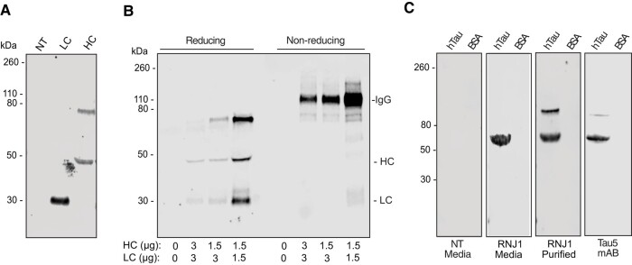
Monoclonal antibodies have emerged as a leading therapeutic agent for the treatment of disease, including Alzheimer's disease. In the last year, two anti-amyloid monoclonal antibodies, lecanemab and aducanumab, have been approved in the USA for the treatment of Alzheimer's disease, whilst several tau-targeting monoclonal antibodies are currently in clinical trials. Such antibodies, however, are expensive and timely to produce and require frequent dosing regimens to ensure disease-modifying effects. Synthetic in vitro-transcribed messenger RNA encoding antibodies for endogenous protein expression holds the potential to overcome many of the limitations associated with protein antibody production. Here, we have generated synthetic in vitro-transcribed messenger RNA encoding a tau-specific antibody as a full-sized immunoglobulin and as a single-chain variable fragment. In vitro transfection of human neuroblastoma SH-SY5Y cells demonstrated the ability of the synthetic messenger RNA to be translated into a functional tau-specific antibody. Furthermore, we show that the translation of the tau-specific single-chain variable fragment as an intrabody results in the specific engagement of intracellular tau. This work highlights the utility of messenger RNA for the delivery of antibody therapeutics, including intrabodies, for the targeting of tau in Alzheimer's disease and other tauopathies.

Keywords: Alzheimer’s disease; antibody; immunotherapy; mRNA; tau.


Conflict of interest statement

The authors report no competing interests.

Figures

Graphical Abstract
Graphical Abstract
Figure 1
Figure 1
Characterization of RNJ1 antibody. (A) Schematic representation of tau-specific antibody, RNJ1, bound to tau at amino acids 1–22 of full-length hTau (2N4R). (B) Enzyme-linked immunosorbent assay of the RNJ1 antibody in comparison with phosphate-buffered saline, the commercially available pan-tau antibody, Tau-5 and the hTau-specific antibody, HJ8.5, confirming that RNJ1 binds both mouse and hTau (mean ± SD, n = 3). (C) WB analysis of sarkosyl-soluble (S) and sarkosyl-insoluble (I) cortical brain homogenates derived from Alzheimer’s disease brain probed with tau-specific antibodies RNJ1 (pan tau) and HJ8.5 (pan tau). Schematic created with BioRender.
Figure 2
Figure 2
IVT synthesis of RNJ1 IgG and scFv mRNA. (A) Schematic of RNJ1 murine IgG HC and kappa LC DNA templates and IVT mRNA. The murine IgG HC and LC pair combine to form the RNJ1 IgG (B) scFv DNA template and the subsequent IVT mRNA. (A, B) Each DNA template is composed of a T7 promoter, a 5′ and 3′ UTR a Kozak sequence and the coding sequence. The IgG HC coding sequence consists of a signal peptide, the RNJ1 variable heavy domain and a mouse IgG1 constant heavy domain. The IgG LC coding sequence consists of a signal peptide, the RNJ1 variable light domain and a mouse IgG kappa constant light domain. The RNJ1 scFv coding sequence consists of the RNJ1 variable heavy and variable light domains joined by a flexible glycine–serine linker. The final IVT-synthesized mRNA contains a 5′ Anti-Reverse Cap Analog cap and a poly(A) tail (AAAAA). (C) Agarose gel loaded of IVT mRNA after 5′ capping, addition of the poly(A) tail and purification (kb, kilobase). Schematics created with BioRender. ARCA, Anti-Reverse Cap Analog; CDS, coding sequence; CH, constant heavy; CL, constant light; SP, signal peptide; VH, variable heavy; VL, variable light.
Figure 3
Figure 3
RNJ1 IgG expression and secretion following mRNA transfection. (A) WB analysis of media collected from non-transfected SH-SY5Y cells or SH-SY5Y cells transfected with IgG HC or IgG LC mRNA. Samples were electrophoresed under reducing conditions and probed with an anti-mouse secondary antibody. (B) WB of media collected from non-transfected SH-SY5Y cells (0 μg of HC and LC) or SH-SY5Y cells transfected with either 1.5 or 3 μg of mRNA at different ratios of IgG HC to IgG LC (1:1 or 1:2). Samples were electrophoresed under reducing or non-reducing conditions and probed with anti-mouse secondary antibody. (C) WB analysis of recombinant hTau probed with media collected from non-transfected SH-SY5Y cells (non-transfected media) or SH-SY5Y cells co-transfected with RNJ1 IgG HC and LC mRNA (RNJ1 media), compared with immunoblots probed with recombinant RNJ1 (RNJ1 purified) or Tau-5 (Tau-5 purified). Bovine serum albumin was used a negative control for binding. All primary antibodies were detected with an anti-mouse secondary antibody. BSA, bovine serum albumin; NT, non-transfected.
Figure 4
Figure 4
Expression of mRNA-encoded RNJ1 scFv intrabody. (A) Representative WB of cell lysates from non-transfected SH-SY5Y cells (0-ng mRNA) or SH-SY5Y cells transfected with an increasing amount of RNJ1 scFv mRNA (150–1200 ng). Immunoblots were probed with an anti-Flag antibody to detect the RNJ1 scFv and an anti-β-actin antibody as a loading control. (B) Quantification of RNJ1 scFv fluorescent intensity, normalized to β-actin fluorescence and plotted as a percentage (%) of the highest mRNA dose (mean ± SD; n = 3), one-way ANOVA with Tukey’s multiple comparison test (* = P < 0.05). (C) Quantification of calculated transfection efficiency (%) from SH-SY5Y cells transfected with 0.8-pmol RNJ1 scFv mRNA or 0.8-pmol RNJ1 scFv pDNA for 24 h (mean ± SD; n = 3), unpaired t-test. (D) Representative WB of cell lysates from SH-SY5Y cells transfected with either RNJ1 scFv mRNA or RNJ1 scFv pDNA for 24, 48 and 72 h. Immunoblots were probed with an anti-Flag antibody to detect the RNJ1 scFv and an anti-β-actin antibody as a loading control. (E) Quantification of RNJ1 scFv fluorescent intensity in D, normalized to β-actin expression (mean ± SD; n = 3), two-way ANOVA with Tukey’s multiple comparison test (*P < 0.05, **P < 0.01 and ***P < 0.001).
Figure 5
Figure 5
RNJ1 intrabody binds intracellular tau. (A) Representative immunofluorescent imaging (60×) of fixed tau-GFP SH-SY5Y cells either non-transfected (-scFv) or transfected with RNJ1 scFv mRNA or RNJ1 scFv pDNA. hTau was detected by its GFP fusion, and an anti-Flag antibody was used to detect the RNJ1 scFv. The nuclear marker, 4′,6-diamidino-2-phenylindole (DAPI), was used to identify all cells. (i and ii) Representative Z-stack images (60×) with orthogonal views showing overlap of RNJ1 scFv fluorescence and tau fluorescence in the cell cytoplasm (white) following (i) RNJ1 scFv mRNA transfection or (ii) RNJ1 scFv pDNA transfection. Scale bars represent 10 µm. (B) Pearson’s correlation coefficient between tau and RNJ1 scFv delivered as pDNA or mRNA. (C) Representative WB of the cell lysates from wild-type SH-SY5Y cells or Tau-GFP SH-SY5Y cells non-transfected or transfected with RNJ1 scFv mRNA, before (input) and after GFP-specific immunoprecipitation. Immunoblot was probed with an anti-Flag antibody to detect the RNJ1 scFv or Tau-5 to detect Tau. IP, immunoprecipitation.

References

    1. Bajracharya R, Cruz E, Götz J, Nisbet RM. Ultrasound-mediated delivery of novel tau-specific monoclonal antibody enhances brain uptake but not therapeutic efficacy. J Control Release. 2022;349:634–648. - PubMed
    1. d'Abramo C, Acker CM, Jimenez HT, Davies P. Tau passive immunotherapy in mutant P301L mice: Antibody affinity versus specificity. PLoS One. 2013;8(4):e62402. - PMC - PubMed
    1. Castillo-Carranza DL, Sengupta U, Guerrero-Munoz MJ, et al. Passive immunization with tau oligomer monoclonal antibody reverses tauopathy phenotypes without affecting hyperphosphorylated neurofibrillary tangles. J Neurosci. 2014;34(12):4260–4272. - PMC - PubMed
    1. Umeda T, Eguchi H, Kunori Y, et al. Passive immunotherapy of tauopathy targeting pSer413-tau: A pilot study in mice. Ann Clin Transl Neurol. 2015;2(3):241–255. - PMC - PubMed
    1. Ittner A, Bertz J, Suh LS, Stevens CH, Gotz J, Ittner LM. Tau-targeting passive immunization modulates aspects of pathology in tau transgenic mice. J Neurochem. 2015;132(1):135–145. - PubMed

LinkOut - more resources