[Preprint]. 2024 Mar 29:2024.03.28.587274.
doi: 10.1101/2024.03.28.587274.

Three Modes of Viral Adaption by the Heart

Affiliations

Three Modes of Viral Adaption by the Heart

Cameron D Griffiths et al. bioRxiv. .

Update in

  • Three modes of viral adaption by the heart.
    Griffiths CD, Shah M, Shao W, Borgman CA, Janes KA. Griffiths CD, et al. Sci Adv. 2024 Nov 15;10(46):eadp6303. doi: 10.1126/sciadv.adp6303. Epub 2024 Nov 13. Sci Adv. 2024. PMID: 39536108 Free PMC article.

Abstract

Viruses elicit long-term adaptive responses in the tissues they infect. Understanding viral adaptions in humans is difficult in organs such as the heart, where primary infected material is not routinely collected. In search of asymptomatic infections with accompanying host adaptions, we mined for cardio-pathogenic viruses in the unaligned reads of nearly one thousand human hearts profiled by RNA sequencing. Among virus-positive cases (~20%), we identified three robust adaptions in the host transcriptome related to inflammatory NFκB signaling and post-transcriptional regulation by the p38-MK2 pathway. The adaptions are not determined by the infecting virus, and they recur in infections of human or animal hearts and cultured cardiomyocytes. Adaptions switch states when NFκB or p38-MK2 are perturbed in cells engineered for chronic infection by the cardio-pathogenic virus, coxsackievirus B3. Stratifying viral responses into reversible adaptions adds a targetable systems-level simplification for infections of the heart and perhaps other organs.


Conflict of interest statement

Competing interests: The authors declare that they have no competing interests.

Figures
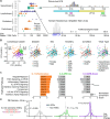
Fig. 1.
Fig. 1.. Transcriptomic identification and classification of human heart samples harboring cardio-pathogenic viruses.
(A) Evidence for vasculotropic, lymphotropic, cardiotoxic, and cardiotropic viruses (1) in human transcriptomic (HTX) datasets (Table 1). The percentage of samples in which each class of virus was not detected (n.d.) is shown in gray. Representative alignments to the parvovirus B19 and human herpesvirus 4 genomes are shown above relevant gene products for each virus: NS, 7.5-kDa, and 11-kDa nonstructural proteins, VP1/2 structural proteins, and Epstein-Barr virus nuclear antigen leader protein (EBNA-LP). (B) Principal component (PC) projection of the 1000 most-variable genes among virus-positive heart samples (n = 189). Samples are colored according to their discrete consensus cluster (fig. S3); HTX source (Table 1); sex; inferred African (AFR), Admixed American (AMR), East Asian (EAS), European (EUR), or South Asian (SAS) ancestry; cases with or without dilated cardiomyopathy (DCM); and type of cardio-pathogenic virus (1). (C) MSigDB analysis of hallmark pathway enrichments for the three consensus clusters: C-1, Inflammatory; C-2, ARE-Up; and C-3, ARE-Down. The normalized enrichment score is shown for differentially increased (positive) or decreased (negative) genes relative to virus-negative controls. The false discovery rate-corrected P value for the enrichment (Penrichment) is inset. The full set of enrichments is listed in table S4. (D) The differentially expressed (DE) genes of C-2 and C-3 show biased proportions of transcripts with AU-rich elements (AREs). The distributions of ARE-containing genes from the ARED-Plus (129) database were plotted along with non-ARE DE genes for each cluster, and the DE genes were assessed for ARE enrichment by the hypergeometric test with all genes (DE and non-DE) as the reference and Bonferroni correction for multiple-hypothesis testing.
Fig. 2.
Fig. 2.. Cellular and molecular phenotypes of the three viral adaptions.
(A) Deconvolution of bulk heart samples into constituent cell types with CIBERSORTx (36). The signature matrix (table S5) was created from a left ventricle single-nucleus RNA-seq atlas of the heart (121). Differences in absolute cell type composition between a virus-positive consensus cluster (C-1:Inflammatory, C-2:ARE-Up, C-3:ARE-Down) and the virus-negative reference (Neg) were assessed by multiple t tests with the Benjamini-Hochberg correction for multiple hypothesis testing. (B) The C-1:Inflammatory cluster is elevated for transcriptional biomarkers of heart failure (45). Z-scores of DESeq2-normalized transcript counts are shown for each heart sample, divided by cluster. (C) The C-1:Inflammatory cluster shows altered abundances of genes involved in matrix remodeling (45). For (B) and (C), differences between C-1 and C-2,3 were assessed by multiple t-tests with the Benjamini-Hochberg correction for multiple hypothesis testing. *P < 0.05, **P < 0.01, ***P < 0.001.
Fig. 3.
Fig. 3.. Inflammatory, ARE-Up, and ARE-Down adaptions in an engineered model of chronic cardiomyocyte infection.
(A) Structural proteins (VP1, VP2, VP3, VP4) of the cardiotoxic virus, coxsackievirus B3 (CVB3). Capsid structure is reprinted from PDB 1COV (55). (B) Enlarged view of the VP4:VP2 cleavage site of CVB3. The Ala67 site of VP4 (VP4A67, red) is highlighted along with the putative catalytic Asp11 of VP2 (VP2D11, orange). (C) Multiple sequence alignment of the residues flanking Ala67 among enteroviruses with an N:S cleavage site for VP4:VP2 (arrowhead). The consensus sequence for 30 N:S-containing enteroviruses is shown. (D) The CVB3 A67G mutant is not productively infectious. 293T cells were transfected with wildtype CVB3 or the A67G mutant (n = 4 biological replicates), and equal titers of released viral RNA were tested for serial infectivity by plaque assay followed by rank-sum test. One replicate of each condition is shown. (E and F) VP4:VP2-complemented CVB3 A67G infections yield similar transcriptional changes as wildtype CVB3 infections of AC16 cardiomyocytes ectopically expressing CVB3 receptor (AC16-CAR) (E) and induced pluripotent stem cell (iPSC)-derived cardiomyocytes (F). Data are shown as log2 fold change, along with the Pearson correlations (R) of n = 8093 (E) and 9461 (F) differentially expressed (DE) genes. (G) Principal component (PC) projection of the 816 genes shared among the 3000 most variable for AC16 clones (triangles) and HTX samples (circles). (H) Statistical overlap of DE genes among AC16 consensus clusters (C-A,B,C) and HTX consensus clusters (C-1,2,3).
Fig. 4.
Fig. 4.. Perturbations of inflammatory and ARE-adapted states.
(A) Differential changes in inflammatory hallmarks for CVB3-expressing AC16 clones engineered with constitutively active IKK-EE or dominant-negative IκBα super-repressor (IκBα-SR). The normalized enrichment score (NES) was calculated for each inflammatory hallmark pathway relative to empty vector control. The hallmark pathways are ordered as in Fig. 1C. (B) Reversion of ARE-Down and ARE-Up states in AC16 clones engineered with constitutively active MKK6-EE or treated with the p38 inhibitor (p38i) BIRB796 (5 μM). The distribution of differentially expressed genes with or without AREs for each adaption was compared as in Fig. 1D. (C) Principal component (PC) paths between centroids of adapted states. RNA sequencing data from empty vector controls of each clone are shown and organized by adaption. (D and E) Displacement of adapted states with IKK-EE, and convergence toward ARE-Down with IκBα-SR. (F and G) Convergence toward ARE-Down with p38i. Clones (n = 3 per adaption) were acutely transduced or treated, quiesced, and profiled by RNA sequencing. For (D) and (E), the relocalization of perturbed centroids (arrowheads) toward the empty vector controls of other states (circles) was assessed by paired two-sided t test. Bonferroni-corrected P values are colored according to the adaption the centroid moves toward. For IKK-EE, the Inflammatory adaption (black P value) moves away from the ARE-Down adaption.
Fig. 5.
Fig. 5.. Rapid and divergent viral adaptions to various cardio-pathogens.
(A) MSigDB analysis of hallmark pathway enrichments among differentially expressed (DE) genes for induced pluripotent stem cell (iPSC)-derived cardiomyocytes infected with coxsackievirus B3 (CVB3) [n = 4 samples (control) or 8 samples (wildtype CVB3 MOI = 10 for 8 hours or CVB3 A67G MOI = 3 for 16 hours, each in quadruplicate)]. (B) Acute CVB3 infection of iPSC cardiomyocytes triggers the Inflammatory adaption. (C) AU-rich element (ARE) analysis of DE genes from AC16 cells overexpressing the CVB3 receptor CXADR (AC16-CAR) infected with CVB3 [n = 2 samples (control) or 4 samples (wildtype CVB3 MOI = 10 for 8 hours or CVB3 A67G MOI = 3 for 16 hours, each in duplicate)]. (D) Acute CVB3 infection of AC16-CAR cells triggers the ARE-Down adaption. (E) MSigDB analysis of hallmark pathway enrichments among DE genes for autopsied hearts from individuals who died from SARS-CoV-2. Data from n = 32 cases and 5 controls were obtained from phs002258.v1.p1 (66). (F) Acute SARS-CoV-2 infection elicits the Inflammatory adaption in human hearts. (G) ARE analysis of DE genes from hamsters infected with SARS-CoV–2 or influenza A for 31 days post-infection after histological pathology had resolved. Data from n = 5 infections and 3 controls were obtained from GSE203001 (67). (H) Resolved SARS-CoV-2 and influenza infections elicit the ARE-Up adaption in hamster hearts. For (A) and (E), the hallmark pathways are ordered and displayed as in Fig. 1C by their normalized enrichment score (NES) with false discovery rate-corrected P value for the enrichment in the inset. For (B), (D), (F), and (H), the excess overlap of DE genes between the indicated study and the three viral adaptions was calculated and assessed by the hypergeometric test with Bonferroni correction. For (C) and (G), DE gene density is displayed according to log2 fold change (FC) and analyzed as in Fig. 1D.
Fig. 6.
Fig. 6.. Proposed relationship between heart adaptions after infection.
NFκB is acutely activated after infection (dashed vertical), leading to an Inflammatory state that may evolve over time to either the ARE-Up or ARE-Down adaption depending on the trajectory of p38–MK2 signaling (solid). It is unclear whether ARE-Down proceeds through an ARE-Up intermediate (dashed horizontal), and ARE-Down might bypass an Inflammatory intermediate (dashed vertical).

References

    1. Tschope C. et al., Myocarditis and inflammatory cardiomyopathy: current evidence and future directions. Nat. Rev. Cardiol. 18, 169–193 (2021). - PMC - PubMed
    1. Kuhl U. et al., Viral persistence in the myocardium is associated with progressive cardiac dysfunction. Circulation 112, 1965–1970 (2005). - PubMed
    1. Why H. J. et al., Clinical and prognostic significance of detection of enteroviral RNA in the myocardium of patients with myocarditis or dilated cardiomyopathy. Circulation 89, 2582–2589 (1994). - PubMed
    1. Pankuweit S., Klingel K., Viral myocarditis: from experimental models to molecular diagnosis in patients. Heart Fail. Rev. 18, 683–702 (2013). - PubMed
    1. Abdel-Aty H. et al., Diagnostic performance of cardiovascular magnetic resonance in patients with suspected acute myocarditis: comparison of different approaches. J. Am. Coll. Cardiol. 45, 1815–1822 (2005). - PubMed

Publication types