[Preprint]. 2024 Mar 28:rs.3.rs-4108866.
doi: 10.21203/rs.3.rs-4108866/v1.

Temporal Single-Cell Sequencing Analysis Reveals That GPNMB-Expressing Macrophages Potentiate Muscle Regeneration

Affiliations

Temporal Single-Cell Sequencing Analysis Reveals That GPNMB-Expressing Macrophages Potentiate Muscle Regeneration

Yu-Fan Chen. Res Sq. .

Update in

Abstract

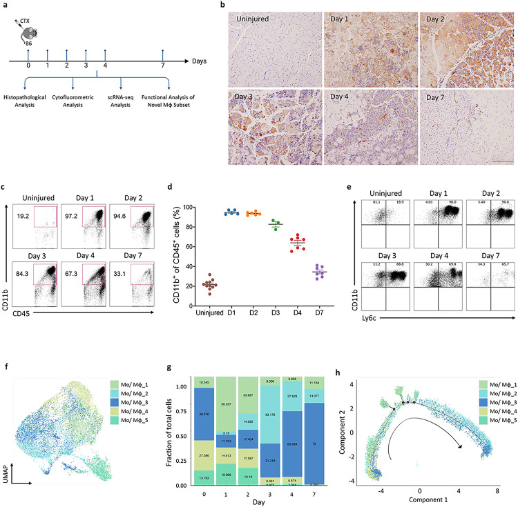
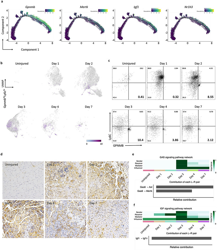
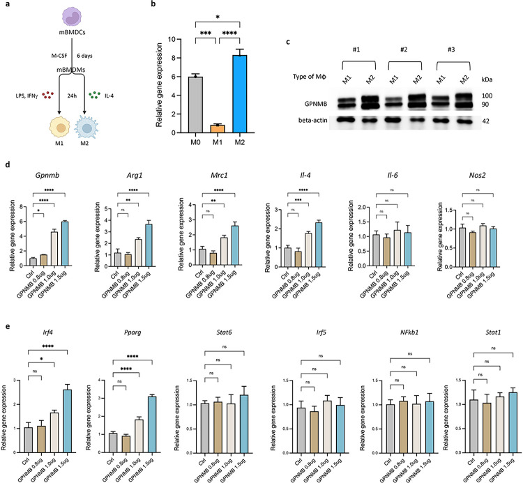
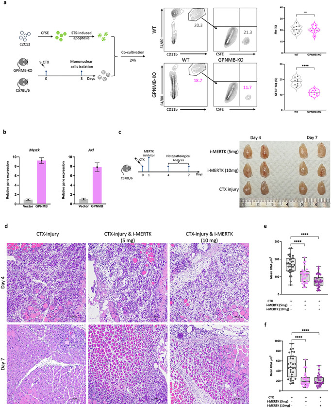
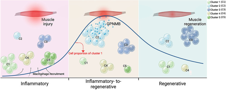
Macrophages play a crucial role in coordinating the skeletal muscle repair response, but their phenotypic diversity and the transition of specialized subsets to resolution-phase macrophages remain poorly understood. To address this issue, we induced injury and performed single-cell RNA sequencing on individual cells in skeletal muscle at different time points. Our analysis revealed a distinct macrophage subset that expressed high levels of Gpnmb and that coexpressed critical factors involved in macrophage-mediated muscle regeneration, including Igf1, Mertk, and Nr1h3. Gpnmb gene knockout inhibited macrophage-mediated efferocytosis and impaired skeletal muscle regeneration. Functional studies demonstrated that GPNMB acts directly on muscle cells in vitro and improves muscle regeneration in vivo. These findings provide a comprehensive transcriptomic atlas of macrophages during muscle injury, highlighting the key role of the GPNMB macrophage subset in regenerative processes. Targeting GPNMB signaling in macrophages could have therapeutic potential for restoring skeletal muscle integrity and homeostasis.


Conflict of interest statement

Declarations CONFLICT OF INTEREST The authors declare no competing interests.

Figures

Figure 1
Figure 1. Identification of five distinct macrophage subsets during skeletal muscle regeneration via single-cell RNA-seq.
(a) Schematic showing the experimental timeline for CTX-induced skeletal muscle injury in mice, delineating the key analyses performed at various days post-injury. (b) Immunohistochemical detection of Cd68+ macrophages within injured muscle at specified time intervals post-injury. The magnification scale bar represents 100 μm. (c) Flow cytometric analysis showed a surge in CD11b+ cells immediately after injury, which diminishes starting from day 3. (d) Graphical representation of CD11b+ cell percentages, corresponding to flow cytometry results depicted in (c). (e) The transition of macrophage phenotypes from proinflammatory Ly6chi early post-injury to anti-inflammatory Ly6clo by day 4, as assessed by flow cytometry. (f) UMAP plot illustrating the distribution of five identified monocyte/macrophage subsets across the time series. (g) Bar graph showing the relative frequencies of each monocyte/macrophage subset at the designated time points post-injury. (h) Pseudotime analysis projecting the potential developmental trajectories of the monocyte/macrophage subsets identified.
Figure 2
Figure 2. Temporal single-cell sequencing unveils GPNMB+ macrophage signaling as a dominant subset of tissue regeneration.
(a) Trajectory plots tracing the gene expression profiles mirroring the Gpnmb pattern within the monocyte/macrophage subsets suggest synchronous gene expression events that may underpin shared functional pathways in regeneration. (b) UMAP series depicting the dynamic expression of the GpnmbhiLy6clo macrophage subset throughout the regenerative timeline. The UMAPs consecutively illustrate the prevalence and distribution of the GpnmbhiLy6clo population at each time point following skeletal muscle injury, highlighting the shifts in the macrophage landscape. (c) Flow cytometric quantification capturing the temporal prevalence of the GpnmbhiLy6clo population. The analysis delineates the frequency of this subset at progressive intervals post-injury, reflecting its involvement in the regenerative process. (d) Immunohistochemical staining of TA muscle sections across a temporal spectrum, identifying the presence of GPNMB-expressing cells. The staining intensity indicates the cellular localization and temporal expression pattern of GPNMB during the regeneration phases. (e and f) Heatmap analysis summarizing the relative engagement of cell populations in GAS and IGF signaling pathways. Upper panel offers insights into the predominant cellular functions at each time point, while the lower panel details the contribution of specific ligand-receptor pairs to the composite signaling communication network.
Figure 3
Figure 3. Ectopic GPNMB enhances M2 macrophage polarization by modulating key regulatory genes.
(a) Experimental scheme for differentiating murine bone marrow-derived cells (mBMDCs) into M1 and M2 macrophages, followed by LPS, IFNγ, and IL-4 treatment. (b) Quantitative PCR analysis shows significantly higher GPNMB expression in M2 polarized macrophages than in M0 and M1. (c) Western blot confirming the increased protein levels of GPNMB in M2 macrophages across three independent experiments. (d)Ectopic expression of GPNMB in mBMDMs leads to the upregulation of M2-associated markers Arg1, Mrc1, and IL-4, with no significant effect on the M1 markers IL-6 and Nos2. (e)Overexpression of GPNMB results in heightened expression of M2-related transcription factors Irf4 and Pparg, suggesting a potential pathway for GPNMB-mediated macrophage polarization.
Figure 4
Figure 4. Impaired muscle regeneration in GPNMB-knockout mice post-CTX injury.
(a)Schematic of the experimental design for assessing muscle regeneration in GPNMB-KO and C57BL/6 control mice following CTX injection. (b and c)Representative images of tibialis anterior muscles from GPNMB-KO and WT mice at uninjured, day 4, and day 7 post-injury, demonstrating differences in muscle morphology. (d) Hematoxylin and eosin staining of TA muscle sections revealed histological changes during regeneration, with GPNMB-KO mice showing reduced tissue repair compared to controls. (e) Quantitative analysis of cross-sectional area of muscle fibers in uninjured, day 4, and day 7 post-injury muscle sections confirmied statistically significant impairment of regeneration in GPNMB-KO mice.
Figure 5
Figure 5. Diminished macrophage efferocytosis and impaired muscle regeneration following GPNMB-knockout and MERTK inhibition.
(a) This panel illustrates the experimental setup and subsequent flow cytometry analysis for evaluating macrophage efferocytosis. On day 3 post-CTX-induced muscle injury, mononuclear cells were isolated from both WT and GPNMB-KO mice and co-cultured with CFSE-labeled, staurosporine (STS)-induced apoptotic C2C12 myoblasts for 24 hours. Flow cytometry was then employed to assess the phagocytosis of apoptotic cells by CD11b+F4/80+ macrophages. The rightmost graph in Panel A provides a quantitative comparison between the WT and GPNMB-KO groups, showing a significant decrease in efferocytosis efficiency in the GPNMB-KO macrophages, as reflected by their reduced uptake of CFSE-labeled apoptotic bodies. (b) Quantitative PCR analysis reveals that overexpression of GPNMB in primary macrophages significantly increases the mRNA levels of key efferocytosis genes, Mertk and Axl. (c) Left panel, schematic of the experimental design depicting the treatment of C57BL/6 mice with a MERTK inhibitor post-CTX injury to evaluate its effect on muscle regeneration. Right panel, gross morphology of TA muscles from mice treated with MERTK inhibitor doses shows dose-dependent effects on muscle appearance. (d) Histological examination of muscle regeneration by H&E staining at days 4 and 7 post-CTX injury with or without MERTK inhibitor treatment, highlighting the impact of MERTK signaling on tissue repair. (e and f) Statistical analysis of muscle fiber CSA and length at days 4 and 7 post-injury, with MERTK inhibition leading to a marked reduction in both parameters, signifying compromised regenerative capacity.
Figure 6
Figure 6. GPNMB stimulation promotes muscle regeneration and myogenic differentiation of murine myoblasts.
(a) Following GPNMB overexpression, quantitative real-time PCR was utilized to monitor the temporal expression of key myogenic regulatory factors in C2C12 cells, including Myf5, Myf2a, Myod, and Myog. (b)Immunofluorescent staining for MyHC of C2C12 myoblast cultured in differentiation medium with (100 ng/mL or 200 ng/mL) or without rGPNMB. C2C12 myoblasts were induced to differentiate for three days; (c) fusion indices were calculated by expressing the number of nuclei within MyHC-positive myotubes with ≥2 nuclei as a percentage of the total nuclei, and (d) the myotube width was measured at 3 different points on the cell. The average width per myotube was calculated. Data are presented as mean±s.d. of three independent experiments. (e)H&E staining of injured mouse TA muscle on days 4 and 7 with (10 μg or 20 μg) or without rGPNMB. Scale bar: 100 μm. (f) and (g)Cross-sectional area of myofibers from injured only and treated with rGPNMB (10 μg and 20 μg) groups on days 4 and 7. Data are presented as mean±s.d. (n=3) of each time point.
Figure 7
Figure 7. GPNMB-expressing macrophage contributes to skeletal muscle regeneration.
The dynamic response of skeletal muscle to injury can be broadly categorized into three primary stages: the inflammatory, the inflammatory-to-regenerative, and the regenerative stage. Following muscle injury, muscle tissues recruit monocyte-derived macrophages, categorized into five distinct clusters (C1, 2, 3, 4, and 5) based on their inflammatory profiles. Our study identifies that macrophages with high GPNMB expression during the transitional phase from inflammation to regeneration are crucial in activating and expanding muscle progenitor cells. This process aids in the transition of macrophages towards an anti-inflammatory phenotype, which is critical for the resolution of inflammation and the subsequent muscle repair and regeneration. The dynamic shifts in macrophage subsets, highlighted by changes in the proportions of Cluster 2, underscore the importance of GPNMB as an effector in muscle regeneration. This figure was generated using Biorender.com.

References

    1. Giordani L. et al. High-dimensional single-cell cartography reveals novel skeletal muscle-resident cell populations. Mol. Cell 74, 609–621.e6 (2019). - PubMed
    1. Wosczyna M. N. & Rando T. A. A muscle stem cell support group: coordinated cellular responses in muscle regeneration. Dev. Cell 46, 135–143 (2018). - PMC - PubMed
    1. Wang X. et al. Prolonged hypernutrition impairs TREM2-dependent efferocytosis to license chronic liver inflammation and NASH development. Immunity 56, 58–77.e11 (2022). - PMC - PubMed
    1. Fidler T. P. et al. The AIM2 inflammasome exacerbates atherosclerosis in clonal haematopoiesis. Nature 592, 296–301 (2021). - PMC - PubMed
    1. Shang M. et al. Macrophage-derived glutamine boosts satellite cells and muscle regeneration. Nature 587, 626–631 (2020). - PMC - PubMed

Publication types

LinkOut - more resources