[Preprint]. 2024 Mar 27:2024.03.26.586863.
doi: 10.1101/2024.03.26.586863.

Intron-lariat spliceosomes convert lariats to true circles: implications for intron transposition

Affiliations

Intron-lariat spliceosomes convert lariats to true circles: implications for intron transposition

Manuel Ares Jr et al. bioRxiv. .

Update in

Abstract

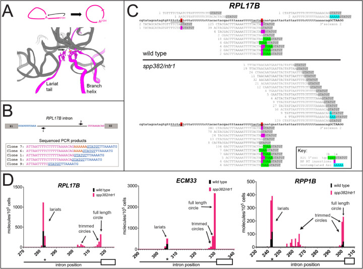
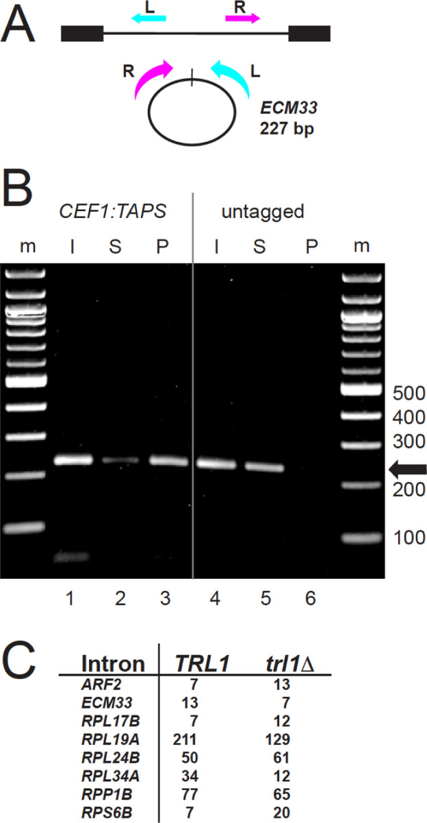
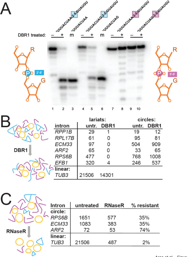
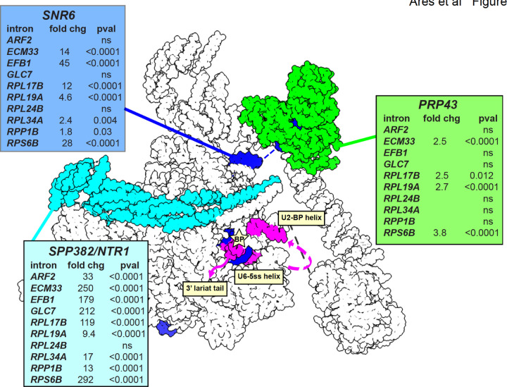
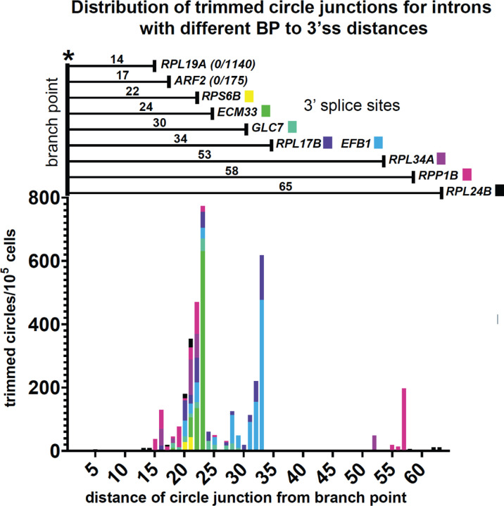
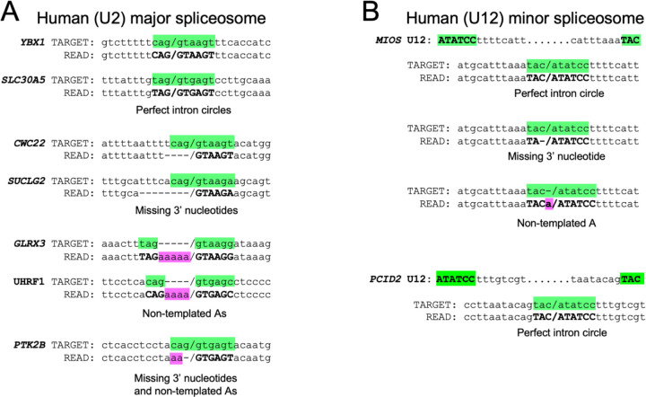
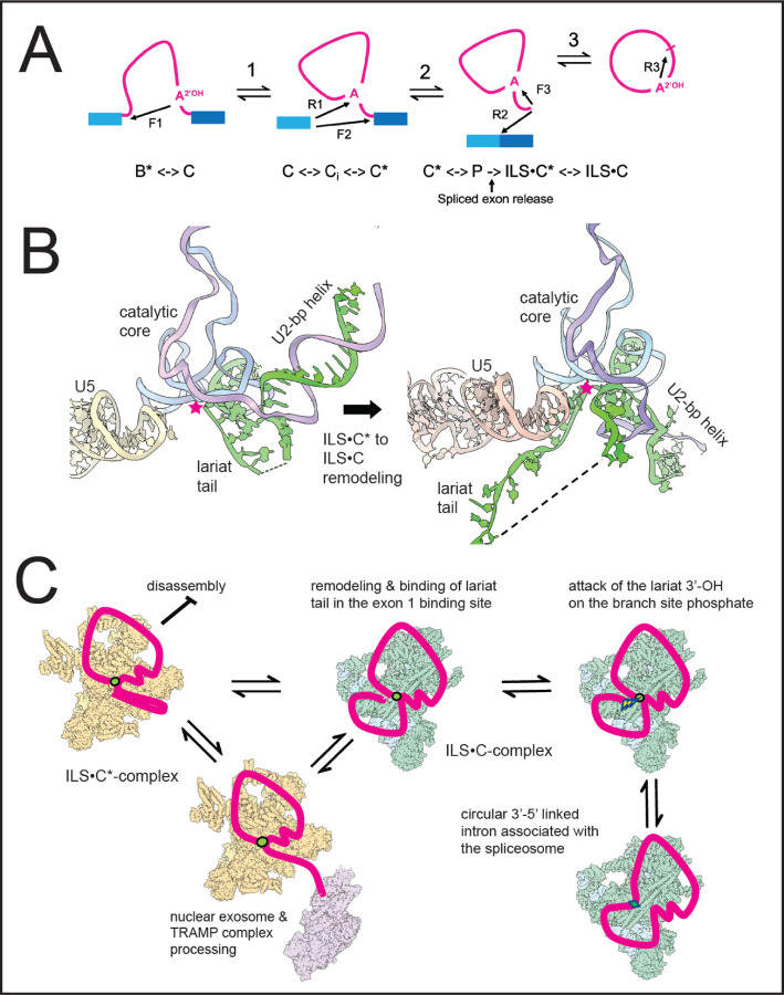
Rare, full length circular intron RNAs distinct from lariats have been reported in several species, but their biogenesis is not understood. We envision and test a hypothesis for their formation using Saccharomyces cerevisiae, documenting full length and novel processed circular RNAs from multiple introns. Evidence implicates a previously undescribed catalytic activity of the intron-lariat spliceosome (ILS) in which the 3'-OH of the lariat tail (with optional trimming and adenylation by the nuclear 3' processing machinery) attacks the branch, joining the intron 3' end to the 5' splice site in a 3'-5' linked circle. Human U2 and U12 spliceosomes produce analogous full length and processed circles. Post-splicing catalytic activity of the spliceosome may promote intron transposition during eukaryotic genome evolution.


Figures

Figure 1.
Figure 1.. A hypothesis, and detection of intron circles in S. cerevisiae.
(A) A possible mechanism for the spliceosome to create intron circles by nucleophilic attack of the lariat tail 3’-OH (arrow, top and bottom) on the branch phosphate (yellow sphere, bottom), forming a 3’−5’ junction between the 3’ss and the 5’ss (top). A remodeled intron-lariat spliceosome (ILS) in which the lariat tail 3’-OH is bound in E1 binding site (bottom, arrow) could carry out a reaction formally similar to the reverse of step 1 (reverse of branch formation) to create the intron circle (bottom). Cartoon derived from C-complex model 5GMK (Wan et al. 2016), using ChimeraX (Meng et al. 2023). Only RNA is shown, U2, U6, and U5 in gray, intron in magenta. (B) RPL17B intron with “inverse” primers to capture circular intron junctions. Sequences of selected cloned PCR products are shown, with the 5’ss underlined and non-genomic As in gold. (C) High throughput sequencing reveals a complex set of processed circular RPL17B introns whose abundance is dramatically increased by the spp382–1 mutation. The intron is in bold. Reads are aligned above (circles) or below (lariats), the 5’ss sequence highlighted in gray, and non-genomic As in blue. Deduplicated (unique) read counts from each library are shown in front of each read. Lariats derived from incorrect 5’ss are in green. (D) Different introns produce distinct distributions of intron circles. Graphs show the junction locations (x-axis) and number of unique reads with junctions at that location (y-axis, calibrated to a spiked-in circular RNA) per 105 yeast cells, from wild type (black bars) or spp382–1 (pink bars) cells. The 3’ part of each intron (line) and its second exon (white box) are shown below the x-axis, with the asterisk indicating the position of the bp. Additional introns and data are in Fig S1 and Table S1.
Figure 2.
Figure 2.. Intron circles are associated with the spliceosome and are not formed by the tRNA ligase Trl1.
(A) Detecting circular ECM33 introns. Inverse PCR primers that create a 227 bp product from full length ECM33 intron circles are shown; the R primer is 3’ of the branch point and cannot detect lariats. (B) Association of ECM33 intron circles with the spliceosome in spp382–1 cells. The spp382–1 mutant was fitted with a TAPS tag at the C-terminus of its CEF1 coding region for affinity purification of spliceosomes. Extracts from this strain and an untagged control were prepared (Input, I) and bound in batch to IgG-sepharose. Unbound (Supernatant, S) and bound (Pellet, P) fractions were prepared. RNA from each fraction was used for RT-PCR to identify ECM33 circles (227 bp product). By primer extension the pellet fraction from the tagged (but not the untagged) extract is enriched in spliceosomal U2 snRNA and free of cytoplasmic SCR1 (Fig S2). (C) Robust detection of intron circles in a strain lacking tRNA ligase. The table shows spike-in normalized circular intron counts for 8 introns from libraries prepared from strains that differ by deletion of TRL1. Detailed data are in Table S2.
Figure 3.
Figure 3.. Enzymatic tests of circular intron structure.
(A) Dbr1 does not require a 3’-nucleotide at the branch and can cleave an internal 2’−5’ linkage. 32P-5’ end-labeled RNA oligonucleotides carrying either a single 2’−5’ internal linkage in the middle of an otherwise 3’−5’ linked RNA, or carrying only 3’−5’ linked nucleotides were incubated (or not) with recombinant DBR1 and separated by electrophoresis on an 8M urea, 20% acrylamide gel. Two different substrate sequences (bpA-G5ss and 3ssG-G5ss) were made with either a 2’−5’ (blue) or 3’−5’ (pink) linkage at the junction marked by the colored square are shown above each reaction. Minus and plus signs indicate enzyme addition. Two 8-mers identical to the expected Dbr1 cleavage products were labeled as markers (m, lanes 3 and 6). Comigration with the marker indicates cleavage at the 2’=5’ linkage. (B) Circle junctions from the spp382–1 mutant are resistant to cleavage with Dbr1, whereas lariat junctions are not. Read counts of lariats and circles for six introns from libraries made with or without prior treatment of the input RNA with Dbr1. (C) Circle junction-containing RNAs from the spp382–1 mutant are much more resistant to RNAseR than a linear pre-mRNA. Read counts of circles for three introns from libraries made with or without prior treatment of the input RNA with RNAseR. Detailed data are in Table S3.
Figure 4.
Figure 4.. Effects of spliceosome disassembly mutants on abundance of different intron circles.
Catalytic inactivation of the spliceosome is mediated by the Prp43 helicase (green) acting in concert with its G-patch protein Spp382/Ntr1 (cyan) on the 3’ end of U6 snRNA (SNR6, blue) to pull U6 from the spliceosome (Toroney et al. 2019). Intron circles were counted in pairs of strains carrying wild type or mutant alleles of each factor, normalized using a spiked in circle and tested for significant change using Fisher’s exact test (detailed data are in Table S4). Each table shows the log odds calculation as fold change and the two-sided p value for each of 10 intron comparisons. ns, not significant. The position of the intron is shown in magenta. Image made using ChimeraX (Meng et al. 2023) and the S. cerevisiae ILS structure model (5Y88, (Wan et al. 2017).
Figure 5.
Figure 5.. Effects of nuclear 3’ processing on abundance and structure of different intron circles.
(A) The distribution of trimmed intron circles for all tested introns has a limit of 15–20 nt of lariat tail remaining, independent of the initial tail length. Two introns with natural BP-3’ss tails of 14 (RPL19A) or 17 (ARF2) nt are not trimmed. Detailed data are in Table S5.
Figure 6.
Figure 6.. Evidence for full length and processed circles from human cells.
(A) Selected reads from major (U2) spliceosomal introns found by remapping the branch point enrichment libraries from Mercer et al. (Mercer et al. 2015) to permuted intron target sequences from the human genome. Junctions between the 5’ and 3’ intron ends are denoted by a forward slash. Splice site nucleotides from the permuted target intron are highlighted in green. Dashes are added to the target sequence to maintain alignment when non-genomic As are present in the read. Nucleotides in the read that match the splice sites are in bold, non-genomic As are highlighted in pink. Dashes are added to the read sequence to maintain alignment where nucleotides from the intron 3’ end are missing. (B) Selected reads from minor (U12) spliceosomal introns found by remapping the branch point enrichment libraries (Mercer et al. 2015) to permuted intron target sequences from the human genome. Highlight and dash placement are the same as for (A). Additional examples with read identifiers are shown in Table S6.
Figure 7.
Figure 7.. Model for the formation of intron circles.
(A) Transesterification reactions catalyzed by the spliceosome. F, forward reaction, R, reverse reaction, numbered 1 through 3. The arrows indicate the direction of nucleophilic attack. F1: Attack of the bp 2’-OH on the phosphate (P) at the 5’ ss. F2: Attack of the 3’-OH at the end of E1 on the P at the 3’ ss. F3: attack of the lariat tail 3’-OH on the BP P to form the intron circle. Below the pathway are spliceosome complexes within which these reactions occur. The active site of the C-complex configuration has the 5’ss P and the bp 2’-OH in the active site for F1, whereas the C*-complex configuration has the U2 BP helix pulled out of the active site to create the 3’ss binding site for F2. ILS, intron lariat spliceosome. ILS is formed by spliced exon release (arrow), in the C* configuration (ILS•C*). For F3 it must remodel to the C configuration (ILS•C). (B) Cartoon showing remodeling of the RNA in the core of the ILS•C* complex to the proposed ILS•C complex and docking of the lariat tail for circularization. (C) Cartoon showing events leading to mixtures of processed and unprocessed full length intron circles. Yellow spliceosomes represent ILS•C*-complex, green represent ILS•C-complexes. The nuclear exosome (purple) represents nuclear 3’ processing machinery interacting with ILS•C* but it is not known which ILS forms permit processing. Intron is in red, key linkages are: green circles: 2’−5’ linkages; green diamonds: 3’−5’ linkages. Lightning bolt indicates nucleophilic attack. Cartoons were made using ChimeraX as above.

Similar articles

References

    1. Arenas JE, Abelson JN. 1997. Prp43: An RNA helicase-like factor involved in spliceosome disassembly. Proc Natl Acad Sci U S A 94: 11798–11802. - PMC - PubMed
    1. Ares M. 2012. Isolation of total RNA from yeast cell cultures. Cold Spring Harb Protoc 2012: 1082–1086. - PubMed
    1. Aupič J, Borišek J, Fica SM, Galej WP, Magistrato A. 2023. Monovalent metal ion binding promotes the first transesterification reaction in the spliceosome. Nat Commun 14: 8482. - PMC - PubMed
    1. Bertram K, Agafonov DE, Liu W-T, Dybkov O, Will CL, Hartmuth K, Urlaub H, Kastner B, Stark H, Lührmann R. 2017. Cryo-EM structure of a human spliceosome activated for step 2 of splicing. Nature 542: 318–323. - PubMed
    1. Bertram K, El Ayoubi L, Dybkov O, Agafonov DE, Will CL, Hartmuth K, Urlaub H, Kastner B, Stark H, Lührmann R. 2020. Structural Insights into the Roles of Metazoan-Specific Splicing Factors in the Human Step 1 Spliceosome. Mol Cell 80: 127–139.e6. - PubMed

Publication types