[Preprint]. 2024 Mar 29:2024.03.28.587234.
doi: 10.1101/2024.03.28.587234.

Wrapping of single-stranded DNA by Replication Protein A and modulation through phosphorylation

Affiliations

Wrapping of single-stranded DNA by Replication Protein A and modulation through phosphorylation

Rahul Chadda et al. bioRxiv. .

Update in

Abstract

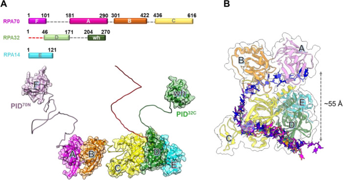
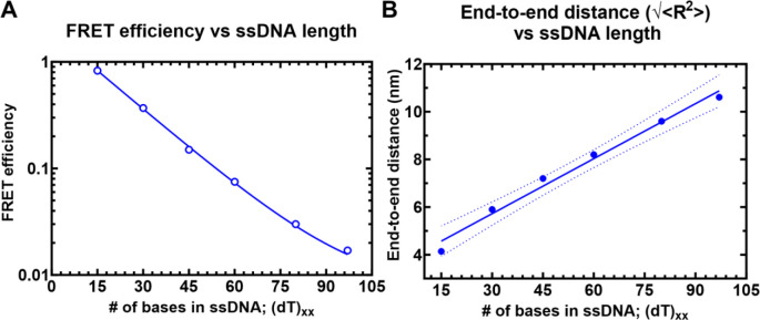
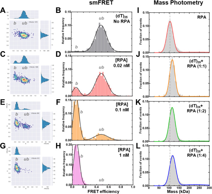
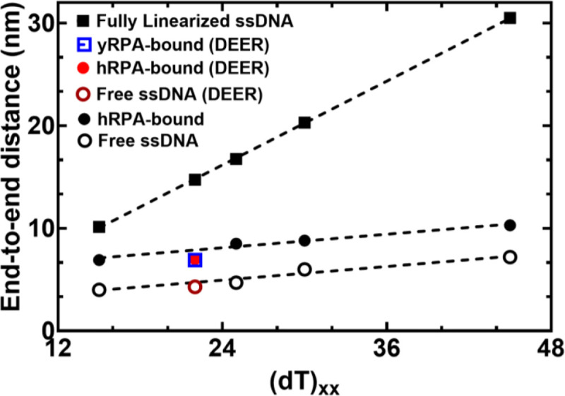
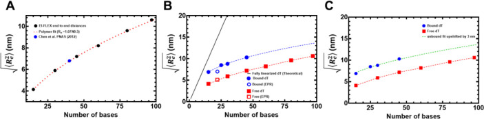
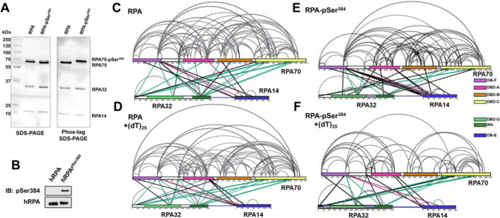
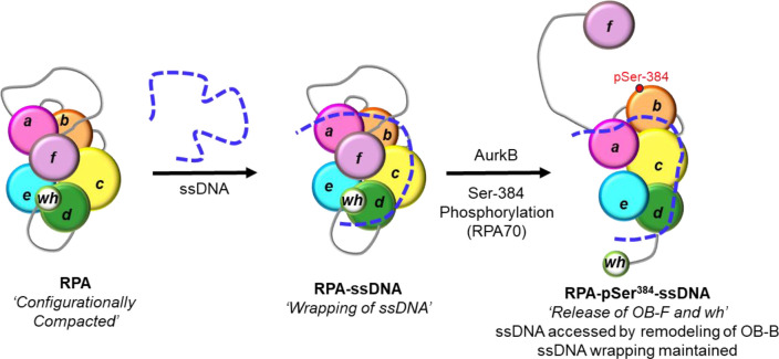
Single-stranded DNA (ssDNA) intermediates, which emerge during DNA metabolic processes are shielded by Replication Protein A (RPA). RPA binds to ssDNA and acts as a gatekeeper, directing the ssDNA towards downstream DNA metabolic pathways with exceptional specificity. Understanding the mechanistic basis for such RPA-dependent specificity requires a comprehensive understanding of the structural conformation of ssDNA when bound to RPA. Previous studies suggested a stretching of ssDNA by RPA. However, structural investigations uncovered a partial wrapping of ssDNA around RPA. Therefore, to reconcile the models, in this study, we measured the end-to-end distances of free ssDNA and RPA-ssDNA complexes using single-molecule FRET and Double Electron-Electron Resonance (DEER) spectroscopy and found only a small systematic increase in the end-to-end distance of ssDNA upon RPA binding. This change does not align with a linear stretching model but rather supports partial wrapping of ssDNA around the contour of DNA binding domains of RPA. Furthermore, we reveal how phosphorylation at the key Ser-384 site in the RPA70 subunit provides access to the wrapped ssDNA by remodeling the DNA-binding domains. These findings establish a precise structural model for RPA-bound ssDNA, providing valuable insights into how RPA facilitates the remodeling of ssDNA for subsequent downstream processes.


Conflict of interest statement

CONFLICT OF INTEREST The authors declare no conflict of interest.

Figures

Figure 1.
Figure 1.. Architecture of the Replication Protein A (RPA) – ssDNA complex.
A) The three subunits of RPA – RPA70, RPA32, and RPA14 house several oligonucleotide/oligosaccharide binding (OB) domains that are classified as either DNA binding domains (DBDs: DBD-A, DBD-B, DBD-C and DBD-D) or protein-interaction domains (OB-F/PID70N and winged helix (wh)/PID32C). A structural model of RPA is shown and was generated using information from known structures of the OB-domains and AlphaFold models of the linkers. B) Crystal structure of Ustilago maydis RPA bound to ssDNA is shown (PDB 4GNX). This structure lacks OB-F, the F-A linker, wh, and the D-wh linker. ssDNA from cryoEM structures of Saccharomyces cerevisiae RPA (PDB 6I52) and Pyrococcus abyssi RPA (8OEL) are superimposed and reveal an end-to-end DNA distance of ~55 Å. Structural data supports partial wrapping of ssDNA around the OB domains.
Figure 2.
Figure 2.. Solution confocal-based FRET measurements accurately report on ssDNA end-to-end distances.
A) FRET efficiencies were calculated based on single molecule measurements of Cy3 (donor) and Cy5 (acceptor) fluorescence. Data were collected on a series of ssDNA substrates (30 pM) of increasing lengths. Energy transfer events were detected as bursts of photons as the molecules transited the confocal volume. The mean of the distribution is plotted in panel A as a function of length of poly-thymidine (dTxx). B) The end-to-end distances were calculated based on a R0 value of 5.4 nm for the Cy3/Cy5 pair and Eq. 3 described in the Methods.
Figure 3.
Figure 3.. Concentration dependence of RPA-ssDNA complexes.
A-H) FRET analysis of (dT)25 ssDNA bound to increasing concentrations of RPA show a shift from the unbound to bound complex. As RPA concentrations are increased, a complete shift to the bound population is observed. I-L) Mass photometry analysis of RPA and RPA-(dT)25 complexes show formation of predominantly single RPA bound (dT)25 complexes.
Figure 4.
Figure 4.. RPA binding produces a modest 2.6 nm increase in end-to-end distance.
The estimated distance between 3’ and 5’ end of ssDNA is plotted as a function of nucleotides which make up the chain. Closed squares represent theoretically calculated end-to-end distances assuming the ssDNA was completely linearized. Open and closed circles represent experimental end-to end distance measurements from smFRET analysis ssDNA −/+ RPA, respectively. The figure also shows that end-to-end distances of yRPA-bound, hRPA-bound, and free (dT)22 ssDNA, measured using DEER spectroscopy, fall on the respective trend lines suggesting good agreement between experimental measurements performed using two independent biophysical approaches.
Figure 5.
Figure 5.. DEER spectroscopy of RPA and RPA-pSer384 bound to ssDNA.
A) Structure of the isoindoline nitroxide spin label and the position on the (dT) oligonucleotide. B) Raw DEER decays and fits are presented for the experimentally determined distance distributions P(r) (C). D) CW EPR spectra of labeled ssDNA in the absence and presence of RPA and RPA-pSer384.
Figure 6.
Figure 6.. Computational predictions of ssDNA wrapping agree well with experimental measurements.
A) Black squares are data, and the red dashed line is a fit to those data using a polymer scaling model. The blue circle is data reporting on the end-to-end distance for (dT)40 made by Chen et al. 2011). B) Blue circles are from RPA-bound (dT) oligonucleotides, red squares are from RPA-free (dT) oligonucleotides. Data include distances measured by both EPR and smFRET experiments. Blue and red dashed lines are polymer model fits to the data, respectively. The black line is the theoretical expected end-to-end distance if the DNA were fully linearized and stretched to its contour length. C) The root mean squared end-to-end distance dependence on the number of bases is essentially identical for the bound and unbound states, as highlighted by the fact that the exact polymer fit upshifted by ~3 nm fully describes the bound dT dependency.
Figure 7.
Figure 7.. Phosphorylation at a single position in RPA70 alters access to the ssDNA with minimal alternations of end-to-end distance.
A) SDS-PAGE and Phos-Tag SDS-PAGE analysis of RPA and RPA-pSer384 proteins show selective shift of the RPA70 band only in the RPA-pSer384 sample confirming 100% incorporation of pSer. B) Western blotting of RPA and RPA-pSer384 with an antibody specific to pSer384 confirms the site-specific phosphorylation. C & D) Cross-linking mass spectrometry (XL-MS) analysis of RPA in the absence or presence of ssDNA (dT)25. XLs arising from RPA32 and RPA14 are colored green and red, respectively. XLs unique to each sample are denoted by dotted lines. F & G) XL-MS comparison between RPA-pSer384 and the RPA-pSer384ssDNA complex. XLs are marked in a comparative manner between tow two samples as explained in C-D.
Figure 8.
Figure 8.. Model for ssDNA wrapping by RPA.
The OB-domains of RPA are depicted along with the connecting disordered linkers. The XL-MS data suggest that the domains are compacted together and ssDNA is wrapped around this architecture. The domains are remodeled upon phosphorylation by Aurora kinase B at position Ser-384 in the RPA70 subunit. This modification releases the OB-F and wh domains and protein RPA interactions with other proteins. However, the wrapping of the ssDNA is not altered, but remodeling allows access to the ssDNA.

References

    1. Caldwell C.C. and Spies M. (2020) Dynamic elements of replication protein A at the crossroads of DNA replication, recombination, and repair. Crit Rev Biochem Mol Biol, 55, 482–507. - PMC - PubMed
    1. Wold M.S. and Kelly T. (1988) Purification and characterization of replication protein A, a cellular protein required for in vitro replication of simian virus 40 DNA. Proc Natl Acad Sci U S A, 85, 2523–2527. - PMC - PubMed
    1. Wold M.S. (1997) Replication protein A: a heterotrimeric, single-stranded DNA-binding protein required for eukaryotic DNA metabolism. Annu Rev Biochem, 66, 61–92. - PubMed
    1. Iftode C., Daniely Y. and Borowiec J.A. (1999) Replication protein A (RPA): the eukaryotic SSB. Crit Rev Biochem Mol Biol, 34, 141–180. - PubMed
    1. Kim C., Paulus B.F. and Wold M.S. (1994) Interactions of human replication protein A with oligonucleotides. Biochemistry, 33, 14197–14206. - PubMed

Publication types