[Preprint]. 2024 Oct 28:2024.03.21.586113.
doi: 10.1101/2024.03.21.586113.

Sir2 and Fun30 regulate ribosomal DNA replication timing via MCM helicase positioning and nucleosome occupancy

Affiliations

Sir2 and Fun30 regulate ribosomal DNA replication timing via MCM helicase positioning and nucleosome occupancy

Carmina Lichauco et al. bioRxiv. .

Update in

Abstract

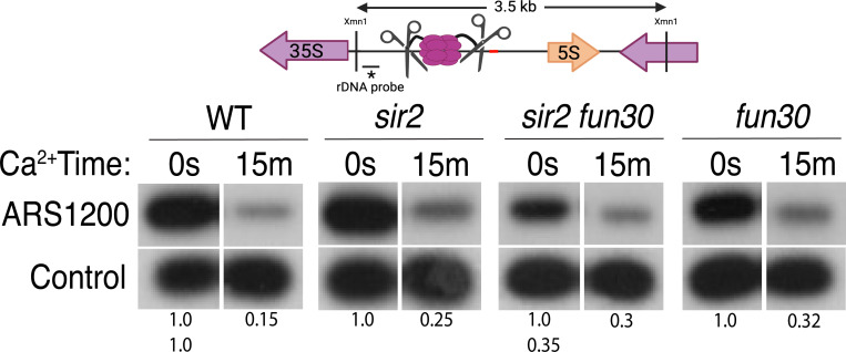
The association between late replication timing and low transcription rates in eukaryotic heterochromatin is well-known, yet the specific mechanisms underlying this link remain uncertain. In Saccharomyces cerevisiae, the histone deacetylase Sir2 is required for both transcriptional silencing and late replication at the repetitive ribosomal DNA arrays (rDNA). We have previously reported that in the absence of SIR2, a derepressed RNA PolII repositions MCM replicative helicases from their loading site at the ribosomal origin, where they abut well-positioned, high-occupancy nucleosomes, to an adjacent region with lower nucleosome occupancy. By developing a method that can distinguish activation of closely spaced MCM complexes, here we show that the displaced MCMs at rDNA origins have increased firing propensity compared to the nondisplaced MCMs. Furthermore, we found that both, activation of the repositioned MCMs and low occupancy of the adjacent nucleosomes critically depend on the chromatin remodeling activity of FUN30. Our study elucidates the mechanism by which Sir2 delays replication timing, and it demonstrates, for the first time, that activation of a specific replication origin in vivo relies on the nucleosome context shaped by a single chromatin remodeler.


Figures

Figure 1.
Figure 1.. rDNA structure, Chromatin Endogenous Cleavage (ChEC), and MCM displacement in sir2.
A. The 1.4 megabase rDNA region on chromosome XII is composed of approximately one hundred and fifty 9.1 kb repeats. Each repeat encodes both the 35S and 5S ribosomal RNAs (rRNA), which are transcribed by RNA Polymerase I (Pol I) and RNA Polymerase III (Pol III), respectively. The ribosomal origin of replication (rARS) is located between the 5’ ends of these genes. The C-pro transcript, which is suppressed by Sir2, initiates approximately two hundred base pairs from the rARS. The unidirectional replication fork block, which functions to prevent collision between replication and transcription machinery, is depicted in green. The probe used in Southern blots in Figure 3 to assess licensing is marked by *. B. In ChEC-seq, micrococcal nuclease (MNase; depicted as scissors) is fused to the protein of interest, in this case MCM2. The double-hexameric MCM helicase complex, MCM2–7, is depicted in purple, and nucleosomes are in blue. Cleavage is induced by addition of calcium to permeabilized cells. Due to the proximity of nucleosome entrance and exit sites, MCM2-ChEC can reveal not only the binding site of the MCM complex but also that of the adjacent nucleosome. C. Depiction of nucleosomes and MCM complex in wild type and sir2 in G1 and G2. Nucleosomes are numbered with respect to C-pro transcription. De-repression of C-pro transcription in sir2 causes RNA PolII to displace the MCM complex to the right. The three different MCM helicase complexes depicted in the bottom right panel are intended to convey the three most prominent locations for the complex in sir2; this is not intended to indicate that there is ever more than one MCM complex in the same rDNA repeat. Created with BioRender.com.
Figure 2.
Figure 2.. S-Seq applied to rDNA replication timing and copy number determination.
A. S-seq replication profile of Chromosome XII. The region in the middle noted as the rDNA has been collapsed. Note that the rDNA replicates much earlier in sir2 (16559) (orange) than in wild type (16535) (blue). B. rDNA replication timing in double mutants between sir2 and various chromatin remodeling enzymes: nhp10 (16729), chd1 (16725), swp82 (17061), isw1 (16724), htl1 (17059), swr1 (16728), isw2 (16673) and irc5 (16723). fun30-S20A S28A (17113 and 17114) is non-phosphorylatable, and fun30-K603R (17345 and 17346) is catalytically inactive. The S to G1 ratio values (mean±SD) were 0.90±.0.02 for WT, 1.04±0.04 for fun30sir2 (16909), 1.01±0.02 for fun30K603R and 1.21±0.03 for sir2 (p<0.001 for WT vs sir2, sir2 vs fun30 sir2 and sir2 vs fun30K603Rsir2 using t-test). C. Effect of sir2 and fun30 mutation on rDNA size, as determined from the fraction of G1 sequencing reads that arise from 450–470 kb on chrXII. The values (% mean±SD) were 9.4±1.6 for WT, 4.6±1.2 for fun30sir2, 4.8±0.3 for fun30K603R and 9.2±1.8 for sir2 (p<0.001 for WT vs sir2, sir2 vs fun30sir2 and sir2 vs fun30K603R sir2 using t-test)
Figure 3.
Figure 3.. Licensing at the rARS, as determined by Southern blot.
Activation of MCM-MNase in G1-arrested WT (16747), sir2 (16769), sir2fun30 (17257) and fun30 (17256) cells at the rARS with calcium will eliminate the 3.5 kb XmnI fragment (upper panel). PIK1, a single-copy gene in which we detect no MCM binding, is used as a loading control. Normalized ARS1200 band intensity at 15m is expressed relative to time 0. Quantitation of the uncut band was used to infer relative rDNA array size in sir2 fun30 mutant at 0.35 relative to WT.
Figure 4.
Figure 4.. Deletion of FUN30 suppresses origin activity at rDNA and promotes it elsewhere in the genome.
A. Schematic of the right end of the rDNA locus depicting Nhe1 cut sites. Digestion with Nhe1 releases multiple copies of a 4.7 kb rARS-containing internal fragment (1N) and a single copy of a 24.4 kb fragment that contains the right-most rDNA origin. B. Diagram of replication structures detected by 2D gel. C. Replicas of 2D gels showing replication at the rDNA locus 30 minutes after release from alpha factor into HU in sir2 fob1 (17564) or sir2 fun30 fob1 (17556) strains with short (29 copies and 35 copies, respectively) rDNA arrays. Numbers indicate the ratio of bubble arc to single copy ARS signal, reflecting the number of activate origins per cell. The average number of activate origins (mean±SD) for sir2 fob1 is 16.9±2.2 vs 4.2±0.7 (P<0.05 by t-test). D. 2D gels showing replication at the rDNA. Cells were arrested in G1 with alpha factor and then released into medium containing 200 mM HU. Quantitation of the ratio of bubble arc to 24.4 kb single copy rARS is shown below. * indicates significant differences (p £ 0.05 as determined by comparing combined signals from all 4 time points by Student’s t-test). Strain numbers used were as follows: wild type (16747), sir2 (16769), sir2 fun30 (17257) and fun30 (17256). E. Replication of ARS305 was examined as in D, but the 1N spot was used for normalization and ratios of bubble arc to 1N spot were normalized to this ratio for the 20 minute time point in wild type. F. EdU incorporation at 111 early origins 1 hour after release from G1 into medium containing 200 mM HU. Total genome-wide read counts in each sample were normalized to the number of reads in the sample with the highest total, thereby normalizing numbers to genome-wide incorporation of EdU. Each dot represents a single origin, with read depths summed across a 5 kb window centered on the MCM binding site within each origin. Points are plotted according to EdU signal in wild type on the x axis and in the mutant in question on the y axis. Suppression of origin activity is reflected in points dropping below the dotted line at 45°. The rARS is circled in green. Strain numbers used were as follows: wild type (17265), sir2 (17271), sir2 fun30 (17279) and fun30 (17281).
Figure 5.
Figure 5.. Disappearance of MCM2-ChEC signal can be used to monitor origin firing.
A. Cells were synchronized in G1 phase using alpha factor for 1.5 hours before being released into media supplemented with hydroxyurea (HU). Cells were harvested at various time points post-release (15–90 minutes) and subjected to MCM2-ChEC analysis. B. Decrease in MCM2-ChEC signal in wild type (16747) for 111 early (orange) and 101 late (blue) origins. MCM2-ChEC signal was quantified over 200 base pair windows centered on the MCM binding site within each origin. Each point is plotted according to MCM2-ChEC signal at the time point in question on the y axis and the corresponding signal in G1 on the x axis, thus decrease in signal appears as a drop below the 45° diagonal. C. MCM signal at the rDNA. MCM2-ChEC signal in G1 appears predominantly at the location indicated by the blue rectangle seen in wild type (16747) and fun30 (17256), whereas it is spread across both the blue and orange rectangles in the absence of Sir2. The displaced MCM2-ChEC signal (orange rectangle) in sir2 (16769) disappears more rapidly than its non-displaced counterpart, and this effect is suppressed by fun30 (17257). Decrease of MCM2-ChEC signal at the rARS is accompanied by increased signal to the right.
Figure 6.
Figure 6.. Nucleosome occupancy assessed by MNase-seq and MCM2-ChEC.
A. Analysis of nucleosome occupancy at rDNA origins using MNase-seq revealed a consistent high occupancy of the +1 nucleosome across WT (16747), sir2 (16769), sir2 fun30 (17257) and fun30 (17256), which served as our normalization reference. Occupancy at the +2 (green box) and +3 (red box) positions was increased by deletion of FUN30 in both sir2 and SIR2. MCM2-ChEC signal was quantified specifically from the 151–200 base pair (nucleosome) size range. B. Analysis of nucleosome occupancy with MCM2-ChEC (see Figure 1B) reveals nucleosome occupancy in that subset of cells and rDNA repeats in which MCM is present. Deletion of FUN30 leads to increased occupancy at the +2 and +3 positions in a sir2 background. MCM2-ChEC signal was quantified specifically from the 151–200 base pair (nucleosome) size range.
Figure 7.
Figure 7.. Model for relationship between MCM location and replication timing at the rDNA.
MCM helicase complex (purple ovals) in wild type abuts the +1 nucleosome (blue cylinder) in G1, making it relatively resistant to activation. Deletion of SIR2 derepresses C-pro transcription (arrow pointing to the right), and RNA PolII pushes MCM complex to a nucleosome free area, where it is more prone to activation. Deletion of FUN30 in a sir2 mutant leads to increased nucleosome occupancy at the +2 position, adjacent to MCM complex, making this complex resistant to activation. Short red stretch of DNA (e.g. between +2 and +3 nucleosomes in top row) indicates ACS. “Ghost” Mcm complex in wild type indicates the small fraction of Mcm that is displaced even in wild type cells (see Figure 5-figure supplement 5). Created with BioRender.com.

References

    1. Ansari A, Gartenberg MR. 1999. Persistence of an alternate chromatin structure at silenced loci in vitro. Proc Natl Acad Sci U S A 96: 343–348. - PMC - PubMed
    1. Azmi IF, Watanabe S, Maloney MF, Kang S, Belsky JA, MacAlpine DM, Peterson CL, Bell SP. 2017. Nucleosomes influence multiple steps during replication initiation. Elife 6. - PMC - PubMed
    1. Bantele SC, Ferreira P, Gritenaite D, Boos D, Pfander B. 2017. Targeting of the Fun30 nucleosome remodeller by the Dpb11 scaffold facilitates cell cycle-regulated DNA end resection. Elife 6. - PMC - PubMed
    1. Batrakou DG, Muller CA, Wilson RHC, Nieduszynski CA. 2020. DNA copy-number measurement of genome replication dynamics by high-throughput sequencing: the sort-seq, sync-seq and MFA-seq family. Nat Protoc 15: 1255–1284. - PubMed
    1. Bell SD, Botchan MR. 2013. The minichromosome maintenance replicative helicase. Cold Spring Harb Perspect Biol 5: a012807. - PMC - PubMed

Publication types