. 2024 Mar 22:18:1375208.
doi: 10.3389/fnins.2024.1375208. eCollection 2024.

Comparative development of the serotonin- and FMRFamide-immunoreactive components of the nervous system in two distantly related ribbon worm species (Nemertea, Spiralia)

Affiliations

Comparative development of the serotonin- and FMRFamide-immunoreactive components of the nervous system in two distantly related ribbon worm species (Nemertea, Spiralia)

Jörn von Döhren. Front Neurosci. .

Abstract

Introduction: Neurodevelopment in larval stages of non-model organisms, with a focus on the serotonin- and FMRFamide-immunoreactive components, has been in the focus of research in the recent past. However, some taxonomic groups remain understudied. Nemertea (ribbon worms) represent such an understudied clade with only few reports on nervous system development mostly from phylogenetically or developmentally derived species. It would be insightful to explore neurodevelopment in additional species to be able to document the diversity and deduce common patterns to trace the evolution of nervous system development.

Methods: Fluorescent immunohistochemical labeling with polyclonal primary antibodies against serotonin and FMRF-amide and a monoclonal antibody against synapsin performed on series of fixed larval stages of two nemertean species Cephalothrix rufifrons (Archinemertea, Palaeonemertea) and Emplectonema gracile (Monostilifera, Hoplonemertea) were analyzed with confocal laser scanning microscopy.

Results: This contribution gives detailed accounts on the development of the serotonin- and FMRFamide-immunoreactive subsets of the nervous system in two nemertean species from the first appearance of the respective signals. Additionally, data on synapsin-like immunoreactivity illustrates the general structure of neuropil components. Events common to both investigated species are the appearance of serotonin-like immunoreactive signals before the appearance of FMRF-like immunoreactive signals and the strict progression of the development of the lateral nerve cords from the anteriorly located, ring-shaped brain toward the posterior pole of the larva. Notable differences are (1) the proboscis nervous system that is developing much earlier in investigated larval stages of E. gracile and (2) distinct early, but apparently transient, serotonergic neurons on the frontal and caudal pole of the larva in E. gracile that seem to be absent in C. rufifrons.

Discussion: According to the results from this investigation and in line with previously published accounts on nervous system development, the hypothetical last common ancestor of Nemertea had a ring-shaped brain arranged around the proboscis opening, from which a pair of ventro-lateral nerve cords develops in anterior to posterior progression. Early frontal and caudal serotonergic neurons that later degenerate or cease to express serotonin are an ancestral character of Nemertea that they share with several other spiralian clades.

Keywords: FMRFamide; Lophotrochozoa; Nemertea; development; larva; nervous system; serotonin; synapsin.


Conflict of interest statement

The author declares that the research was conducted in the absence of any commercial or financial relationships that could be construed as a potential conflict of interest.

Figures

FIGURE 1
FIGURE 1
Current phylogenetic hypothesis and larval types of Nemertea. Phylogeny modified after Andrade et al. (2014) and Kvist et al. (2014). Images of larvae modified from Hindinger et al. (2013), under the terms of CC-BY 2.0.
FIGURE 2
FIGURE 2
Development (DIC, all images from living specimens); (A–C) Emplectonema gracile and (D,E) Cephalothrix rufifrons. (A) Two-cell stage, approximately 12 h after oviposition (lateral view, apical to top-right corner), note excess sperm cells caught in jelly coat (arrowheads); (B) young larva shortly after hatching, approximately 1 day after oviposition (apical is up); (C) older larva 7 days after oviposition (dorsal view, apical is up); (D) young larva at 11 days after oviposition (ventral view, apical is up); (E) older larva 42 days after oviposition (ventral view, apical to top-right corner). at, apical tuft; bm, blastomeres; ch, chorion; cs, central stylet; ct, caudal tuft; jc, jelly coat; lc, lateral cirrus; mo, mouth opening; oc, ocellus; pb, polar bodies; pr, proboscis; rs, reserve stylet.
FIGURE 3
FIGURE 3
Emplectonema gracile larvae, FMRFamide-like immunoreactivity, maximum projection, frontal faces up in all images. (A) 1 day after oviposition, dorsal view (z ≈ 49 μm, γ = 1.5); (B) 3 days after oviposition, dorso-lateral view (z ≈ 48 μm, γ = 0.6), note the caudal connection of the lateral longitudinal nerve cords by the anal commissure (arrowhead); (C) 5 days after oviposition, ventral view (z ≈ 59 μm, γ = 1.2); (D) 6 days after oviposition, ventral view (z ≈ 48 μm, γ = 0.7). bni, brain ring neurites; bno, brain ring neuron; c, caudal; cni, cephalic neurites; f, frontal; ffs, frontal RFa-lir signals; fna, fronto-dorsal neurite aggregation; lni, longitudinal nerve cord neurites; pep, peripheral epidermal plexus; pns, proboscis nervous system; vfs, ventral RFa-lir signal aggregation.
FIGURE 4
FIGURE 4
Emplectonema gracile larvae, serotonin-like immunoreactivity, maximum projection, frontal faces up in all images. (A) 1 day after oviposition, dorsal view (z ≈ 84 μm, γ = 1.5); (B) 2 days after oviposition, dorsal view (z ≈ 66 μm, γ = 0.8); (C) 3 days after oviposition, dorsal view (z ≈ 56 μm, γ = 0.8); (D) 6 days after oviposition, ventral view, (z ≈ 43 μm, γ = 1.1). bni, brain ring neurites; bno, brain ring neuron; c, caudal; cni, cephalic neurites; cso, caudal 5HT-lir neuron; esn, esophageal nervous system; f, frontal; fso, frontal 5HT-lir neuron; fss, frontal 5HT-lir signals; lni, longitudinal nerve cord neurites; lno, longitudinal nerve cord neuron; pep, peripheral epidermal plexus; pns, proboscis nervous system.
FIGURE 5
FIGURE 5
Emplectonema gracile larvae, synapsin-like immunoreactivity, maximum projection, frontal faces up in all images. (A) 2 days after oviposition, dorso-lateral view (z ≈ 42 μm, γ = 1.2); (B) 4 days after oviposition, dorsal view (z ≈ 51 μm, γ = 1.1); (C) 8 days after oviposition, ventral view (z ≈ 53 μm, γ = 0.8), note the caudal connection of the lateral longitudinal nerve cords by the anal commissure (arrowhead). bnp, brain ring neuropil; c, caudal; cnp, cephalic neuropil; enp, esophageal neuropil; f, frontal; lnp, longitudinal nerve cord neuropil; pep, peripheral epidermal plexus; pns, proboscis nervous system.
FIGURE 6
FIGURE 6
Cephalothrix rufifrons larvae, FMRFamide-like immunoreactivity, maximum projection, frontal faces up in panels (A–D) and left in panel (E). (A) 4 days after oviposition, left lateral view, (z ≈ 72 μm, γ = 1.1); (B) 5 days after oviposition, ventro-lateral view (z ≈ 58 μm, γ = 1); (C) 14 days after oviposition, dorsal view (z ≈ 56 μm, γ = 0.8), note the caudal connection of the lateral longitudinal nerve cords by the anal commissure (arrowhead); (D) 21 days after oviposition, ventral view (z ≈ 79 μm, γ = 0.7); (E) 42 days after oviposition, ventro-lateral view (z ≈ 14 μm, γ = 0.8). bni, brain ring neurites; bno, brain ring neuron; c, caudal; cni, cephalic neurites; csn, caudal sensory neuron; dpn, dorsal pharyngeal neurite; f, frontal; ffs, frontal RFa-lir signals; fgs, frontal gland/sensory cell signals; fsn, frontal sensory neuron; lni, longitudinal nerve cord neurites; mni, mouth ring neurites; mno, mouth ring neuron; pep, peripheral epidermal plexus; pni, proboscis neurites; son, suboral neurite; tni, transversal lateral neurite; tno, transversal lateral neuron; vni, ventral neurite.
FIGURE 7
FIGURE 7
Cephalothrix rufifrons larvae, serotonin-like immunoreactivity, maximum projection, frontal faces up in panels (A–D) and left in panel (E). (A) 4 days after oviposition, ventral view (z ≈ 73 μm, γ = 1.2); (B) 7 days after oviposition, dorsal view (z ≈ 56 μm, γ = 0.8); (C) 9 days after oviposition, dorsal view (z ≈ 63 μm, γ = 0.8); (D) 21 days after oviposition, dorsal view (z ≈ 84 μm, γ = 1.2); (E) 42 days after oviposition, ventral view (z ≈ 14 μm, γ = 0.8). bni, brain ring neurites; bno, brain ring neuron; c, caudal; cni, cephalic neurites; f, frontal; lni, longitudinal nerve cord neurites; cno, caudal neuron; lno, longitudinal nerve cord neuron; pep, peripheral epidermal plexus; pni, proboscis neurites.
FIGURE 8
FIGURE 8
Cephalothrix rufifrons larvae, synapsin-like immunoreactivity, maximum projection, frontal faces up in all images. (A) 4 days after oviposition, dorso-lateral view (z ≈ 72 μm, γ = 0.9); (B) 9 days after oviposition, dorsal view (z ≈ 58 μm, γ = 1.1); (C) 21 days after oviposition, ventral view (z ≈ 62 μm, γ = 1.3), note the caudal connection of the lateral longitudinal nerve cords by the anal commissure (arrowhead). bnp, brain ring neuropil; c, caudal; cnp, cephalic neuropil; f, frontal; lnp, longitudinal nerve cord neuropil; mnp, mouth ring neuropil; pep, peripheral epidermal plexus.
FIGURE 9
FIGURE 9
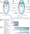
Comparative summary of nervous system development. (A) Emplectonema gracile, ventral view, schematic representation of nervous system in 8 dpo larvae; (B) Cephalothrix rufifrons, ventral view, schematic representation of nervous system in 42 dpo larvae; larvae are not shown to scale, esophageal plexus in E. gracile, peripheral sensory cells and peripheral epidermal plexus in both species omitted for clarity; (C) schematic timeline of development of major components of the nervous system (color coding as in panels A,B, vertical dotted line indicates time of hatching, horizontal dotted lines indicate not completely formed or degenerating structures).

References

    1. Andrade S. C. S., Montenegro H., Strand M., Schwartz M. L., Kajihara H., Norenburg J. L., et al. (2014). A transcriptomic approach to ribbon worm systematics (Nemertea): resolving the pilidiophora problem. Mol. Biol. Evol. 31 3206–3215. 10.1093/molbev/msu253 - DOI - PubMed
    1. Bartolomaeus T. (1993). “Die Leibeshöhlenverhältnisse und Verwandtschaftsbeziehungen der Spiralia (Body cavities and relationships within the Spiralia),” in Verhandlungen der Deutschen Zoologischen Gesellschaft, ed. H.-D. Pfannenstiel (Salzburg: Gustav Fischer Verlag; ), 42.
    1. Battonyai I., Voronezhskaya E. E., Obukhova A., Horváth R., Nezlin L. P., Elekes K. (2018). Neuronal development in the larvae of the invasive biofouler Dreissena polymorpha (Mollusca: Bivalvia), with special attention to sensory elements and swimming behavior. Biol. Bull. 234 192–206. 10.1086/698511 - DOI - PubMed
    1. Beckers P. (2015). The nervous systems of Pilidiophora (Nemertea). Zoomorphology 134 1–24. 10.1007/s00435-014-0246-3 - DOI
    1. Beckers P., Faller S., Loesel R. (2011). Lophotrochozoan neuroanatomy: an analysis of the brain and nervous system of Lineus viridis (Nemertea) using different staining techniques. Front. Zool. 8:17. 10.1186/1742-9994-8-17 - DOI - PMC - PubMed