. 2024 Jun 4;149(23):1833-1851.
doi: 10.1161/CIRCULATIONAHA.123.063965. Epub 2024 Apr 8.

Asparagine Synthetase Marks a Distinct Dependency Threshold for Cardiomyocyte Dedifferentiation

Affiliations

Asparagine Synthetase Marks a Distinct Dependency Threshold for Cardiomyocyte Dedifferentiation

Yike Zhu et al. Circulation. .

Abstract

Background: Adult mammalian cardiomyocytes have limited proliferative capacity, but in specifically induced contexts they traverse through cell-cycle reentry, offering the potential for heart regeneration. Endogenous cardiomyocyte proliferation is preceded by cardiomyocyte dedifferentiation (CMDD), wherein adult cardiomyocytes revert to a less matured state that is distinct from the classical myocardial fetal stress gene response associated with heart failure. However, very little is known about CMDD as a defined cardiomyocyte cell state in transition.

Methods: Here, we leveraged 2 models of in vitro cultured adult mouse cardiomyocytes and in vivo adeno-associated virus serotype 9 cardiomyocyte-targeted delivery of reprogramming factors (Oct4, Sox2, Klf4, and Myc) in adult mice to study CMDD. We profiled their transcriptomes using RNA sequencing, in combination with multiple published data sets, with the aim of identifying a common denominator for tracking CMDD.

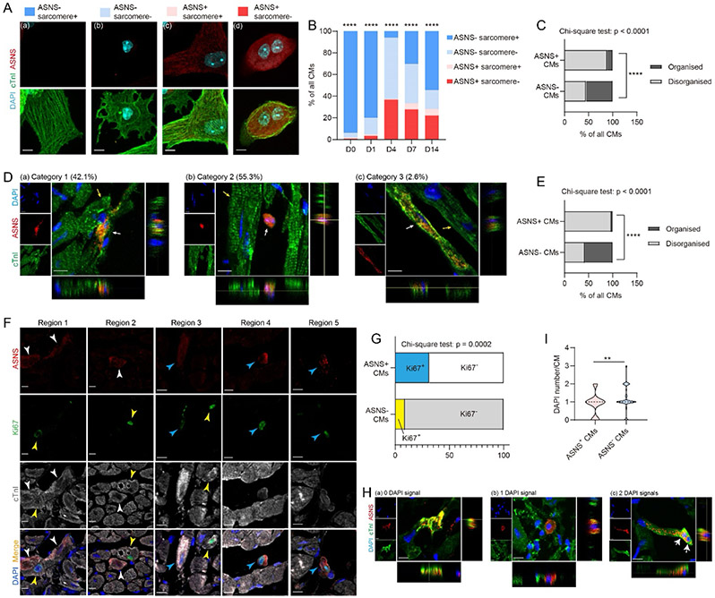
Results: RNA sequencing and integrated analysis identified Asparagine Synthetase (Asns) as a unique molecular marker gene well correlated with CMDD, required for increased asparagine and also for distinct fluxes in other amino acids. Although Asns overexpression in Oct4, Sox2, Klf4, and Myc cardiomyocytes augmented hallmarks of CMDD, Asns deficiency led to defective regeneration in the neonatal mouse myocardial infarction model, increased cell death of cultured adult cardiomyocytes, and reduced cell cycle in Oct4, Sox2, Klf4, and Myc cardiomyocytes, at least in part through disrupting the mammalian target of rapamycin complex 1 pathway.

Conclusions: We discovered a novel gene Asns as both a molecular marker and an essential mediator, marking a distinct threshold that appears in common for at least 4 models of CMDD, and revealing an Asns/mammalian target of rapamycin complex 1 axis dependency for dedifferentiating cardiomyocytes. Further study will be needed to extrapolate and assess its relevance to other cell state transitions as well as in heart regeneration.

Keywords: cell dedifferentiation; cell self-renewal; myocytes, cardiac; regeneration.


Conflict of interest statement

Disclosures None.

Figures

Figure 1.
Figure 1.. Cultured adult mouse CMs (ACMs) dedifferentiate and partially re-differentiate in vitro
(A) Representative images of isolated ACMs in vitro cultured from D0 to D14 (n = 4 independent CM isolations). Scale bar = 200 μm. (B) Immunofluorescence (IF) images showing the breakdown and re-construction of ACM sarcomeres from D0 to D14, staining for α-Actinin, cTnI and MyBPC3. Scale bar = 10 μm. (C) Heatmap of module-trait relationships by WGCNA. Each row represents a gene module, and each column represents one timepoint (n = 4 for each timepoint). Correlation coefficients are labelled in each cell, with corresponding P-values displayed also within parentheses. Units are coloured based on correlation of the module across D0-D14 ACMs: red for positive (high expression), and green for negative (low expression) correlations. Curves on the right column reflect the expressional dynamics of genes in each module from D0 to D14. (D) GO terms enriched in gene modules that had expressional dynamics of interest: red, purple, yellow, blue, and turquoise modules. (E) Top transcription factor DNA binding motifs of ETS family enriched in the turquoise gene module, obtained by HOMER motif analysis.
Figure 2.
Figure 2.. CM-specific transduction of OSKM by AAV9 induces loss of cardiac function and hallmarks of CMDD in vivo
(A) Constructs of AAV9 comprising cTnT-driven Oct4, Sox2, Klf4 and Myc, fused to corresponding His6, Flag, HA and V5 signal peptides. (B) Three dose gradients (1×, 2×, 4×) and (C) experimental timeline of AAV9 injections. AAV-cTnT-BFP was injected as control. (D) Quantification of OSKM mRNA expression abundance in purified CMs by RT-qPCR. (E) Representative Western blot and (F) quantification for OSKM expression in purified CMs. (G) Cardiac functional and structural changes during the experimental timeline by echocardiography. n = 10 to 14 mice per group per timepoint. EF% - ejection fraction; IVS;d - interventricular septum thickness at end-diastole; LVID;d - left ventricular internal diameter at end-diastole. (H) IF images showing α-SMA on heart sections. α-SMA was restricted to vessel smooth muscle cells (VSMCs) in Control group (left), but present in both VSMCs and CMs in OSKM hearts (right). Scale bar = 100 μm and 20 μm, at low and high magnification images, respectively. (I) IF images showing the loss of Connexin 43 at CM-CM junctions in OSKM hearts. Scale bar = 50 μm and 10 μm, in low and high magnification images, respectively. (J) mRNA expression of Gja1 in purified CMs by RT-qPCR. (K-M) Representative images of CMs stained for Ki67, pH3 and Aurora kinase B (left) and quantification of positively stained CMs per heart section (right), respectively. Three heart sections were analysed per mouse heart. Each data point represents an independent mouse heart. The white arrows in (M) indicate a pair of nuclei, between which Aurora kinase B in the midbody remnant (red signal) indicate cytokinesis. Scale bar = 10 μm. (N) Volcano plot and (O) heatmap showing top differentially expressed (DE) genes (ranked by fold change) from RNA-seq. Red represent DE genes (n = 2 for Control-CMs, n = 3 for OSKM-CMs, Log2FC > 1 or < −1, FDR < 0.05). (P) GO terms (solid bars) and KEGG pathways (hollow bars) enriched in upregulated (right, orange) and downregulated (left, blue) genes in OSKM-CMs. Data were presented as mean ± SD. Shapiro–Wilk test was used to examine data normality. Data were analysed with one-way ANOVA test (F, J, L), two-way ANOVA test (G), or Kruskal-Wallis test (D, K, M) followed by multiple comparisons. Multiple comparisons: n.s. (non-significant), *p < 0.05, **p < 0.01, ***p < 0.001, ****p < 0.0001, compared to AAV-Control.
Figure 3.
Figure 3.. Asns is a molecular marker of CMDD shared by both cultured ACMs and OSKM-CMs.
(A) Overlapping DE genes between the cultured-ACMs and OSKM/Control-CMs datasets, and their respective enriched GO terms. From top to bottom: DE genes in OSKM/Control-CMs dataset overlap with the red, brown, purple, yellow, blue and turquoise modules from the cultured-ACMs dataset. (B) A schematic diagram depicting the criteria and cut-off by which Asns was screened out as a marker gene. (C) Asns mRNA expression in cultured ACMs by RT-qPCR. n = 4 biological replicates. (D) Representative Western blot and (F) quantification (n = 3) for ASNS protein expression in cultured ACMs. (E) IF images showing ASNS+ CMs from D0 to D14. Scale bar = 100 μm and 50 μm, in low and high magnification images, respectively. (F) Quantification of ASNS+ CMs from D0 to D14. n = 3 biological replicates, and more than 200 CMs analysed for each timepoint per replicate. (G) Representative Western blot and (H) quantification for ASNS protein expression in purified CMs from Control and OSKM hearts. (I) Asns mRNA expression in purified Control-CMs and OSKM-CMs by RT-qPCR. (J) Representative images of ASNS+ CMs on OSKM-heart sections. Scale bar = 10 μm. (K) Quantification of ASNS+ CMs per heart section. 2-3 heart sections were analysed per mouse. Each data point represents an independent mouse heart. Data were presented as mean ± SD. Shapiro–Wilk test was used to examine data normality. Data were analysed with one-way ANOVA test (C, D, H, I, K) or Kruskal-Wallis test (F) followed by multiple comparisons. One-way ANOVA/Kruskal-Wallis test: #p < 0.05, ####p < 0.0001; Multiple comparisons: n.s. (non-significant), *p < 0.05, **p < 0.01, ***p < 0.001, ****p < 0.0001, compared to D0 (C, D, F) or AAV-Control (H, I, K).
Figure 4.
Figure 4.. The subpopulation of ASNS+ CMs display unique key features of CMDD
(A) Representative IF images showing four categories of cultured ACMs based on sarcomeric organisation and ASNS expression. Scale bar = 10 μm. (B) Percentage of cultured ACMs in each category from D0 to D14. More than 100 CMs were analysed for each timepoint per isolation. n = 3 independent isolations. (C) Percentage of cultured ACMs in each category, by pooling ACMs from all timepoints. n = 3 independent isolations. (D) Representative IF images containing ASNS+ (white arrows) and ASNS CMs (yellow arrows), classified into (a) Cat#1, (b) Cat#2 and (c) Cat#3. Scale bar = 10 μm. (E) Percentage of ASNS+ and ASNS CMs displaying organised and disorganised sarcomeres by pooling OSKM and Control groups, from Cat#2 and #3 in (D). (F) Representative IF images showing four categories of OSKM-CMs: (i) ASNS Ki67 (not indicated by any arrows); (ii) ASNS Ki67+ (yellow arrows); (iii) ASNS+ Ki67 (white arrows); (iv) ASNS+ Ki67+ (blue arrows). Scale bar = 10 μm. (G) Percentage of Ki67+ and Ki67 CMs in the subpopulations of ASNS+ and ASNS CMs. More than 200 CMs from 5 OSKM-heart sections were analysed. (H) Representative IF images showing ASNS+ CMs containing (a-c) 0, 1 or 2 DAPI signals, respectively. The white arrows in (c) indicate two DAPI signals. Scale bar = 10 μm. (I) Average nuclear number (indicated by DAPI staining) in ASNS+ and ASNS CMs (n = 38 CMs) by pooling both OSKM and Control groups. Data were presented as mean ± SD. Shapiro–Wilk test was used to examine data normality. Data were analysed with Chi-square test (B, C, G) and Mann–Whitney U test (I). *p < 0.05, **p < 0.01, ****p < 0.0001.
Figure 5.
Figure 5.. Asns is an essential mediator of CMDD regulating asparagine metabolism, while playing distinct roles in vitro and in vivo
(A) A diagram depicting AAV9 comprising Asns shRNA, LacZ shRNA (negative control) and the experimental timeline. (B) Asns mRNA expression in cultured ACMs by RT-qPCR. (C) Western blot and (D) quantification of ASNS protein expression in cultured ACMs. (E) PI staining and (F) quantification of PI+ cultured ACMs on D4. More than 600 CMs were analysed per isolation per group. Scale bar = 200 μm. (G) The rate of CM loss from D0 to D4 by counting the number of cultured ACMs. (H) Heatmaps showing (a) the mean and (b) individual values of amino acids quantified by metabolomic profiling of cultured ACMs on D0 and D4. WT – LacZ-shRNA, KD – Asns-shRNA. n = 5 to 6 per group. Amino acid quantification was normalised to ACM count. (I) Quantification of PI+ CMs on D4, after Asn, Gln and ASPG treatment. (J) mRNA expression of Asns and other cell cycle genes (Cdk1, Mki67, Aukrb) in cultured ACMs by RT-qPCR. (K) The experimental timeline of Asns KD in 4Fheart mice. (L) RT-qPCR and (M) Western blot validating the efficiency of Asns KD at mRNA and protein expression level in heart tissues. Numbers 1 to 4 represent 4 individual mice. (N-P) Representative images (left) and percentage (right) of CMs stained positive for Ki67, pH3 and Aurora kinase B, respectively. Three heart sections were analysed per mouse heart. Each data point represents an independent mouse heart. The white arrows in (P) indicate a pair of nuclei, between which Aurora kinase B in the midbody remnant (red signal) indicate cytokinesis. Scale bar = 10 μm. (Q) Quantification of amino acids, Asn, Gln, Glu and Asp (normalised to total protein) in heart tissues, measured by LC-MS. Data were presented as mean ± SD. Shapiro–Wilk test was used to examine data normality. Data were analysed with one-way ANOVA test (B, J <Asns, Cdk1, Mki67>, I, K, L, Q) or Kruskal-Wallis test (J <Aurkb>) followed by multiple comparisons. Multiple comparisons: n.s. (non-significant), *p < 0.05, **p < 0.01, ***p < 0.001, ****p < 0.0001. Student’s t test was used for (D, F, G, P), and Mann–Whitney U test was used for (N, O), *p < 0.05, **p < 0.01.
Figure 6.
Figure 6.. Asns deficiency inhibited heart regeneration in the neonatal mouse myocardial infarction (MI) model.
(A) Experimental timeline of MI in neonatal mouse and sample collection. (B) IF staining of ASNS in the border zone of MI heart on D7 post MI. Scale bar = 1000 μm and 50 μm in low and high magnification images, respectively. (C) The experimental timeline of MI and AAV9-shRNA injection in neonatal mouse and sample collection. (D) IF staining results showing GFP reporter on D7 post surgery. Scale bar = 1000 μm and 50 μm in low and high magnification images, respectively. (E) IF staining and (F) quantification of ASNS expression at the border zone of MI heart on D7. BZ-border zone; IZ-infarct zone. Scale bar = 10 μm. (G) Quantification and (H) representative images of CMs stained positive for pH3. Scale bar = 10 μm. (I) Quantification and (J) representative images of CMs stained positive for Aurora kinase B. Scale bar = 10 μm. (K-L) Picrosirius red staining of heart sections collected on D21 post surgery. Scale bar = 1000 μm. (M) Quantification of fibrotic scars in heart sections collected on D21 post surgery. Data were presented as mean ± SD. Shapiro–Wilk test was used to examine data normality. Student’s t test was used for (F, G, I, M), **p < 0.01, ***p < 0.001.
Figure 7.
Figure 7.. Overexpression (OE) of Asns augments the hallmarks of CMDD in OSKM hearts in vivo.
(A) The experimental timeline of Asns OE in OSKM (1x)-CMs in vivo, by AAV9 delivery. (B) Cardiac functional and structural changes measured by echocardiography. (C-E) Representative images (top) and percentage (bottom) of CMs stained positive for Ki67, pH3 and Aurora kinase B, respectively. Three heart sections were analysed per mouse heart. Each data point represents an independent mouse heart. The white arrows in (E) indicate a pair of nuclei, between which Aurora kinase B in the midbody remnant (red signal) indicate cytokinesis. Scale bar = 10 μm. (F) IF staining images showing ASNS and αSMA on mouse heart sections. Scale bar = 50 μm. (G-H) GO terms enriched in downregulated genes in OSKM1x AsnsOE heart, also indicated by the blue dots in (I) the volcano plot showing DE genes between OSKM1x AsnsOE heart and OSKM1xBFPOE heart by RNA-seq (n = 2 for OSKM1xBFPOE, n = 4 for OSKM1x AsnsOE, Log2FC > 1 or < −1, FDR< 0.05). (J) GO terms enriched in upregulated genes in OSKM1x AsnsOE hearts, as indicated by red dots in (I). (K) Quantification of amino acids, Asn, Gln, Glu and Asp (normalised to total protein) in heart tissues, measured by LC-MS. Data were presented as mean ± SD. Shapiro–Wilk test was used to examine data normality. One-way ANOVA followed by multiple comparisons was used for (B , C-E). Multiple comparisons: n.s. (non-significant), *p < 0.05, **p < 0.01, as OSKM1xBFPOE group was compared with BFP group (black labelling in (B)) and OSKM1xAsnsOE group (orange labelling in (B)). Student’s t test was used for (K <a-c> ), and Mann–Whitney U test was used for (K <d>), *p < 0.05.
Figure 8.
Figure 8.. Asns is an upstream regulator of the mTORC1 pathway in CMDD.
(A) Western blot and (B) quantification showing GLUL, p-S6K1 and p-4EBP1 in 4F+AsnsKD mouse heart tissues. Numbers 1 to 4 represent 4 individual mice. (C) Representative Western blot and (D) quantification showing GLUL, p-S6K1 and p-4EBP1 in cultured ACMs with Asns KD. (E) Protein-protein interaction network of mouse Asns, Glul and mTORC1 pathway related genes (Mtor, Rheb, Rps6kb1, Eif4ebp1), from the STRING database. (F) A diagram depicting the molecular mechanisms by which Asns regulate CMDD in cultured ACMs and OSKM-CMs. Data were presented as mean ± SD. Shapiro–Wilk test was used to examine data normality. Data were analysed with one-way ANOVA test (B, D) followed by multiple comparisons was used for. Multiple comparisons: n.s. (non-significant), *p < 0.05, **p < 0.01, ***p < 0.001, ****p < 0.0001. Student’s t test was also used for (D), when comparing D0-LacZKD v.s. D0-AsnsKD, D0-ZacZKD v.s. D4-LacZKD and D4-LacZKD v.s. D4-AsnsKD: n.s. (no significant difference), *p < 0.05, **p < 0.01.

Comment in

References

    1. Heallen T, Morikawa Y, Leach J, Tao G, Willerson JT, Johnson RL, Martin JF. Hippo signaling impedes adult heart regeneration. Dev. 2013;140:4683–4690. - PMC - PubMed
    1. Ikeda S, Mizushima W, Sciarretta S, Abdellatif M, Zhai P, Mukai R, Fefelova N, Oka SI, Nakamura M, Del Re DP, Farrance I, Park JY, Tian B, Xie LH, Kumar M, Hsu CP, Sadayappan S, Shimokawa H, Lim DS, Sadoshima J. Hippo Deficiency Leads to Cardiac Dysfunction Accompanied by Cardiomyocyte Dedifferentiation during Pressure Overload. Circ Res. 2019;124:292–305. - PMC - PubMed
    1. Xin M, Kim Y, Sutherland LB, Murakami M, Qi X, McAnally J, Porrello ER, Mahmoud AI, Tan W, Shelton JM, Richardson JA, Sadek HA, Bassel-Duby R, Olson EN. Hippo pathway effector Yap promotes cardiac regeneration. Proc Natl Acad Sci U S A. 2013;110:13839–13844. - PMC - PubMed
    1. D’Uva G, Aharonov A, Lauriola M, Kain D, Yahalom-Ronen Y, Carvalho S, Weisinger K, Bassat E, Rajchman D, Yifa O, Lysenko M, Konfino T, Hegesh J, Brenner O, Neeman M, Yarden Y, Leor J, Sarig R, Harvey RP, Tzahor E. ERBB2 triggers mammalian heart regeneration by promoting cardiomyocyte dedifferentiation and proliferation. Nat Cell Biol. 2015;17:627–638. - PubMed
    1. Aharonov A, Shakked A, Umansky KB, Savidor A, Genzelinakh A, Kain D, Lendengolts D, Revach OY, Morikawa Y, Dong J, Levin Y, Geiger B, Martin JF, Tzahor E. ERBB2 drives YAP activation and EMT-like processes during cardiac regeneration. Nat Cell Biol [Internet]. 2020;22:1346–1356. Available from: 10.1038/s41556-020-00588-4 - DOI - PubMed

MeSH terms

Substances