. 2024 May 8;52(8):4483-4501.
doi: 10.1093/nar/gkae240.

Identifying human pre-mRNA cleavage and polyadenylation factors by genome-wide CRISPR screens using a dual fluorescence readthrough reporter

Affiliations

Identifying human pre-mRNA cleavage and polyadenylation factors by genome-wide CRISPR screens using a dual fluorescence readthrough reporter

Zuyao Ni et al. Nucleic Acids Res. .

Abstract

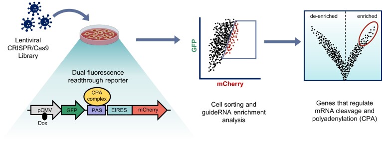
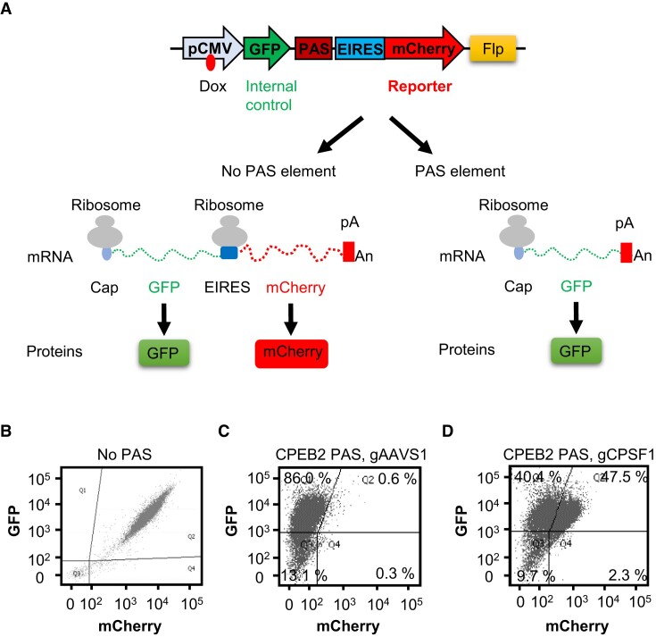
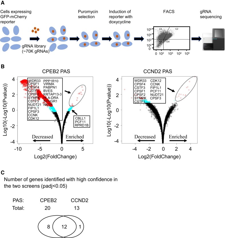
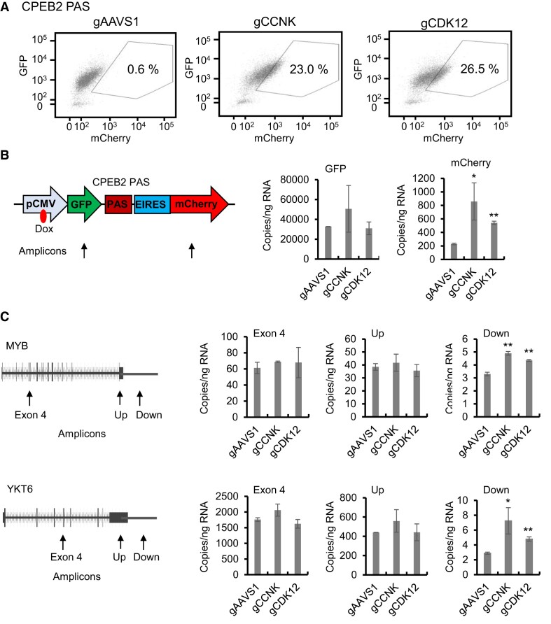
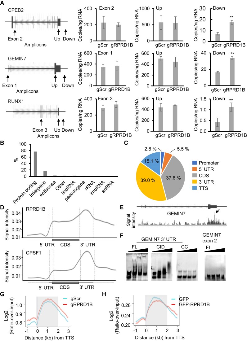
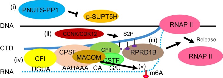
Messenger RNA precursors (pre-mRNA) generally undergo 3' end processing by cleavage and polyadenylation (CPA), which is specified by a polyadenylation site (PAS) and adjacent RNA sequences and regulated by a large variety of core and auxiliary CPA factors. To date, most of the human CPA factors have been discovered through biochemical and proteomic studies. However, genetic identification of the human CPA factors has been hampered by the lack of a reliable genome-wide screening method. We describe here a dual fluorescence readthrough reporter system with a PAS inserted between two fluorescent reporters. This system enables measurement of the efficiency of 3' end processing in living cells. Using this system in combination with a human genome-wide CRISPR/Cas9 library, we conducted a screen for CPA factors. The screens identified most components of the known core CPA complexes and other known CPA factors. The screens also identified CCNK/CDK12 as a potential core CPA factor, and RPRD1B as a CPA factor that binds RNA and regulates the release of RNA polymerase II at the 3' ends of genes. Thus, this dual fluorescence reporter coupled with CRISPR/Cas9 screens reliably identifies bona fide CPA factors and provides a platform for investigating the requirements for CPA in various contexts.


Figures

Graphical Abstract
Graphical Abstract
Figure 1.
Figure 1.
A dual fluorescence readthrough reporter to measure the effects of 3′ end processing. (A) Schematic illustration of the dual fluorescence readthrough reporter. In the construct without a PAS, a doxycycline-inducible CMV promoter drives transcription through both fluorescent reporters, GFP and mCherry. Ribosomes dock at the 5′ end cap of the bicistronic mRNA to translate GFP or at the EIRES to translate the uncapped mCherry portion of the transcript. In the construct with a PAS, the transcription is terminated upstream of mCherry, whereas the GFP portion of the transcript remains intact. The expression ratio between the mCherry and GFP reporters measures transcriptional readthrough effects that reflect 3′ end processing. Dox: doxycycline. (B) Constant mCherry/GFP ratio in the dual fluorescence readthrough reporter without a PAS. (C) A distal PAS of the CPEB2 gene decreases the mCherry/GFP ratio in the dual fluorescence readthrough reporter. (D) CRISPR/Cas9-mediated depletion of CPSF1 increases the mCherry/GFP ratio in the dual fluorescence readthrough reporter containing the distal PAS of the CPEB2 gene. Expression in (B)–(D) was induced by the addition of 2 μg/ml doxycycline for 4 days in HEK293 cells, and each dot represents the expression in one living cell. GFP and mCherry expressions in 10 000 cells were analyzed by FACS in each experiment.
Figure 2.
Figure 2.
CRISPR/Cas9 screens identified CPA factors. (A) CRISPR/Cas9 screening pipeline. Flp-In T-REx HEK293 cells expressing the GFP-mCherry readthrough reporter with an inserted PAS were infected with the pooled lentiviral human TKOv3 gRNA library followed by puromycin selection. After doxycycline-induced expression of the two reporters, cells were sorted by FACS. Genomic DNA was isolated from unsorted cells and sorted cells that exhibited an increased mCherry/GFP ratio, and the gRNAs were amplified and subjected to sequencing. (B) CRISPR/Cas9 screen results for HEK293 cell lines expressing the GFP and mCherry reporters separated by the distal PAS from the CPEB2 (left) and CCND2 (right) genes. Scatterplots (volcano plots) display pairwise comparisons of the gRNAs between sorted cells that exhibited an increased mCherry/GFP ratio and unsorted cells. Red circles: Padj < 0.05. Blue circles: Padj < 0.25, but >0.05. Black circles: Padj > 0.25. Experiments were performed in biological quadruplicate. Padj values were determined using the Benjamini–Hochberg adjustment for multiple comparisons. (C) Genes identified in both screens with high confidence. The gRNAs of 12 genes were enriched with high confidence (Padj < 0.05) in sorted cells with increased mCherry/GFP ratio as compared with unsorted cells in both screens.
Figure 3.
Figure 3.
CCNK and CDK12 in 3′ end processing. (A) CRISPR/Cas9-mediated depletion of CCNK or CDK12 increases the mCherry/GFP ratio in HEK293 cells expressing the dual fluorescence readthrough reporter containing the distal PAS of the CPEB2 gene. 10000 cells were analyzed by FACS. (B) Depletion of CCNK or CDK12 specifically increases the expression of mCherry RNA relative to that of GFP RNA in HEK293 cells expressing the dual fluorescence readthrough reporter containing the distal PAS of the CPEB2 gene. RNA expression levels were measured by RT-qPCR using primers targeting the indicated reporter regions in cells infected with lentiviruses transducing the indicated gRNAs. (C) Depletion of CCNK or CDK12 increases RNA expression downstream of the PAS in endogenous genes. RNA expression from the MYB and YKT6 genes was measured by RT-qPCR using primers targeting the indicated gene regions in cells infected with lentiviruses transducing the indicated gRNAs. Up: upstream of PAS. Down: downstream of PAS. The above results represent two biological replicates. *P< 0.05, **P< 0.01 as compared to the gAAVS1 control.
Figure 4.
Figure 4.
Involvement of RPRD1B in 3′ end processing. (A) Depletion of RPRD1B increases RNA expression downstream of the PAS of endogenous genes. RNA expression levels were measured by RT-qPCR using primers targeting the indicated gene regions after CRISPR/Cas9-mediated stable depletion using gRNAs targeting Scrambled (gScr) or RPRD1B (gRPRD1B). The results represent three biological replicates. **P< 0.01 as compared to Scrambled control. Up: upstream of PAS. Down: downstream of PAS. (B) RPRD1B iCLIP-seq peak annotation. Annotation of peaks to all type of genes is shown. Total number of peaks is 465247. (C) RPRD1B iCLIP-seq peak distribution. A pie chart was generated using the iCLIP-seq peaks at a threshold of FDR < 0.05. CDS: coding sequences; UTR: untranscribed regions; TTS: transcript termination sites. Absolute counts are shown. (D) RPRD1B binds preferentially to RNA at the 3′ends of genes. Standardized metaplot profiles showed the normalized PureCLIP peak densities of GFP-RPRD1B and GFP-CPSF1 along mRNA transcripts. Results from two biological replicates are shown. (E) RPRD1B binding profile on RNA from the GEMIN7 gene. Arrow: the highest peak of the RNA-binding region at the GEMIN7 gene. (F) RPRD1B directly binds to an RNA 3′end. Biotin-labeled RNA probes spanning the RPRD1B crosslinking sites in the 3′UTR or the irrelevant exon 2 region of the GEMIN7 gene were incubated with increasing concentrations (0–1 μg) of recombinant RPRD1B in EMSA assays. FL: full length RPRD1B. CID: CTD-interaction domain of RPRD1B. CC: coiled-coil domain of RPRD1B. (G) Knockout of RPRD1B increases RNAP II occupancy at the 3′ends of genes. Metagene profiles generated from RNAP II ChIP-seq results using N-20 antibodies in HEK293 cells after CRISPR/Cas9-mediated stable knockouts with gRNAs targeting Scrambled (gScr) or RPRD1B (gRPRD1B) are shown. (H) Over-expression of RPRD1B reduces the RNAP II occupancy at the 3′ ends of genes. Metagene profiles generated from ChIP-seq experiments performed as in (G) in HEK293 cells over-expressing either GFP-RPRD1B or GFP as a control are shown. In (G) and (H), density per million of ChIP fragments from 11 340 genes from two biological replicates is shown. TTS: transcript termination sites. Shadow: 3′UTR.
Figure 5.
Figure 5.
Identified CPA factors implicated in pre-mRNA 3′ end processing. Eukaryotic mRNA 3′ end processing is a multi-step process (126,127). In this study, we identified a variety of CPA factors implicated in multiple steps of 3′ end processing. (i) PPP1R10/PNUTS acts as a scaffold of a PP1 phosphatase complex (128), whose dephosphorylation of the elongation factor SUPT5H is important for 3′ end processing (31). (ii) CCNK/CDK12 kinase complex is responsible for S2-phophorylation of the RNAP II CTD (106,107), which exhibits its highest phosphorylation levels at the 3′ ends of genes (109). (iii) CID containing proteins PCF11 and RPRD1B link the S2-phophorylated RNAP II CTD (94,98) to the 3′ ends of mRNA (99,100), resulting in the dislodgement of RNAP II at the 3′ ends of genes. (iv) The canonical core CPA machinery comprising CPSF, CSTF, CFI, CFII and the scaffold protein SYMPK bind to the PAS and adjacent RNA sequences (4,17). (v) CBLL1-containing MACOM synthesizes the m6A that has been implicated in 3′ end processing (32,33).

Similar articles

Cited by

References

    1. Colgan D.F., Manley J.L.. Mechanism and regulation of mRNA polyadenylation. Genes Dev. 1997; 11:2755–2766. - PubMed
    1. Di Giammartino D.C., Nishida K., Manley J.L.. Mechanisms and consequences of alternative polyadenylation. Mol. Cell. 2011; 43:853–866. - PMC - PubMed
    1. Proudfoot N.J. Ending the message: poly(A) signals then and now. Genes Dev. 2011; 25:1770–1782. - PMC - PubMed
    1. Xiang K., Tong L., Manley J.L.. Delineating the structural blueprint of the pre-mRNA 3′-end processing machinery. Mol. Cell. Biol. 2014; 34:1894–1910. - PMC - PubMed
    1. Yang Q., Doublié S.. Structural biology of poly(A) site definition. Wiley Interdiscip. Rev. RNA. 2011; 2:732–747. - PMC - PubMed

Publication types

Substances