. 2024 May 8;15(5):e0011924.
doi: 10.1128/mbio.00119-24. Epub 2024 Apr 9.

Neisseria gonorrhoeae scavenges host sialic acid for Siglec-mediated, complement-independent suppression of neutrophil activation

Affiliations

Neisseria gonorrhoeae scavenges host sialic acid for Siglec-mediated, complement-independent suppression of neutrophil activation

Amaris J Cardenas et al. mBio. .

Abstract

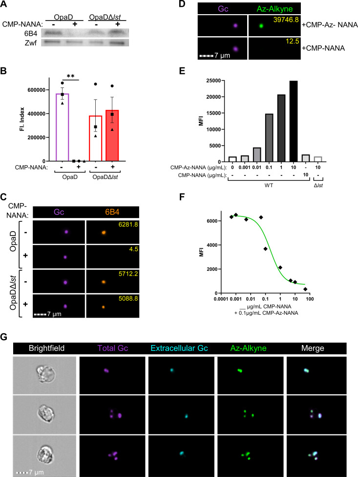
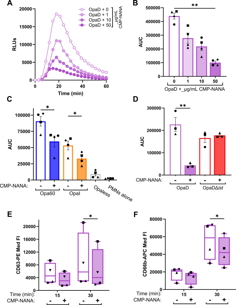
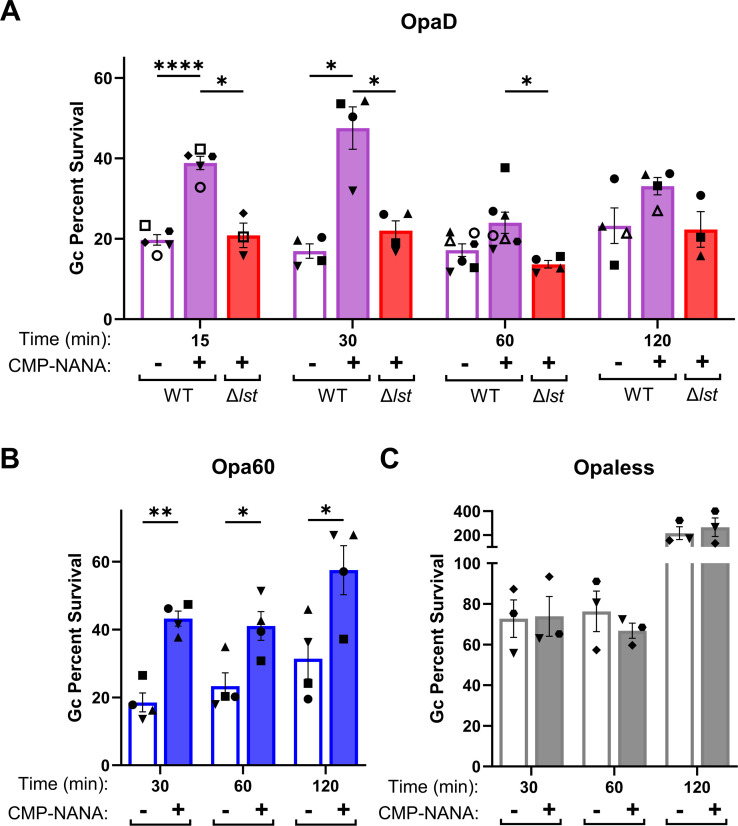
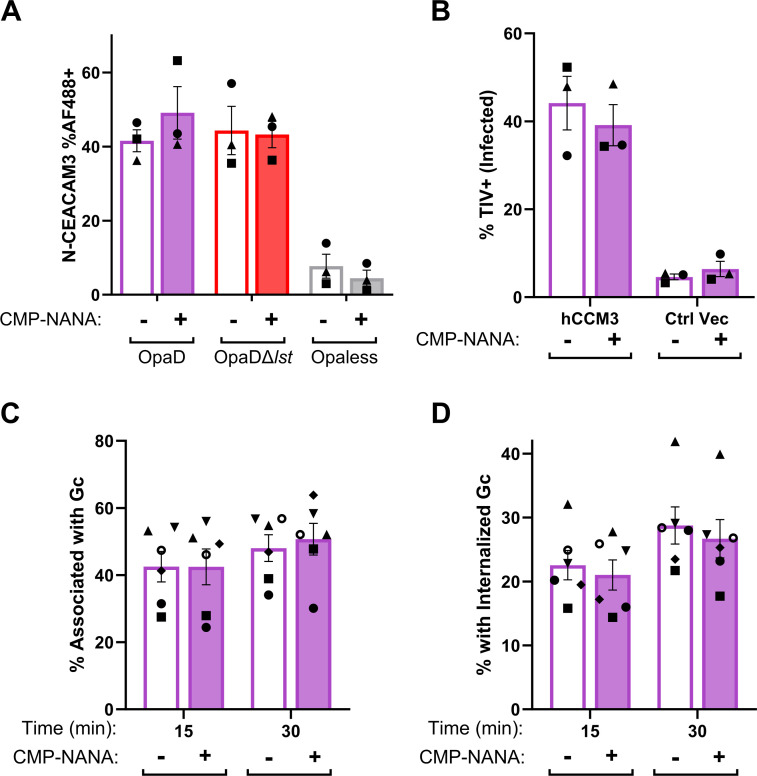
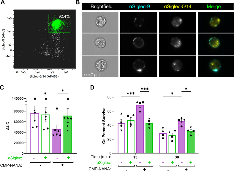
Gonorrhea, caused by the bacterium Neisseria gonorrhoeae (Gc), is characterized by neutrophilic influx to infection sites. Gc has developed mechanisms to resist killing by neutrophils that include modifications to its surface lipooligosaccharide (LOS). One such LOS modification is sialylation: Gc sialylates its terminal LOS sugars with cytidine-5'-monophosphate-N-acetylneuraminic acid, which is scavenged from the host using LOS sialyltransferase (Lst) since Gc cannot make its sialic acid. Sialylation enables sensitive strains of Gc to resist complement-mediated killing in a serum-dependent manner. However, little is known about the contribution of sialylation to complement-independent, direct Gc-neutrophil interactions. In the absence of complement, we found sialylated Gc expressing opacity-associated (Opa) proteins decreased the oxidative burst and granule exocytosis from primary human neutrophils. In addition, sialylated Opa+ Gc survived better than vehicle treated or Δlst Gc when challenged with neutrophils. However, Gc sialylation did not significantly affect Opa-dependent association with or internalization of Gc by neutrophils. Previous studies have implicated sialic acid-binding immunoglobulin-type lectins (Siglecs) in modulating neutrophil interactions with sialylated Gc. Blocking neutrophil Siglecs with antibodies that bind to their extracellular domains eliminated the ability of sialylated Opa+ Gc to suppress the oxidative burst and resist neutrophil killing. These findings highlight a new role for sialylation in Gc evasion of human innate immunity, with implications for the development of vaccines and therapeutics for gonorrhea.

Importance: Neisseria gonorrhoeae, the bacterium that causes gonorrhea, is an urgent global health concern due to increasing infection rates, widespread antibiotic resistance, and its ability to thwart protective immune responses. The mechanisms by which Gc subverts protective immune responses remain poorly characterized. One way N. gonorrhoeae evades human immunity is by adding sialic acid that is scavenged from the host onto its lipooligosaccharide, using the sialyltransferase Lst. Here, we found that sialylation enhances N. gonorrhoeae survival from neutrophil assault and inhibits neutrophil activation, independently of the complement system. Our results implicate bacterial binding of sialic acid-binding lectins (Siglecs) on the neutrophil surface, which dampens neutrophil antimicrobial responses. This work identifies a new role for sialylation in protecting N. gonorrhoeae from cellular innate immunity, which can be targeted to enhance the human immune response in gonorrhea.

Keywords: Neisseria gonorrhoeae; degranulation; gonorrhea; infection; lipooligosaccharide; neutrophils; reactive oxygen species; sialylation.


Conflict of interest statement

The authors declare no conflict of interest.

Figures

Fig 1
Fig 1
Analysis and quantification of Lst-dependent sialylation of gonococcal lipooligosaccharide. (A) OpaD and OpaDΔlst were grown with 50 µg/mL CMP-Neu5Ac (NANA) (+) or vehicle (Veh) (–). Bacterial lysates were Western blotted with mAb 6B4, or anti-Zwf (glucose 6-phosphate dehydrogenase) as loading control, followed by HRP-conjugated secondary antibodies. The blot is representative of n = 3 biological replicates. (B-C) OpaD and OpaDΔlst were incubated with CMP-NANA or vehicle as in A, then stained with Tag-IT Violet (TIV). Bacteria were incubated with 6B4 followed by AlexaFluor647-coupled secondary antibody, fixed, and analyzed by imaging flow cytometry. (B) Average fluorescence index (Fl: mean fluorescence intensity (MFI) x percent positive) from n = 3 biological replicates with 20,000 events collected per condition; each symbol indicates one matched biological replicate. Statistical analysis by one-way ANOVA with Tukey’s multiple comparisons test. **P < .01. (C) One representative image from imaging flow cytometry for each treatment condition and each fluorescence channel is shown. AF647-6B4 signal is a false-colored orange, and yellow numbers indicate the object’s fluorescence intensity value. (D-G) Gc were incubated with either CMP-NANA and/or CMP-Az-NANA at 100 ng/mL. Bacteria were stained with TIV, fixed, and then subjected to copper-catalyzed click chemistry azide-alkyne cycloaddition using fluorescein isothiocyanate (FITC)-alkyne. (D) Representative images of CMP-NANA and CMP-Az-NANA sialylated OpaD, each of which was subjected to alkyne-FITC cycloaddition. Yellow numbers are as in C. (E) OpaD or OpaDΔlst were incubated with indicated concentrations of CMP-Az-NANA or CMP-NANA. The MFI of each condition was quantified from flow cytometry, n = 1. (F) WT OpaD was incubated with 0.1 µg/mL of CMP-Az-NANA along with the indicated concentration of unmodified CMP-NANA. Results are graphed as the MFI of each condition and fit with a non-linear regression line. (G) OpaD sialylated with CMP-Az-NANA was stained with TIV and used to infect IL-8-treated, adherent primary human neutrophils for 30 min. After fixation, extracellular Gc was labeled with anti-PorB and AF647-coupled secondary antibody (false-colored cyan) before permeabilization with 0.1% saponin followed by copper click chemistry. Representative images of infected neutrophils with intracellular (TIV+AF647−) and extracellular (TIV+AF647+) Gc with azide-sialylated LOS that is clicked with the alkyne-FITC probe (FITC+). In C and D, images from the indicated fluorescence channels were exported, false-colored, and cropped for presentation purposes. In G, brightfield and fluorescence channels from each cell were exported from the imaging flow cytometer as a combined image. White lines were added between the images from each channel for clarity.
Fig 2
Fig 2
Sialylation dampens neutrophil activation in response to Opa+ bacteria. (A-B) OpaD incubated with the indicated concentrations of CMP-NANA or vehicle (0 µg/mL) were exposed to primary human neutrophils in the presence of luminol at a multiplicity of infection (MOI) of 100. Neutrophil oxidative burst was measured as relative light units (RLUs) of luminol-dependent chemiluminescence every 3 min over 1 h. (A) displays one representative graph of n = 4; (B) is the average ±SEM area under the curve (AUC) for each condition across replicates (symbols indicate matched biological replicates). (C) Opa60 Gc (blue) or OpaI Gc (orange) were incubated with 50 µg/mL CMP-NANA (+) or vehicle (−) before neutrophil exposure; AUCs of n = 4 biological replicates. Non-stimulatory Opaless Gc (dark gray) exposed neutrophils or neutrophils (PMNs) in luminol alone (light gray) serve as negative controls. (D) WT (purple) or Δlst (red) OpaD were treated with or without CMP-NANA and then added to neutrophils as above; AUCs of n = 3 biological replicates. Statistical analyses by one-way ANOVA with Tukey’s multiple comparisons test. *P < .05; **P < .01. (E-F) OpaD treated with CMP-NANA or vehicle as above was added to adherent, IL-8 primed neutrophils for 15 or 30 min at an MOI of 1. Neutrophils were stained for viability (Zombie Near Infrared) with a PE-coupled antibody against primary granule protein CD63 (E) and an APC-coupled antibody against secondary granule protein CD66b (F) on the cell surface. After fixation, cells were analyzed via spectral flow cytometry. Data are presented as median fluorescence (Med Fl) of PE+ (E) and APC+ (F) live cells, gated using unstained and isotype controls. Results are from n = 4 biological replicates (symbol matched). Statistical analyses were performed by two-way ANOVA with Šídák’s multiple comparisons test. *P < .05.
Fig 3
Fig 3
Sialylated, Opa+ bacteria have a survival advantage at early times of neutrophil challenge. Adherent, IL-8 primed neutrophils were infected with sialylated (filled) or non-sialylated (empty) (A) OpaD (purple) or OpaDΔlst (red), at a multiplicity of infection (MOI) of 1. Gc viability was determined by the enumeration of CFUs from lysed neutrophils at the indicated times post-infection, expressed as a percentage of CFU at 0 min. (B-C) Sialylated and non-sialylated Opa60 Gc (blue) (B) and Opaless Gc (gray) (C) were exposed to neutrophils and bacterial viability was measured as in A. Results are presented as the average ± SEM for n > 3 (A) n = 4 (B) or n = 3 (C) biological replicates, matched by symbol within each data set. Statistical comparisons were by mixed effect analysis (A) or two-way ANOVA (B-C) with Holm-Šídák’s multiple comparisons test with the following pairwise significances: *P < .05; **P <  .01; ****P < .0001.
Fig 4
Fig 4
Sialylation does not affect the binding of OpaD bacteria to CEACAM3. (A) OpaD grown with CMP-NANA or vehicle was incubated with GST-tagged recombinant N-terminal domain of CEACAM3 (N-CEACAM3). The binding of N-CEACAM3 to Gc was detected using a mouse anti-GST antibody, followed by a goat anti-mouse IgG AF488-conjugated antibody. Gc were fixed, stained with DAPI, and analyzed by imaging flow cytometry, to calculate the percent of singlet Gc that are AF488+. Opaless Gc that does not bind CEACAM is shown as a negative control (gray). (B) CHO cells transfected with human CEACAM3 (hCCM3) or empty control vector (Ctrl Vec) were infected with TIV-labeled OpaD, and treated with CMP-NANA or vehicle. After 30 min, cells were collected, stained with a pan-CEACAM antibody followed by an AF555-conjugated secondary antibody, and fixed. The percent of singlet CHO cells that are TIV+ (infected) was calculated using imaging flow cytometry. Results are from n = 3 biological replicates (symbol matched). Statistical comparisons were by two-way ANOVA with Tukey’s multiple comparisons test; not significant. (C-D) OpaD with or without sialylated LOS were labeled with TIV and used to infect adherent, IL-8 primed neutrophils. At the indicated time points, infected cells were fixed and extracellular Gc was detected with an anti-PorB antibody, followed by an AF488-coupled secondary antibody, without permeabilization. Cells were then analyzed using imaging flow cytometry to report the percent of neutrophils with associated (%TIV+) Gc (C) or internalized (%TIV+ AF488-) Gc (D). Results are from n = 6 biological replicates (symbol matched). Statistical comparisons were by two-way ANOVA with Tukey’s multiple comparisons test; not significant.
Fig 5
Fig 5
Blockade of neutrophil Siglec-9 and Siglec-5/14 reverses the ability of sialylated gonococci to suppress neutrophil activation and antibacterial activity. (A-B) Uninfected and adherent, IL-8 primed human neutrophils were incubated with antibodies that recognize human Siglec-9-APC and/or Siglec-5/Siglec-14-AF488, without cell permeabilization before fixation and processing via imaging flow cytometry. (A) Dot plot of focused singlet neutrophils with anti-Siglec-9(APC) and anti-Siglec-5/14(AF488). The X-axis indicates AF488 fluorescence intensity and the y-axis indicates APC fluorescence intensity. Double positive (green) gate based on single stains; number within the gate is the percent of the stained condition. (B) Representative images of neutrophils stained with anti-Siglec-9-APC (cyan) and anti-Siglec-5/14-AF488 (yellow); colored fluorescence in merged. Brightfield and fluorescence channels from each cell were exported from the imaging flow cytometer as a combined image. White lines were added between the images from each channel for clarity. Results are representative of n = 3 biological replicates by spectral flow cytometry and confocal microscopy (not shown) and demonstrate n = 1 replicate with imaging flow cytometry. (C-D) Primary human neutrophils were incubated with anti-human Siglec-9 and Siglec-5/-14 antibodies (green) or media alone (purple) for 30 min. (C) Neutrophils were exposed to sialylated (filled bars) or non-sialylated (open bars) OpaD at an MOI of 100 in the presence of luminol. ROS production was measured over 1 h by luminol-dependent chemiluminescence and presented as an area under the curve for each condition across n = 6 biological replicates (symbol matched). Statistical comparisons by one-way ANOVA with Tukey’s multiple comparisons test. *P < .05. (D) Neutrophils were treated with anti-human Siglec-9 and Siglec-5/-14 antibodies (green) or media alone (purple) as in C. Cells were then infected with sialylated (filled) or non-sialylated (empty) Gc at an MOI of 1. Survival of Gc is presented as a percent of the bacteria at time 0 min, as in Fig. 3. Statistical analyses by two-way ANOVA with Holm-Šídák’s multiple comparisons test. n = 4 biological replicates (symbol matched); *P ≤ .05, ***P < 0.0001.

Update of

Similar articles

Cited by

References

    1. World Health Organization . 2024. Gonorrhoea (Neisseria gonorrhoeae infection). Available from: https://www.who.int/news-room/fact-sheets/detail/gonorrhoea-(neisseria-g...
    1. Workowski KA, Bachmann LH, Chan PA, Johnston CM, Muzny CA, Park I, Reno H, Zenilman JM, Bolan GA. 2021. Sexually transmitted infections treatment guidelines, 2021. MMWR Recomm Rep 70:1–187. doi:10.15585/mmwr.rr7004a1 - DOI - PMC - PubMed
    1. Rice PA, Shafer WM, Ram S, Jerse AE. 2017. Neisseria gonorrhoeae: drug resistance, mouse models, and vaccine development. Annu Rev Microbiol 71:665–686. doi:10.1146/annurev-micro-090816-093530 - DOI - PubMed
    1. Yamasaki R, Bacon BE, Nasholds W, Schneider H, Griffiss JM. 1991. Structural determination of oligosaccharides derived from lipooligosaccharide of Neisseria gonorrhoeae F62 by chemical, enzymatic, and two-dimensional NMR methods. Biochemistry 30:10566–10575. doi:10.1021/bi00107a028 - DOI - PubMed
    1. John CM, Griffiss JM, Apicella MA, Mandrell RE, Gibson BW. 1991. The structural basis for pyocin resistance in Neisseria gonorrhoeae lipooligosaccharides. J Biol Chem 266:19303–19311. doi:10.1016/S0021-9258(18)54998-X - DOI - PubMed

Publication types

MeSH terms

Substances