

Role of Hsa_circ_0000880 in the Regulation of High Glucose-Induced Apoptosis of Retinal Microvascular Endothelial Cells
- PMID: 38587436
- PMCID: PMC11005064
- DOI: 10.1167/tvst.13.4.12
Role of Hsa_circ_0000880 in the Regulation of High Glucose-Induced Apoptosis of Retinal Microvascular Endothelial Cells
Abstract
Purpose: Circular RNAs (circRNAs) have been verified to participate in multiple biological processes and disease progression. Yet, the role of circRNAs in the pathogenesis of diabetic retinopathy (DR) is still poorly understood and deserves further study. This study aimed to investigate the role of circRNAs in the regulation of high glucose (HG)-induced apoptosis of retinal microvascular endothelial cells (RMECs).
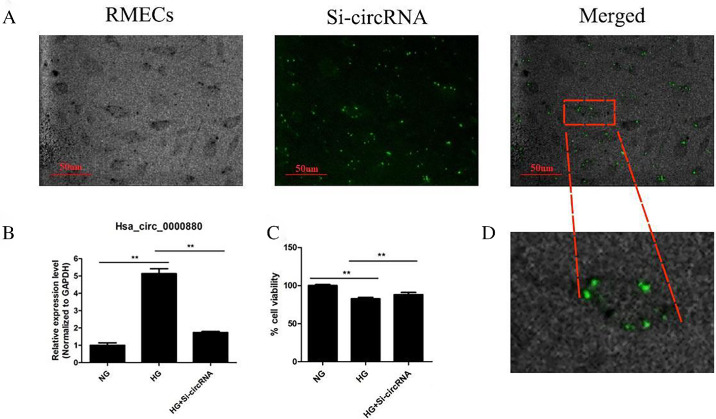
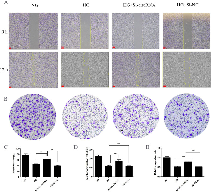
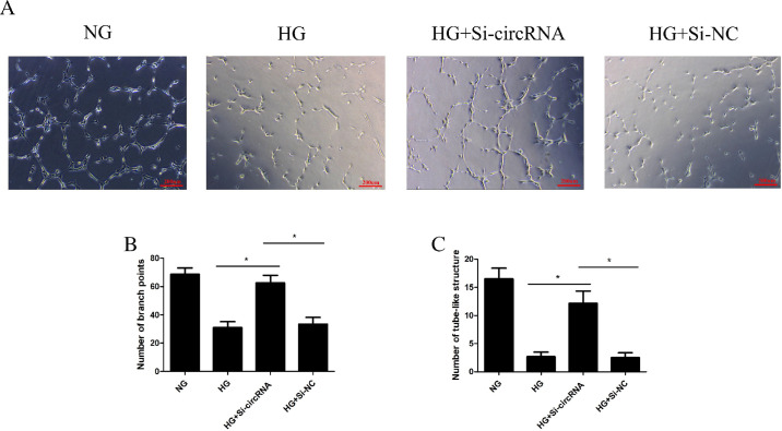
Methods: Epiretinal membranes from patients with DR and nondiabetic patients with idiopathic macular epiretinal membrane were collected for this study. The circRNA microarrays were performed using high-throughput sequencing. Hierarchical clustering, functional enrichment, and network regulation analyses were used to analyze the data generated by high-throughput sequencing. Next, RMECs were subjected to HG (25 mM) conditions to induce RMECs apoptosis in vitro. A series of experiments, such as Transwell, the Scratch wound, and tube formation, were conducted to explore the regulatory effect of circRNA on RMECs. Fluorescence in situ hybridization (FISH), immunofluorescence staining, and Western blot were used to study the mechanism underlying circRNA-mediated regulation.
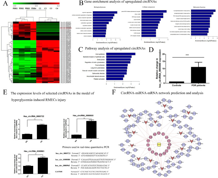
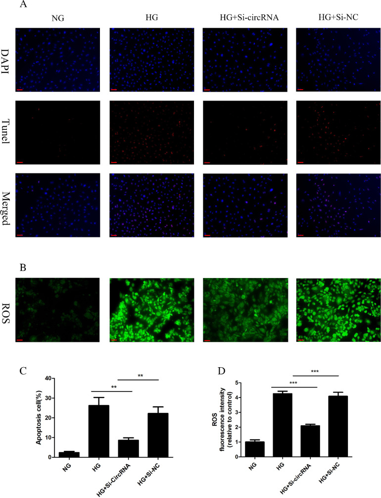
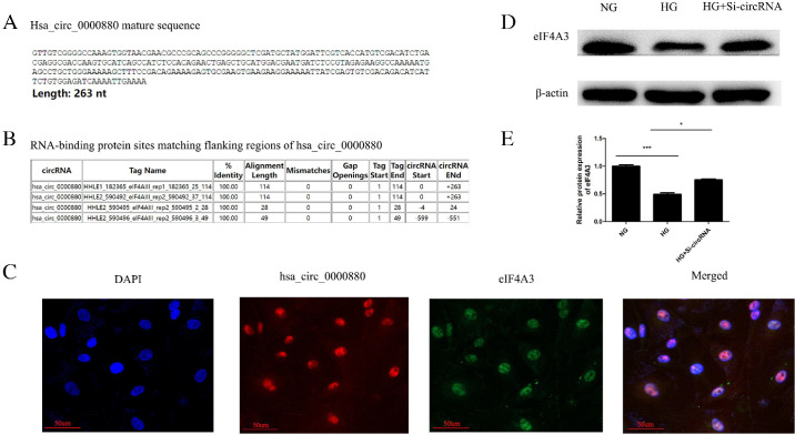
Results: A total of 53 differentially expressed circRNAs were found in patients with DR. Among these, hsa_circ_0000880 was significantly upregulated in both the diabetic epiretinal membranes and in an in vitro DR model of HG-treated RMECs. Hsa_circ_0000880 knockout facilitated RMECs vitality and decreased the paracellular permeability of RMECs under hyperglycemia. More importantly, silencing of hsa_circ_0000880 significantly inhibited HG-induced ROS production and RMECs apoptosis. Hsa_circ_0000880 acted as an endogenous sponge for eukaryotic initiation factor 4A-III (EIF4A3). Knockout of hsa_circ_0000880 reversed HG-induced decrease in EIF4A3 protein level.
Conclusions: Our findings suggest that hsa_circ_0000880 is a novel circRNA can induce RMECs apoptosis in response to HG conditions by sponging EIF4A3, offering an innovative treatment approach against DR.
Translational relevance: The circRNAs participate in the dysregulation of microvascular endothelial function induced by HG conditions, indicating a promising therapeutic target for DR.
Conflict of interest statement
Disclosure:
Figures
Similar articles
-
Hsa_circ_0030042 regulates abnormal autophagy and protects atherosclerotic plaque stability by targeting eIF4A3.Theranostics. 2021 Mar 12;11(11):5404-5417. doi: 10.7150/thno.48389. eCollection 2021. Theranostics. 2021. PMID: 33859754 Free PMC article.
-
Circular RNA hsa_circ_0000467 promotes colorectal cancer progression by promoting eIF4A3-mediated c-Myc translation.Mol Cancer. 2024 Jul 31;23(1):151. doi: 10.1186/s12943-024-02052-5. Mol Cancer. 2024. PMID: 39085875 Free PMC article.
-
Down-Regulation of circCOL1A2 Suppresses the Dysfunction of Diabetes-Related Retinal Microvascular Endothelial Cells via miR-646/FGF7 Axis.Curr Eye Res. 2022 Nov;47(11):1525-1533. doi: 10.1080/02713683.2022.2110264. Epub 2022 Aug 22. Curr Eye Res. 2022. PMID: 35924466
-
hsa_circ_0101119 facilitates the progression of cervical cancer via an interaction with EIF4A3 to inhibit TCEAL6 expression.Mol Med Rep. 2021 Sep;24(3):654. doi: 10.3892/mmr.2021.12293. Epub 2021 Jul 19. Mol Med Rep. 2021. PMID: 34278492 Free PMC article.
-
hsa_circ_0000047 targeting miR-6720-5p/CYB5R2 axis alleviates inflammation and angiogenesis in diabetic retinopathy.Arch Physiol Biochem. 2024 Oct;130(5):537-545. doi: 10.1080/13813455.2023.2190055. Epub 2023 Mar 27. Arch Physiol Biochem. 2024. PMID: 36971486
Cited by
-
Integrative Analysis and Experimental Validation Reveal FCGR1A and ITGAL as Key Inflammatory Biomarkers in Proliferative Diabetic Retinopathy.J Inflamm Res. 2025 May 13;18:6229-6243. doi: 10.2147/JIR.S519725. eCollection 2025. J Inflamm Res. 2025. PMID: 40386178 Free PMC article.
-
Role of non-coding RNAs in the pathogenesis of viral myocarditis.Virulence. 2025 Dec;16(1):2466480. doi: 10.1080/21505594.2025.2466480. Epub 2025 Feb 20. Virulence. 2025. PMID: 39950847 Free PMC article. Review.
References
-
- Cheung N, Mitchell P, Wong TY.. Diabetic retinopathy. Lancet. 2010; 376(9735): 124–136. - PubMed
-
- Dal Canto E, Ceriello A, Rydén L, et al. .. Diabetes as a cardiovascular risk factor: an overview of global trends of macro and micro vascular complications. Eur J Prev Cardiol. 2019; 26: 25–32. - PubMed
-
- Cerani A, Tetreault N, Menard C, et al. .. Neuron-derived semaphorin 3A is an early inducer of vascular permeability in diabetic retinopathy via neuropilin-1. Cell Metab. 2013; 18: 505–518. - PubMed
-
- Roy S, Bae E, Amin S, Kim D.. Extracellular matrix, gap junctions, and retinal vascular homeostasis in diabetic retinopathy. Exp Eye Res. 2015; 133: 58–68. - PubMed
MeSH terms
Substances
LinkOut - more resources
Full Text Sources
Medical
