

In Vivo Assessment of Retinal Phenotypes in Axenfeld-Rieger Syndrome
- PMID: 38587439
- PMCID: PMC11005067
- DOI: 10.1167/iovs.65.4.20
In Vivo Assessment of Retinal Phenotypes in Axenfeld-Rieger Syndrome
Abstract
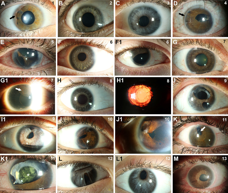
Purpose: Axenfeld-Rieger syndrome (ARS) is characterized by ocular anomalies including posterior embryotoxon, iridocorneal adhesions, corectopia/iris hypoplasia, and developmental glaucoma. Although anterior segment defects and glaucoma contribute to decreased visual acuity, the role of potential posterior segment abnormalities has not been explored. We used high-resolution retinal imaging to test the hypothesis that individuals with ARS have posterior segment pathology.
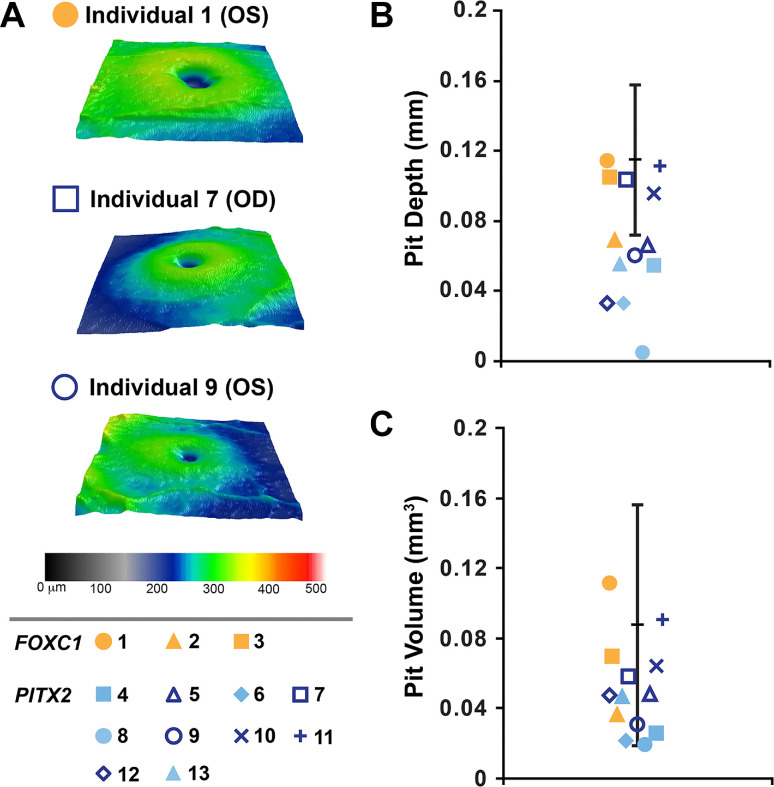
Methods: Three individuals with FOXC1-ARS and 10 with PITX2-ARS completed slit-lamp and fundus photography, optical coherence tomography (OCT), OCT angiography, and adaptive optics scanning light ophthalmoscopy (AOSLO). Quantitative metrics were compared to previously published values for individuals with normal vision.
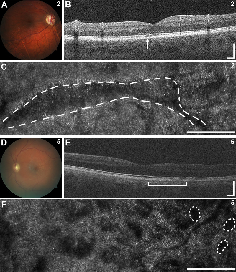
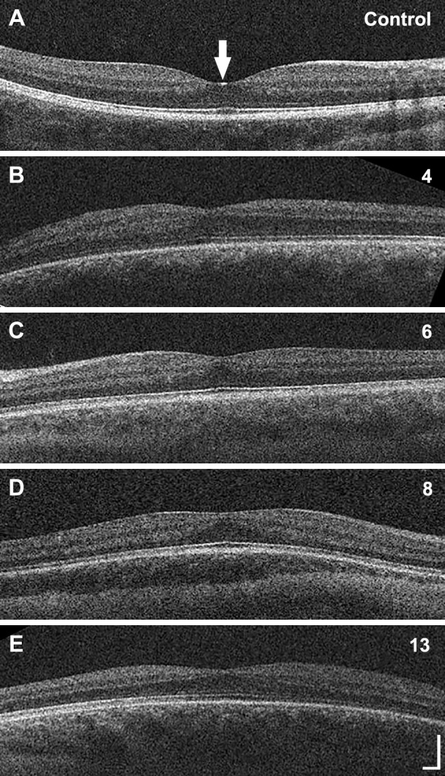
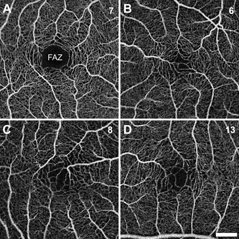
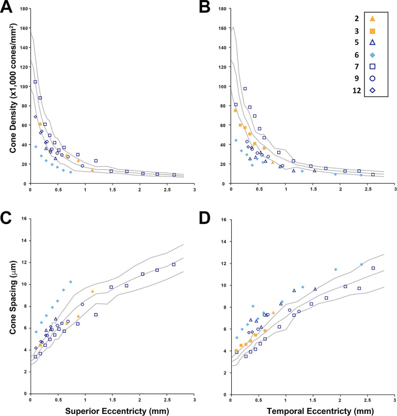
Results: All individuals demonstrated typical anterior segment phenotypes. Average ganglion cell and inner plexiform layer thickness was lower in PITX2-ARS, consistent with the glaucoma history in this group. A novel phenotype of foveal hypoplasia was noted in 40% of individuals with PITX2-ARS (but none with FOXC1-ARS). Moreover, the depth and volume of the foveal pit were significantly lower in PITX2-ARS compared to normal controls, even excluding individuals with foveal hypoplasia. Analysis of known foveal hypoplasia genes failed to identify an alternative explanation. Foveal cone density was decreased in one individual with foveal hypoplasia and normal in six without foveal hypoplasia. Two individuals (one from each group) demonstrated non-foveal retinal irregularities with regions of photoreceptor anomalies on OCT and AOSLO.
Conclusions: These findings implicate PITX2 in the development of the posterior segment, particularly the fovea, in humans. The identified posterior segment phenotypes may contribute to visual acuity deficits in individuals with PITX2-ARS.
Conflict of interest statement
Disclosure:
Figures
Similar articles
-
Axenfeld-Rieger syndrome combined with a foveal anomaly in a three-generation family: a case report.BMC Ophthalmol. 2021 Mar 29;21(1):154. doi: 10.1186/s12886-021-01899-2. BMC Ophthalmol. 2021. PMID: 33781219 Free PMC article.
-
Cone Density Changes After Repeated Low-Level Red Light Treatment in Children With Myopia.JAMA Ophthalmol. 2025 Jun 1;143(6):480-488. doi: 10.1001/jamaophthalmol.2025.0835. JAMA Ophthalmol. 2025. PMID: 40272813 Free PMC article.
-
Optic nerve head and fibre layer imaging for diagnosing glaucoma.Cochrane Database Syst Rev. 2015 Nov 30;2015(11):CD008803. doi: 10.1002/14651858.CD008803.pub2. Cochrane Database Syst Rev. 2015. PMID: 26618332 Free PMC article.
-
Optical coherence tomography (OCT) for detection of macular oedema in patients with diabetic retinopathy.Cochrane Database Syst Rev. 2015 Jan 7;1(1):CD008081. doi: 10.1002/14651858.CD008081.pub3. Cochrane Database Syst Rev. 2015. PMID: 25564068 Free PMC article.
-
Peripheral iridotomy for pigmentary glaucoma.Cochrane Database Syst Rev. 2016 Feb 12;2(2):CD005655. doi: 10.1002/14651858.CD005655.pub2. Cochrane Database Syst Rev. 2016. PMID: 26871761 Free PMC article.
References
-
- Or L, Barkana Y, Hecht I, Weiner C, Einan-Lifshitz A, Pras E. FOXC1 variant in a family with anterior segment dysgenesis and normal-tension glaucoma. Exp Eye Res. 2020; 200: 108220. - PubMed
-
- Weisschuh N, Dressler P, Schuettauf F, Wolf C, Wissinger B, Gramer E. Novel mutations of FOXC1 and PITX2 in patients with Axenfeld-Rieger malformations. Invest Ophthalmol Vis Sci. 2006; 47: 3846–3852. - PubMed
-
- Zhou L, Wang X, An J, Zhang Y, He M, Tang L. Genotype-phenotype association of PITX2 and FOXC1 in Axenfeld-Rieger syndrome. Exp Eye Res. 2023; 226: 109307. - PubMed
MeSH terms
Supplementary concepts
Grants and funding
LinkOut - more resources
Full Text Sources
Medical
