. 2024 Apr 1;65(4):19.
doi: 10.1167/iovs.65.4.19.

Comparison of Brn3a and RBPMS Labeling to Assess Retinal Ganglion Cell Loss During Aging and in a Model of Optic Neuropathy

Affiliations

Comparison of Brn3a and RBPMS Labeling to Assess Retinal Ganglion Cell Loss During Aging and in a Model of Optic Neuropathy

Miranda Meng et al. Invest Ophthalmol Vis Sci. .

Abstract

Purpose: Retinal ganglion cell (RGC) loss provides the basis for diagnosis and stage determination of many optic neuropathies, and quantification of RGC survival is a critical outcome measure in models of optic neuropathy. This study examines the accuracy of manual RGC counting using two selective markers, Brn3a and RBPMS.

Methods: Retinal flat mounts from 1- to 18-month-old C57BL/6 mice, and from mice after microbead (MB)-induced intraocular pressure (IOP) elevation, are immunostained with Brn3a and/or RBPMS antibodies. Four individuals masked to the experimental conditions manually counted labeled RGCs in three copies of five images, and inter- and intra-person reliability was evaluated by the intraclass correlation coefficient (ICC).

Results: A larger population (approximately 10% higher) of RGCs are labeled with RBPMS than Brn3a antibody up to 6 months of age, but differences decrease to approximately 1% at older ages. Both RGC-labeled populations significantly decrease with age. MB-induced IOP elevation is associated with a significant decrease of both Brn3a- and RBPMS-positive RGCs. Notably, RGC labeling with Brn3a provides more consistent cell counts than RBPMS in interpersonal (ICC = 0.87 to 0.11, respectively) and intra-personal reliability (ICC = 0.97 to 0.66, respectively).

Conclusions: Brn3a and RBPMS markers are independently capable of detecting significant decreases of RGC number with age and in response to IOP elevation despite RPBMS detecting a larger number of RGCs up to 6 months of age. Brn3a labeling is less prone to manual cell counting variability than RBPMS labeling. Overall, either marker can be used as a single marker to detect significant changes in RGC survival, each offering distinct advantages.


Conflict of interest statement

Disclosure: M. Meng, None; B. Chaqour, None; N. O'Neill, None; K. Dine, None; N. Sarabu, None; G.-S. Ying, None; K.S. Shindler, None; A.G. Ross, None

Figures

Figure 1.
Figure 1.
Effects of aging on RGC marker expression. (A) Representative fluorescence micrographs of flat mounted wild-type mouse retina co-stained with antibodies against Brn3a and RBPMS markers of RGCs. Displayed images were taken half of the retinal radius from the center of the retina at 1, 6, 12, and 18 months of age. (B, C) The number of Brn3a (B) and RPBMS (C) labeled RGCs per sampled retinal area at different postnatal stages is shown. An average of all retinal fields quantified (central, mid-peripheral, and peripheral) are shown as mean ± standard deviation (n = 6–10). Significance was determined by 1-way ANOVA and Tukey's multiple comparisons test. * P < 0.05, ** P < 0.01, *** P < 0.001.
Figure 2.
Figure 2.
Effects of MB-induced IOP elevation on RGC marker expression. (A) Mice were injected in the anterior chamber with MBs in one eye and BSS in the contralateral eye. (B) IOP was elevated in the MB-injected compared to the BSS-injected eyes. (C) Representative Brn3a- and RBPMS- immunofluorescently labeled flat mounted mouse retinas sampled from the mid-peripheral retina from BSS- and MB-injected mouse eyes taken at 40 times magnification. (D) Total number of Brn3a- and RBPMS-labeled RGCs per area in the control BSS-injected and MB-injected mouse eyes. An average of all retinal fields quantified (central, mid-peripheral, and peripheral) are shown as means ± standard deviation (n = 8–12). Data represented as mean ± SEM. ** P < 0.01 and *** P < 0.001 by an unpaired one-sided t-test.
Figure 3.
Figure 3.
Retinal images used to assess variability of manual counts. Retinal flat mount from wild-type mice stained with either Brn3a (A) or RBPMS (B) antibodies. Each original image was duplicated and flipped either horizontally (middle row) or vertically (bottom row). This change of the orientation of the images was performed to determine the reliability of the cell count of the same image by individual counters who did not know that the same images were being counted in different orientations.
Figure 4.
Figure 4.
Average RGC counts show masked investigators are fairly consistent in identifying both Brn3a- and RPBMS-positive RGCs. Graphs show the average (mean ± standard deviation) number of (A) Brn3a- and (B) RBPMS-positive cell counts in retinal flat mount images. RGC count in an image is compared to those of the same image oriented horizontally or vertically by counters masked to the orientation changes (n = 4 counters/image). For each image, there is no significant difference between the number of RGCs counted in the original orientations versus either flipped orientation. Standard deviations show there is modest variability of RGC counts between individual counters in all five RPBMS labeled images, but only three of five Brn3a labeled images.
Figure 5.
Figure 5.
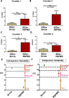
Quantitative analysis of interpersonal and intrapersonal variability of manual counting of Brn3a and RBPMS-labeled RGC. (AD) The standard deviation of RGC counts was calculated for counts of the three different orientations of each individual image obtained from each masked investigator, in order to assess differences in intrapersonal quantification of RGCs labeled with different cell markers. Graphs show the average standard deviations of Brn3a and RBPMS-positive cell counts (n = 5 images/counter for each RGC marker). Standard deviations were significantly higher for RBPMS-labeled images as compared with the standard deviation of Brn3a cell counts from three of four investigators (BD), with a similar strong trend seen with counter 1 (A). Data represented as mean ± standard deviation (n = 5); comparisons by unpaired student's t-test. (E, F) The relative reliability of both interpersonal (E) and intrapersonal (F) cell counts was further analyzed for both Brn3a (green bars) and RPBMS (red bars) labeled cells using the intraclass correlation coefficient (ICC). Lines indicate cutoffs for “poor” (<0.50), “moderate” (≥0.50), “good” (≥0.75), and “excellent” (≥0.90) reliability between counts. ICC shows that there is very good interpersonal reliability and excellent intrapersonal reliability of manual counts of Brn3a-positive cells; whereas, there is poor interpersonal reliability and only moderate intrapersonal reliability of manual counts of RPBMS-positive cells. Data represented as mean ± SEM.

Similar articles

Cited by

References

    1. Wu GF, Parker Harp CR, Shindler KS. Optic neuritis: a model for the immuno-pathogenesis of central nervous system inflammatory demyelinating diseases. Curr Immunol Rev. 2015; 11: 85–92. - PMC - PubMed
    1. Newman NJ, Yu-Wai-Man P, Biousse V, Carelli V.. Understanding the molecular basis and pathogenesis of hereditary optic neuropathies: towards improved diagnosis and management. Lancet Neurol. 2023; 22: 172–188. - PubMed
    1. Chapelle AC, Rakic JM, Plant GT.. The occurrence of intraretinal and subretinal fluid in anterior ischemic optic neuropathy: pathogenesis, prognosis, and treatment. Ophthalmology. 2023; 130: 1191–1200. - PubMed
    1. Soucy JR, Aguzzi EA, Cho J, et al. .. Retinal ganglion cell repopulation for vision restoration in optic neuropathy: a roadmap from the RReSTORe Consortium. Mol Neurodegener. 2023; 18: 64. - PMC - PubMed
    1. Kang EY, Liu PK, Wen YT, et al. .. Role of oxidative stress in ocular diseases associated with retinal ganglion cells degeneration. Antioxidants (Basel). 2021; 10. - PMC - PubMed