. 2024 Jul;51(9):2744-2757.
doi: 10.1007/s00259-024-06704-y. Epub 2024 Apr 8.

Targeting CXCR4/CXCL12 axis via [177Lu]Lu-DOTAGA.(SA.FAPi)2 with CXCR4 antagonist in triple-negative breast cancer

Affiliations

Targeting CXCR4/CXCL12 axis via [177Lu]Lu-DOTAGA.(SA.FAPi)2 with CXCR4 antagonist in triple-negative breast cancer

Guangfa Bao et al. Eur J Nucl Med Mol Imaging. 2024 Jul.

Abstract

Purpose: Radiopharmaceutical therapies targeting fibroblast activation protein (FAP) have shown promising efficacy against many tumor types. But radiopharmaceuticals alone in most cases are insufficient to completely eradicate tumor cells, which can partially be attributed to the protective interplay between tumor cells and cancer-associated fibroblasts (CAFs). The C-X-C chemokine receptor type 4/C-X-C motif chemokine 12 (CXCR4/CXCL12) interaction plays an important role in orchestrating tumor cells and CAFs. We hereby investigated the feasibility and efficacy of [177Lu]Lu-DOTAGA.(SA.FAPi)2, a FAP-targeting radiopharmaceutical, in combination with AMD3100, a CXCR4 antagonist, in a preclinical murine model of triple-negative breast cancer (TNBC).

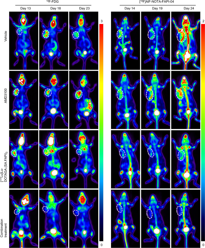
Methods: Public database was first interrogated to reveal the correlation between CAFs' scores and the prognosis of TNBC patients, as well as the expression levels of FAP and CXCR4 in normal tissues and tumors. In vitro therapeutic efficacy regarding cell proliferation, migration, and colony formation was assessed in BALB/3T3 fibroblasts and 4T1 murine breast cancer cells. In vivo therapeutic efficacy was longitudinally monitored using serial 18F-FDG, [18F]AlF-NOTA-FAPI-04, and [68Ga]Ga-DOTA-Pentixafor PET/CT scans and validated using tumor sections through immunohistochemical staining of Ki-67, α-SMA, CXCR4, and CXCL12. Intratumoral abundance of myeloid-derived suppressive cells (MDSCs) was analyzed using flow cytometry in accordance with the PET/CT schedules. Treatment toxicity was evaluated by examining major organs including heart, lung, liver, kidney, and spleen.

Results: CAFs' scores negatively correlated with the survival of TNBC patients (p < 0.05). The expression of CXCR4 and FAP was both significantly higher in tumors than in normal tissues. The combination of [177Lu]Lu-DOTAGA.(SA.FAPi)2 and AMD3100 significantly suppressed cell proliferation, migration, and colony formation in cell culture, and exhibited synergistic effects in 4T1 tumor models along with a decreased number of MDSCs. PET/CT imaging revealed lowest tumor accumulation of 18F-FDG and [18F]AlF-NOTA-FAPI-04 on day 13 and day 14 after treatment started, both of which gradually increased at later time points. A similar trend was observed in the IHC staining of Ki-67, α-SMA, and CXCL12.

Conclusion: The combination of [177Lu]Lu-DOTAGA.(SA.FAPi)2 and AMD3100 is a feasible treatment against TNBC with minimal toxicity in main organs.

Keywords: CXCR4 antagonist; CXCR4/CXCL12; Triple-negative breast cancer (TNBC); [177Lu]Lu-DOTAGA.(SA.FAPi)2.


Conflict of interest statement

The authors declare no competing interests.

Figures

Fig. 1
Fig. 1
Correlation between the CAFs’ scores and the survival of TNBC patients. Data were generated from the TCGA (A, n = 134) and a GEO dataset (GSE58812-series) (B, n = 107). The CAFs’ scores were calculated using two R packages (CAF-EPIC, CAF-MCPcounter) and via a website (TIDE, http://tide.dfci.harvard.edu). Patients were then stratified into CAF high (red) or low (blue) groups using Cox regression model. Statistical parameters including p value, hazard rate (HR), and confidence interval (CI) are included in the figure
Fig. 2
Fig. 2
The gene expression of CXCR4 and FAP in normal tissues (n = 11) and TNBC tumors (n = 101). Data were obtained from the TCGA database and evaluated by R software; significance was determined using Student t test
Fig. 3
Fig. 3
Effect of AMD3100 on BALB/3T3 fibroblast proliferation after [177Lu]Lu-DOTAGA.(SA.FAPi)2 treatment. Immunofluorescent EdU (red) and Hoechst (blue) staining of BALB/3T3 fibroblasts on days 1, 3, and 5 after treatment. Cells were non-treated, treated with [177Lu]Lu-DOTAGA.(SA.FAPi)2, AMD3100, or combination of both. Scale bar = 50 µm. The percentage of EdU positive cells were quantified and analyzed using one‐way ANOVA analysis. Error bars represent the SD. *p < 0.05, **p < 0.01, ***p < 0.001, ****p < 0.0001
Fig. 4
Fig. 4
Therapeutic efficacy of AMD3100 and [177Lu]Lu-DOTAGA.(SA.FAPi)2 in 4T1 tumor model. A Treatment schedule and dosages. AMD3100 was intraperitoneally injected daily at 5 mg/kg/injection for 2 weeks. [177Lu]Lu-DOTAGA.(SA.FAPi)2 was intravenously injected at 18.5 MBq/injection on days 3 and 8. PET/CT scans with the three tracers were performed on days 13-15, 18-20, 23-25, 28-30, and 33-35. B Tumor growth curves, group size: vehicle (n = 9), AMD3100 (n = 9), [177Lu]Lu-DOTAGA.(SA.FAPi)2 (n = 10), combination treatment (n = 9). C Body weight change of 4T1 model suffered different treatment. D Kaplan–Meier survival curves of 4T1 model in groups of vehicle control (n = 9), AMD3100 (n = 9), [.177Lu]Lu-DOTAGA.(SA.FAPi)2 (n = 10), combination treatment (n = 9). Data in B and C are presented as mean ± SD in line diagram, statistical difference was evaluated using one‐way ANOVA analysis, and survival difference in (D) was evaluated using Log‐rank test. *p < 0.05, **p < 0.01, ***p < 0.001, ****p < 0.0001
Fig. 5
Fig. 5
18F-FDG and [18F]AlF-NOTA-FAPI-04 PET/CT imaging during and after treatment. 18F-FDG and [18F]AlF-NOTA-FAPI-04 PET/CT imaging were performed on days 13-14, 18-19, and 23-24, respectively, from the initiation treatment (n = 4). 4T1-bearing mice were treated with vehicle, [177Lu]Lu-DOTAGA.(SA.FAPi)2, AMD3100, or combination of [177Lu]Lu-DOTAGA.(SA.FAPi)2 and AMD3100. Tumors were indicated by the white circle
Fig. 6
Fig. 6
Tumoral immunohistochemical staining of Ki-67 on days 14, 19, 24, 29, and 34 from the initiation treatment. Mice were treated with vehicle control, [177Lu]Lu-DOTAGA.(SA.FAPi)2, AMD3100, or combination of both. The percentage of Ki-67 positive nuclei was quantified by ImageJ software and presented as mean ± SD in line diagram. Scale bar = 25 µm
Fig. 7
Fig. 7
Representative flow cytometry images (A) and quantification of MDSCs’ numbers (CD11b+Gr-1+) (B-D). Tumor tissues were collected from mice of untreated control, AMD3100- or [177Lu]Lu-DOTAGA.(SA.FAPi)2-treated, or combination of both on days 14 (B), 19 (C), and 24 (D) from the initiation the treatment (n = 3). The percentage of MDSCs’ numbers in total tumoral single cell suspension was quantified. Significance of differences was determined using one‐way ANOVA analysis. *p < 0.05, **p < 0.01, ***p < 0.001, ****p < 0.0001

References

    1. Leon-Ferre RA, Goetz MP. Advances in systemic therapies for triple negative breast cancer. BMJ. 2023;381:e071674. doi: 10.1136/bmj-2022-071674. - DOI - PubMed
    1. Epstein RJ. The CXCL12-CXCR4 chemotactic pathway as a target of adjuvant breast cancer therapies. Nat Rev Cancer. 2004;4(11):901–909. doi: 10.1038/nrc1473. - DOI - PubMed
    1. Jiang K, et al. SDF-1/CXCR4 axis facilitates myeloid-derived suppressor cells accumulation in osteosarcoma microenvironment and blunts the response to anti-PD-1 therapy. Int Immunopharmacol. 2019;75:105818. doi: 10.1016/j.intimp.2019.105818. - DOI - PubMed
    1. Chen X, Song E. Turning foes to friends: targeting cancer-associated fibroblasts. Nat Rev Drug Discov. 2019;18(2):99–115. doi: 10.1038/s41573-018-0004-1. - DOI - PubMed
    1. Feig C, et al. Targeting CXCL12 from FAP-expressing carcinoma-associated fibroblasts synergizes with anti-PD-L1 immunotherapy in pancreatic cancer. Proc Natl Acad Sci U S A. 2013;110(50):20212–20217. doi: 10.1073/pnas.1320318110. - DOI - PMC - PubMed

MeSH terms

LinkOut - more resources