

Rate and Predictors of Misclassification of Active Diabetic Macular Edema as Detected by an Automated Retinal Image Analysis System
- PMID: 38587776
- PMCID: PMC11109071
- DOI: 10.1007/s40123-024-00929-8
Rate and Predictors of Misclassification of Active Diabetic Macular Edema as Detected by an Automated Retinal Image Analysis System
Abstract
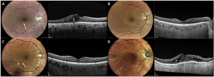
Introduction: The aim of this work is to estimate the sensitivity, specificity, and misclassification rate of an automated retinal image analysis system (ARIAS) in diagnosing active diabetic macular edema (DME) and to identify factors associated with true and false positives.
Methods: We conducted a cross-sectional study of prospectively enrolled patients with diabetes mellitus (DM) referred to a tertiary medical retina center for screening or management of DME. All patients underwent two-field fundus photography (macula- and disc-centered) with a true-color confocal camera; images were processed by EyeArt V.2.1.0 (Woodland Hills, CA, USA). Active DME was defined as the presence of intraretinal or subretinal fluid on spectral-domain optical coherence tomography (SD-OCT). Sensitivity and specificity and their 95% confidence intervals (CIs) were calculated. Variables associated with true (i.e., DME labeled as present by ARIAS + fluid on SD-OCT) and false positives (i.e., DME labeled as present by ARIAS + no fluid on SD-OCT) of active DME were explored.
Results: A total of 298 eyes were included; 92 eyes (31%) had active DME. ARIAS sensitivity and specificity were 82.61% (95% CI 72.37-89.60) and 84.47% (95% CI 78.34-89.10). The misclassification rate was 16%. Factors associated with true positives included younger age (p = 0.01), shorter DM duration (p = 0.006), presence of hard exudates (p = 0.005), and microaneurysms (p = 0.002). Factors associated with false positives included longer DM duration (p = 0.01), worse diabetic retinopathy severity (p = 0.008), history of inactivated DME (p < 0.001), and presence of hard exudates (p < 0.001), microaneurysms (p < 0.001), or epiretinal membrane (p = 0.06).
Conclusions: The sensitivity of ARIAS was diminished in older patients and those without DME-related fundus lesions, while the specificity was reduced in cases with a history of inactivated DME. ARIAS performed well in screening for naïve DME but is not effective in surveillance inactivated DME.
Keywords: Artificial intelligence; Diabetic macular edema; Diabetic retinopathy; Surveillance.
© 2024. The Author(s).
Conflict of interest statement
Lamberto La Franca, Carola Rutigliani and Lisa Checchin have nothing to disclose. Francesco Bandello consultant for: Allergan Inc (Irvine, California, USA), Bayer Shering-Pharma (Berlin, Germany), Hoffmann-La-Roche (Basel, Switzerland), Novartis (Basel, Switzerland), Sanofi-Aventis (Paris, France), Thrombogenics (Heverlee, Belgium), Zeiss (Dublin, USA), Boehringer-Ingelheim, Fidia Sooft, Ntc Pharma, Sifi. Rosangela Lattanzio consultant for: AbbVie (North Chicago, Illinois, USA), Bayer Shering-Pharma (Berlin, Germany), Novartis (Basel, Switzerland), Sifi. Maria Vittoria Cicinelli and Francesco Bandello are Editorial Board members of
Figures
References
LinkOut - more resources
Full Text Sources
