

Transient interdomain interactions in free USP14 shape its conformational ensemble
- PMID: 38588275
- PMCID: PMC11001199
- DOI: 10.1002/pro.4975
Transient interdomain interactions in free USP14 shape its conformational ensemble
Abstract
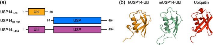
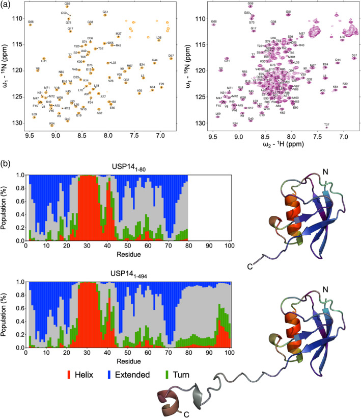
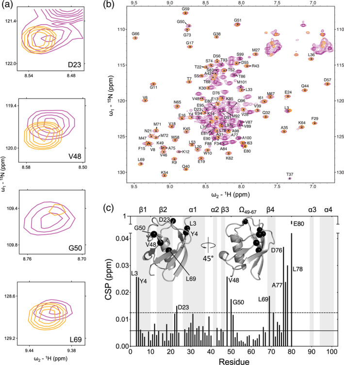
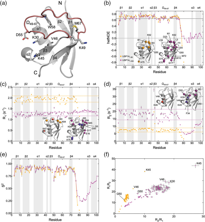
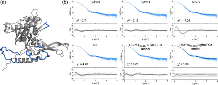
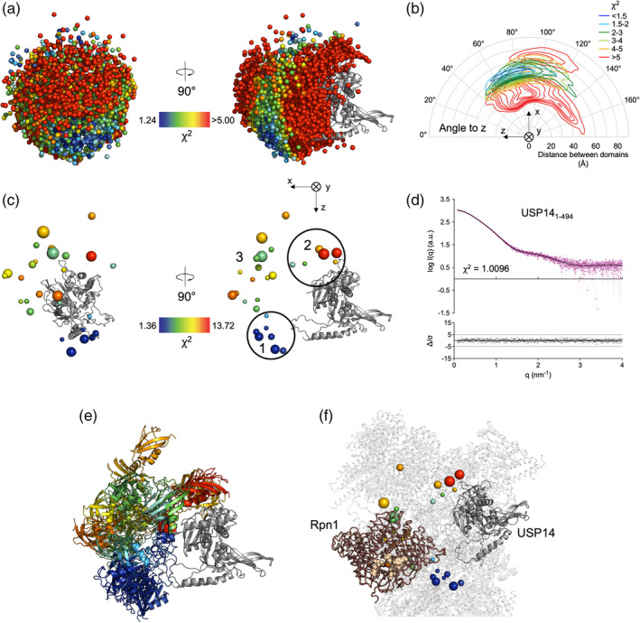
The deubiquitinase (DUB) ubiquitin-specific protease 14 (USP14) is a dual domain protein that plays a regulatory role in proteasomal degradation and has been identified as a promising therapeutic target. USP14 comprises a conserved USP domain and a ubiquitin-like (Ubl) domain separated by a 25-residue linker. The enzyme activity of USP14 is autoinhibited in solution, but is enhanced when bound to the proteasome, where the Ubl and USP domains of USP14 bind to the Rpn1 and Rpt1/Rpt2 units, respectively. No structure of full-length USP14 in the absence of proteasome has yet been presented, however, earlier work has described how transient interactions between Ubl and USP domains in USP4 and USP7 regulate DUB activity. To better understand the roles of the Ubl and USP domains in USP14, we studied the Ubl domain alone and in full-length USP14 by nuclear magnetic resonance spectroscopy and used small angle x-ray scattering and molecular modeling to visualize the entire USP14 protein ensemble. Jointly, our results show how transient interdomain interactions between the Ubl and USP domains of USP14 predispose its conformational ensemble for proteasome binding, which may have functional implications for proteasome regulation and may be exploited in the design of future USP14 inhibitors.
Keywords: DUB; NMR; SAXS; molecular modeling; protein dynamics.
© 2024 The Authors. Protein Science published by Wiley Periodicals LLC on behalf of The Protein Society.
Conflict of interest statement
The authors declare no conflicts of interest.
Figures
Similar articles
-
Dynamic networks connect the USP14 active site region with the proteasome interaction surface.Protein Sci. 2025 Apr;34(4):e70077. doi: 10.1002/pro.70077. Protein Sci. 2025. PMID: 40095364 Free PMC article.
-
UBL domain of Usp14 and other proteins stimulates proteasome activities and protein degradation in cells.Proc Natl Acad Sci U S A. 2018 Dec 11;115(50):E11642-E11650. doi: 10.1073/pnas.1808731115. Epub 2018 Nov 28. Proc Natl Acad Sci U S A. 2018. PMID: 30487212 Free PMC article.
-
The role of UBL domains in ubiquitin-specific proteases.Biochem Soc Trans. 2012 Jun 1;40(3):539-45. doi: 10.1042/BST20120004. Biochem Soc Trans. 2012. PMID: 22616864 Review.
-
Proteins containing ubiquitin-like (Ubl) domains not only bind to 26S proteasomes but also induce their activation.Proc Natl Acad Sci U S A. 2020 Mar 3;117(9):4664-4674. doi: 10.1073/pnas.1915534117. Epub 2020 Feb 18. Proc Natl Acad Sci U S A. 2020. PMID: 32071216 Free PMC article.
-
Small-Molecule Inhibitors Targeting Proteasome-Associated Deubiquitinases.Int J Mol Sci. 2021 Jun 9;22(12):6213. doi: 10.3390/ijms22126213. Int J Mol Sci. 2021. PMID: 34207520 Free PMC article. Review.
Cited by
-
Dynamic networks connect the USP14 active site region with the proteasome interaction surface.Protein Sci. 2025 Apr;34(4):e70077. doi: 10.1002/pro.70077. Protein Sci. 2025. PMID: 40095364 Free PMC article.
-
USP14 is crucial for proteostasis regulation and α-synuclein degradation in human SH-SY5Y dopaminergic cells.Heliyon. 2025 Jan 23;11(3):e42031. doi: 10.1016/j.heliyon.2025.e42031. eCollection 2025 Feb 15. Heliyon. 2025. PMID: 39916840 Free PMC article.
References
-
- Ahlner A, Carlsson M, Jonsson B‐H, Lundström P. PINT: a software for integration of peak volumes and extraction of relaxation rates. J Biomol NMR. 2013;56:191–202. - PubMed
-
- Cavanagh J, Fairbrother WJ, Palmer AG, Rance M, Skelton NJ. Protein NMR spectroscopy: principles and practice. 2nd ed. Academic Press, San Diego; 2006.
MeSH terms
Substances
Grants and funding
LinkOut - more resources
Full Text Sources
Research Materials
Miscellaneous
