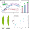
Dehydration-induced corrugated folding in Rhapis excelsa plant leaves
- PMID: 38588439
- PMCID: PMC11047117
- DOI: 10.1073/pnas.2320259121
Dehydration-induced corrugated folding in Rhapis excelsa plant leaves
Abstract
Plant leaves, whose remarkable ability for morphogenesis results in a wide range of petal and leaf shapes in response to environmental cues, have inspired scientific studies as well as the development of engineering structures and devices. Although some typical shape changes in plants and the driving force for such shape evolution have been extensively studied, there remain many poorly understood mechanisms, characteristics, and principles associated with the vast array of shape formation of plant leaves in nature. Here, we present a comprehensive study that combines experiment, theory, and numerical simulations of one such topic-the mechanics and mechanisms of corrugated leaf folding induced by differential shrinking in Rhapis excelsa. Through systematic measurements of the dehydration process in sectioned leaves, we identify a linear correlation between change in the leaf-folding angle and water loss. Building on experimental findings, we develop a generalized model that provides a scaling relationship for water loss in sectioned leaves. Furthermore, our study reveals that corrugated folding induced by dehydration in R. excelsa leaves is achieved by the deformation of a structural architecture-the "hinge" cells. Utilizing such connections among structure, morphology, environmental stimuli, and mechanics, we fabricate several biomimetic machines, including a humidity sensor and morphing devices capable of folding in response to dehydration. The mechanisms of corrugated folding in R. excelsa identified in this work provide a general understanding of the interactions between plant leaves and water. The actuation mechanisms identified in this study also provide insights into the rational design of soft machines.
Keywords: biomechanics; biomimetics; dehydration; leaf folding; plant morphology.
Conflict of interest statement
Competing interests statement:The authors declare no competing interest.
Figures


Similar articles
-
Programmable Morphing Hydrogels for Soft Actuators and Robots: From Structure Designs to Active Functions.Acc Chem Res. 2022 Jun 7;55(11):1533-1545. doi: 10.1021/acs.accounts.2c00046. Epub 2022 Apr 12. Acc Chem Res. 2022. PMID: 35413187
-
CIN-like TCP13 is essential for plant growth regulation under dehydration stress.Plant Mol Biol. 2022 Feb;108(3):257-275. doi: 10.1007/s11103-021-01238-5. Epub 2022 Jan 20. Plant Mol Biol. 2022. PMID: 35050466 Free PMC article.
-
Decline of leaf hydraulic conductance with dehydration: relationship to leaf size and venation architecture.Plant Physiol. 2011 Jun;156(2):832-43. doi: 10.1104/pp.111.173856. Epub 2011 Apr 21. Plant Physiol. 2011. PMID: 21511989 Free PMC article.
-
Leaf morphogenesis: The multifaceted roles of mechanics.Mol Plant. 2022 Jul 4;15(7):1098-1119. doi: 10.1016/j.molp.2022.05.015. Epub 2022 Jun 2. Mol Plant. 2022. PMID: 35662674 Review.
-
Plant-inspired adaptive structures and materials for morphing and actuation: a review.Bioinspir Biomim. 2016 Dec 20;12(1):011001. doi: 10.1088/1748-3190/12/1/011001. Bioinspir Biomim. 2016. PMID: 27995902 Review.
Cited by
-
Nonliving dehydrated leaves-inspired surface anti-wrinkling.Sci Adv. 2025 Jul 4;11(27):eadx7398. doi: 10.1126/sciadv.adx7398. Epub 2025 Jul 4. Sci Adv. 2025. PMID: 40614186 Free PMC article.
-
Mechanics of pressurized cellular sheets.J R Soc Interface. 2025 Feb;22(223):20240653. doi: 10.1098/rsif.2024.0653. Epub 2025 Feb 12. J R Soc Interface. 2025. PMID: 39933593 Free PMC article.
References
-
- Jonsson K., Ma Y., Routier-Kierzkowska A.-L., Bhalerao R. P., Multiple mechanisms behind plant bending. Nat. Plants 9, 13–21 (2023). - PubMed
-
- Briggs D., Walters S. M., Plant Variation and Evolution (Cambridge University Press, 2016).
-
- Givnish T., “On the adaptive significance of leaf form” in Topics in Plant Population Biology (Springer, 1979), pp. 375–407.
-
- Guo K., Huang C., Miao Y., Cosgrove D. J., Hsia K. J., Leaf morphogenesis: The multifaceted roles of mechanics. Mol. Plant 15, 1098–1119 (2022). - PubMed
MeSH terms
Substances
Grants and funding
LinkOut - more resources
Full Text Sources
Medical

