. 2024 Apr 15:292:120603.
doi: 10.1016/j.neuroimage.2024.120603. Epub 2024 Apr 6.

Fetal brain MRI atlases and datasets: A review

Affiliations

Fetal brain MRI atlases and datasets: A review

Tommaso Ciceri et al. Neuroimage. .

Abstract

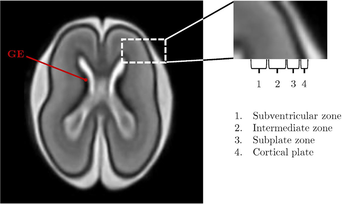
Fetal brain development is a complex process involving different stages of growth and organization which are crucial for the development of brain circuits and neural connections. Fetal atlases and labeled datasets are promising tools to investigate prenatal brain development. They support the identification of atypical brain patterns, providing insights into potential early signs of clinical conditions. In a nutshell, prenatal brain imaging and post-processing via modern tools are a cutting-edge field that will significantly contribute to the advancement of our understanding of fetal development. In this work, we first provide terminological clarification for specific terms (i.e., "brain template" and "brain atlas"), highlighting potentially misleading interpretations related to inconsistent use of terms in the literature. We discuss the major structures and neurodevelopmental milestones characterizing fetal brain ontogenesis. Our main contribution is the systematic review of 18 prenatal brain atlases and 3 datasets. We also tangentially focus on clinical, research, and ethical implications of prenatal neuroimaging.

Keywords: Brain atlas; Brain dataset; Fetus; Magnetic resonance imaging.


Conflict of interest statement

Declaration of competing interest The authors declare no competing interests.

Figures

Fig. 1.
Fig. 1.
T2w image showing the brain structure of a 21-week fetus. The image zoom depicts the fetal telencephalic wall and its sub-structures: subventricular zone, intermediate zone, subplate zone, and cortical plate. The fetal brain depicted here is adapted from the publicly available atlas by Uus et al. (2023). GE: ganglionic eminence.
Fig. 2.
Fig. 2.
PRISMA flow diagram. It reports the study selection process used in this review.
Fig. 3.
Fig. 3.
Categorization of atlases. The number of atlases per category is reported at the bottom right of each box. No single-subject atlas has been identified. Probabilistic atlases are categorized on the basis of the reference image used to characterize the brain anatomy (i.e., anatomical, diffusion, and surface images). DWI: diffusion weighted imaging.
Fig. 4.
Fig. 4.
Gestational age domain of publicly available fetal MRI atlases or datasets. The graph describes gestational age, in terms of weeks, covered by each fetal MRI atlas or datasets included in this review. Examples of normal appearing fetal cortical surfaces at different GAs are reported along the x-axis. DWI: diffusion weighted imaging.
Fig. 5.
Fig. 5.
Example of different atlas reference images in a fetal brain atlas. From left to right: structural anatomical, structural diffusion, and surface images of a 28-week fetus. The structural anatomical and structural diffusion atlas reference images are generated from the publicly available atlas of Uus et al. (2023). The atlas reference image of the surface atlas depicted shows the cortical plate, and is generated via MATLAB from the previously quoted Uus et al. (2023) publicly available atlas.
Fig. 6.
Fig. 6.
T2-weighted images of the fetal brain covering weeks 22 to 36. The maps are generated from the Uus et al. (2023) publicly available atlas.
Fig. 7.
Fig. 7.
Fiber Orientation Distribution (FOD) maps of the fetal brain covering 22 to 36 weeks. Maps are generated from the Uus et al. (2023) publicly available atlas.
Fig. 8.
Fig. 8.
Cortical surface of the fetal brain covering 22 to 38 weeks. The surfaces depicted in the figure are generated from Gholipour et al. (2017) publicly available atlas.
Fig. 9.
Fig. 9.
Example of manual segmentation (Orange: external cerebrospinal fluid; yellow: GM; bright green: WM; dark green: ventricles; blue: cerebellum; purple: deep GM: red: brainstem/spinal cord) on a 28.7-week fetus. Image adapted from Payette et al. (2023) publicly available dataset.

References

    1. Ashburner J, 2007. A fast diffeomorphic image registration algorithm. Neuroimage 38 (1), 95–113. 10.1016/j.neuroimage.2007.07.007. - DOI - PubMed
    1. Avants BB, Epstein CL, Grossman M, Gee JC, 2008. Symmetric diffeomorphic image registration with cross-correlation: evaluating automated labeling of elderly and neurodegenerative brain. Med. Image Anal. 12 (1), 26–41. 10.1016/j.media.2007.06.004. - DOI - PMC - PubMed
    1. Avants BB, Tustison NJ, Wu J, Cook PA, Gee JC, 2011. An open source multivariate framework for n-tissue segmentation with evaluation on public data. Neuroinformatics 9 (4), 381–400. 10.1007/s12021-011-9109-y. - DOI - PMC - PubMed
    1. Ball G, Aljabar P, Nongena P, Kennea N, Gonzalez-Cinca N, Falconer S, Chew ATM, Harper N, Wurie J, Rutherford MA, Counsell SJ, Edwards AD, 2017. Multimodal image analysis of clinical influences on preterm brain development. Ann. Neurol. 82 (2), 233–246. 10.1002/ana.24995. - DOI - PMC - PubMed
    1. Barkovich AJ, Guerrini R, Kuzniecky RI, Jackson GD, Dobyns WB, 2012. A developmental and genetic classification for malformations of cortical development: update 2012. Brain 135 (Pt 5), 1348–1369. 10.1093/brain/aws019. - DOI - PMC - PubMed

Publication types