. 2024 Apr 17;38(5-6):273-288.
doi: 10.1101/gad.351350.123.

Haploinsufficiency of phosphodiesterase 10A activates PI3K/AKT signaling independent of PTEN to induce an aggressive glioma phenotype

Affiliations

Haploinsufficiency of phosphodiesterase 10A activates PI3K/AKT signaling independent of PTEN to induce an aggressive glioma phenotype

Nicholas Nuechterlein et al. Genes Dev. .

Abstract

Glioblastoma is universally fatal and characterized by frequent chromosomal copy number alterations harboring oncogenes and tumor suppressors. In this study, we analyzed exome-wide human glioblastoma copy number data and found that cytoband 6q27 is an independent poor prognostic marker in multiple data sets. We then combined CRISPR-Cas9 data, human spatial transcriptomic data, and human and mouse RNA sequencing data to nominate PDE10A as a potential haploinsufficient tumor suppressor in the 6q27 region. Mouse glioblastoma modeling using the RCAS/tv-a system confirmed that Pde10a suppression induced an aggressive glioma phenotype in vivo and resistance to temozolomide and radiation therapy in vitro. Cell culture analysis showed that decreased Pde10a expression led to increased PI3K/AKT signaling in a Pten-independent manner, a response blocked by selective PI3K inhibitors. Single-nucleus RNA sequencing from our mouse gliomas in vivo, in combination with cell culture validation, further showed that Pde10a suppression was associated with a proneural-to-mesenchymal transition that exhibited increased cell adhesion and decreased cell migration. Our results indicate that glioblastoma patients harboring PDE10A loss have worse outcomes and potentially increased sensitivity to PI3K inhibition.

Keywords: PDE10A; PI3K/ATK pathway; RCAS/tv-a; glioblastoma; mesenchymal cell state.


Figures

Figure 1.
Figure 1.
Nominating PDE10A as a potential glioblastoma tumor suppressor gene. (A) Unbiased cytoband-level log rank analysis identifies loss of cytoband 6q27 as the most prognostic in TCGA glioblastomas. (B) Kaplan–Meier curves show 6q27 loss is prognostic in the TCGA, NYU, and REMBRANDT cohorts. (C) Five genes in 6q27 had sgRNAs that caused cellular proliferation on a genome-wide CRISPR screen of glioblastoma stem-like cells (GSCs) and neural stem cells (NSCs). (D) PDE10A, AFDN, and PDCD2 have gene dosage effects on gene expression. (E) Only PDE10A showed lower expression in human glioblastoma compared with normal brain tissue. (F) In RCAS/tv-a mouse gliomas, only PDE10A showed significantly lower expression levels in mouse glioblastoma compared with normal mouse brain tissue. (HR) Hazard ratio, (OS) overall survival.
Figure 2.
Figure 2.
PDE10A expression is developmentally regulated in human and mouse central nervous systems. (A,B) Primary mouse glioma precursor cells with PDE10A knockdowns showed increased neurosphere formation. (C) Real-time qPCR for neurospheres confirms that both hairpins for PDE10A reduce associated mRNA. (D) PDE10A knockdown led to increased SOX2 protein expression in mouse neurospheres. (E) SOX2 gene expression was negatively correlated with PDE10A expression in the TCGA and CGGA glioblastoma data sets. (F) PDE10A was more highly expressed in postmitotic differentiated neurons compared with progenitor and differentiated glial cells in mice. (G) PDE10A was also highly expressed in postmitotic neurons in humans.
Figure 3.
Figure 3.
PDE10A suppression induces an aggressive phenotype in mouse glioma. (AC) Mice harboring PDGF-driven gliomas with PDE10A knockdown suffered significantly worse survival, higher WHO histological grade, and higher mitotic activity in vivo compared with their control counterparts.
Figure 4.
Figure 4.
PDE10A suppression facilitates a proneural-to-mesenchymal transition in glioma. (A) UMAP plot of single-nucleus RNA sequencing of mouse glioma cells in vivo demonstrating cell type clustering. Cells in clusters expressing high levels of RCAS and hPDGFA were labeled tumor cells (GBM1 and GBM2); other common CNS cell types were identified by standard gene markers. (B) Tumor cluster GBM2 was highly enriched for tumor cells from both PDE10A knockdown gliomas compared with the scrambled control. (C) Tumor cluster GBM2 was also highly enriched for VEGFA expression (the top marker of this cluster). (D) PDE10A knockdown mouse gliomas were enriched for the VEGFA-associated histological features palisading necrosis (PSN) and microvascular proliferation (MVP). (E) The proportion of mesenchymal-like tumor cells in the knockdowns is higher in PDE10A knockdown tumor cells compared with the control in vivo. (F) The mesenchymal subtype is overrepresented in glioblastomas with PDE10A loss in the TCGA data set. (G,H) Western blotting showed unchanged total STAT3 levels with increased pSTAT3 levels in cells with PDE10A knockdown compared with the control cells in vitro.
Figure 5.
Figure 5.
PDE10A suppression causes increased cell adhesion and decreased single-cell migration. (A) In vivo single-nucleus RNA sequencing data showed that nearly all top hits of a gene ontology analysis comparing tumor cells from the PDE10A knockdowns with the controls were related to cell–cell adhesion or cell adhesion molecules. (B) Genes related to cell adhesion were up-regulated (e.g., CADM1), and genes involved in focal adhesion complex disassembly (e.g., DNM3) were down-regulated in the PDE10A knockdown tumor cells compared with the control. (C,D) Western blotting confirmed that CADM1 protein levels were higher, while DNM3 levels were lower, for PDE10A knockdown cells in vitro. (E,F) In vitro cell adhesion and cell migration assays showed a significant increase in cell adhesion with a concurrent significant decrease in cell migration in the PDE10A knockdown cells compared with the control.
Figure 6.
Figure 6.
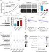
PDE10A suppression activates the PI3K/AKT/pS6 pathway independent of PTEN. (A) Immunohistochemistry of mouse gliomas showed increased pS6 in the PDE10A knockdown tumors compared with the control. (B) Cellular PTEN levels were unaltered with PDE10A knockdown, as determined by Western blotting. (C,D) Western blotting showed unchanged total PI3K levels with increased pPI3K levels in cells with PDE10A knockdown compared with the control cells. (E,F) Western blotting showed unchanged total AKT levels with increased pAKT levels in cells with PDE10A knockdown compared with the control cells. (G,H) Mouse cells with PDE10A knockdown responded strongly to the PI3K inhibitors alpelisib and paxalisib in vitro with increasing dosage. (I) Proposed model of PDE10A as an inhibitor of the PI3K/AKT signaling pathway independent of PTEN activity.

References

    1. Alexander J, LaPlant QC, Pattwell SS, Szulzewsky F, Cimino PJ, Caruso FP, Pugliese P, Chen Z, Chardon F, Hill AJ, et al. 2020. Multimodal single-cell analysis reveals distinct radioresistant stem-like and progenitor cell populations in murine glioma. Glia 68: 2486–2502. 10.1002/glia.23866 - DOI - PMC - PubMed
    1. Andl CD, Al Moustafa AE, Deramaudt TB, O'Neill GM. 2010. Cell adhesion signaling and its impact on tumorigenesis. J Oncol 2010: 257809. - PMC - PubMed
    1. Arora S, Pattwell SS, Holland EC, Bolouri H. 2020. Variability in estimated gene expression among commonly used RNA-seq pipelines. Sci Rep 10: 2734. 10.1038/s41598-020-59516-z - DOI - PMC - PubMed
    1. Ashrafi S, Alam S, Sultana A, Raj A, Emon NU, Richi FT, Sharmin T, Moon M, Park MN, Kim B. 2023. Papaverine: a miraculous alkaloid from opium and its multimedicinal application. Molecules 28: 3149. 10.3390/molecules28073149 - DOI - PMC - PubMed
    1. Baroni LV, Sundaresan L, Heled A, Coltin H, Pajtler KW, Lin T, Merchant TE, McLendon R, Faria C, Buntine M, et al. 2021. Ultra high-risk PFA ependymoma is characterized by loss of chromosome 6q. Neuro Oncol 23: 1360–1370. 10.1093/neuonc/noab034 - DOI - PMC - PubMed

Publication types

Substances