. 2024 Aug;76(8):1303-1316.
doi: 10.1002/art.42858. Epub 2024 May 15.

EphB2 Receptor Promotes Dermal Fibrosis in Systemic Sclerosis

Affiliations

EphB2 Receptor Promotes Dermal Fibrosis in Systemic Sclerosis

Erika S A Egal et al. Arthritis Rheumatol. 2024 Aug.

Abstract

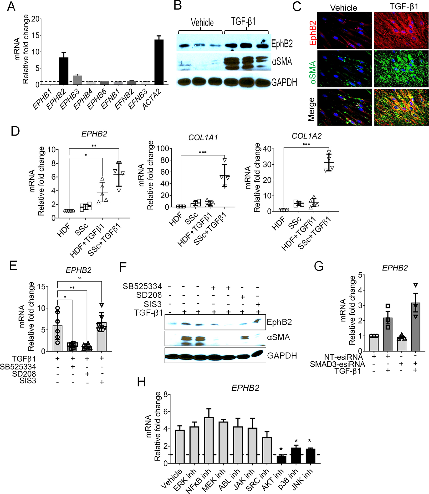
Objective: Erythropoietin-producing hepatocellular (Eph)/Ephrin cell-cell signaling is emerging as a key player in tissue fibrogenesis. The aim of this study was to test the hypothesis that the receptor tyrosine kinase EphB2 mediates dermal fibrosis in systemic sclerosis (SSc).

Methods: We assessed normal and SSc human skin biopsies for EphB2 expression. The in vivo role of EphB2 in skin fibrosis was investigated by subjecting EphB2-knockout mice to both bleomycin-induced and tight skin (Tsk1/+) genetic mouse models of skin fibrosis. EphB2 kinase-dead and overactive point mutant mice were used to evaluate the role of EphB2 forward signaling in bleomycin-induced dermal fibrosis. In vitro studies were performed on dermal fibroblasts from patients with SSc and healthy controls, which was followed by in vivo analysis of fibroblast-specific Ephb2-deficient mice.

Results: Expression of EphB2 is up-regulated in SSc skin tissue and explanted SSc dermal fibroblasts compared with healthy controls. EphB2 expression is elevated in two animal models of dermal fibrosis. In mice, EphB2 drives dermal fibrosis in both the bleomycin and the Tsk1/+ models of skin fibrosis. EphB2 forward signaling is a critical mediator of dermal fibrosis. Transforming growth factor-β (TGF-β) cytokines up-regulate EphB2 in dermal fibroblasts via noncanonical TGF-β/mother against decapentaplegic signaling, and silencing EPHB2 in human dermal fibroblasts is sufficient to dampen TGF-β-induced fibroblast-to-myofibroblast differentiation. Moreover, mice with fibroblast-specific deletion of EphB2 showed impaired fibroblast-to-myofibroblast differentiation and reduced skin fibrosis upon bleomycin challenge.

Conclusion: Our data implicate TGF-β regulation of EphB2 overexpression and kinase-mediated forward signaling in the development of dermal fibrosis in SSc. EphB2 thus represents a potential new therapeutic target for SSc.


Conflict of interest statement

Competitive interest: Nothing to declare

Figures

Figure 1:
Figure 1:. EphB2 expression is upregulated in patients with SSc.
(A) Heat map showing upregulation of EPHB2 (red square) in SSc skin patients compared to normal individuals. (B) EPHB2 transcript levels in the skin of normal individuals and SSc patients. Data was extracted from GEO GSE45485. (C) EPHB2 transcript levels in the skin of normal individuals and SSc patients extracted from GEO GSE130955 (normal = 33, SSc=48). The y-axis in C and B indicates the log2-signal intensity and transformed normalized count of EPHB2. (D) Correlation plot between EPHB2 transcripts count and mRSS from GEO GSE130955. (E) Representative dual immunostainings of EphB2 (red) and αSMA (green) expression in normal (n=10) and SSc skin (n=15) with DAPI nuclear staining (blue). Magnification 20x, scale bar 150μm. Quantification of EphB2 positive myofibroblasts per field. (F) Dual immunostainings show phosphoEphB1/2Y594+596 (red) and αSMA (green) expression in normal (n=10) and SSc skin (n=15) with DAPI nuclear staining (blue). Quantification of phosphoEphB1/2Y594+596 positive myofibroblasts per field. Magnification 20x, scale bar 150μm. (G) EPHB2 mRNA expression was examined in primary dermal fibroblasts derived from five unaffected controls and five SSc patients. Data are mean ± SEM and were analyzed using a Student’s t test, **P < 0.01; ****P < 0.0001.
Figure 2:
Figure 2:. Bleomycin-induced skin fibrosis is reduced in mice lacking Ephb2.
(A) H&E staining of PBS and bleomycin-treated Ephb2+/+ and Ephb2−/− mice (double arrowhead indicates dermal thickness). (B) Masson-trichrome staining of PBS and bleomycin-treated Ephb2+/+ and Ephb2−/− mice. (C) Quantification of dermal thickness of PBS and bleomycin-treated Ephb2+/+ and Ephb2−/− mice (n=6 mice per group). (D) Quantification of hydroxyproline content in lesional skin specimen of PBS- and bleomycin-treated Ephb2−/− and Ephb2+/+ mice (n=4–8 mice per group). (E) mRNA levels of Col1a1, Acta2, Timp1 and Pdgfrβ determined by RT-qPCR. Results were normalized with GAPDH and represent mean ± SEM of duplicate determination from 4–8 mice per group. BLM=Bleomycin. *P<0.05; **P < 0.01 Mann Whitney-U test.
Figure 3:
Figure 3:. EphB2 forward signaling drives bleomycin-induced skin fibrosis in mice:
(A) Representative images of H&E staining from skin sections of bleomycin-treated EphB2-kinase overactive (Ephb2F620D/F620D), EphB2-WT and EphB2-kinase dead (Ephb2K661R/K661R) compared with PBS-treated controls (n=3–4 mice per group). (B) Representative images of Masson’s trichrome staining from skin sections of bleomycin-treated EphB2-kinase overactive (Ephb2F620D/F620D), EphB2-WT and EphB2-kinase dead (Ephb2K661R/K661R) compared with PBS treated controls (n=3–4 mice per group). (C) Phospho-EphB1/2Y594+Y596 immunohistochemistry images from skin sections of bleomycin-treated EphB2-kinase overactive (Ephb2F620D/F620D), EphB2-WT and EphB2-kinase dead (Ephb2K661R/K661R) compared with PBS-treated controls (n=3–4 mice per group). (D) Quantification of dermal thickness of bleomycin-treated EphB2-kinase overactive (Ephb2F620D/F620D), EphB2-WT and EphB2-kinase dead (Ephb2K661R/K661R) compared with PBS-treated controls (n=3–4 mice per group). (E) Hydroxyproline assay in skin of bleomycin-treated EphB2-Kinase overactive (Ephb2F620D/F620D) and EphB2-WT compared with PBS-treated Ephb2F620D/F620D and EphB2-WT (n=3–4 mice per group). (F) Quantitative qPCR assessment of fibrotic genes Ephb2, Col1a1, Col1a2, Tgf-β1, Tgf-β2 and inflammatory markers Tnf-α, Il-6, Cxcl-2, Ccl-3 and Tlr-4 expression in lesional skin of bleomycin-treated EphB2-Kinase overactive (Ephb2F620D/F620D), EphB2-WT and EphB2-kinase dead (Ephb2K661R/K661R) compared with PBS-treated controls. Data represent mean ± SEM, n= 3–4 mice per group. One-Way ANOVA Kruskal-Wallis. *P<0.05 were considered as statistically significant.
Figure 4:
Figure 4:. TGF-β upregulates EphB2 in human dermal fibroblasts in a non-SMAD3 manner.
(A) EPHB1–4,6 and EFNB1–3 and ACTA2 mRNA levels were measured by qPCR in vehicle and TGF-β1 (10ng/ml)-stimulated NHDFs for 24h. Data represent Mean ±SD of three independent experiments. (B) Western blot and (C) immunofluorescence assays depicting an upregulation of EphB2 and αSMA proteins in TGF-β1 stimulated NHDFs compared to vehicle-treated NHDFs for 24h. (D) SSc (n=4) and healthy human dermal fibroblasts (n=5) were stimulated by TGF-β1 for 24h and mRNA expression of EPHB2, COL1A1 and COL1A2 were evaluated by qPCR. (E) qPCR (n=6 per group) and (F) western blot show EPHB2 mRNA and proteins levels upon inhibition of canonical TGF-β/SMAD with SIS3 Smad3 inhibitor, SD208 TGFβR1 inhibitor, and SB525334 TGFβR2 inhibitor in TGF-β1-treated NHDFs. (G) Effects of silencing Smad3 with Mission®-esiRNA on TGF-β-induced EPHB2 mRNA expression in normal human dermal fibroblasts (n=3 per group). Non-targeting (NT) Mission®-esiRNA was used as control. (H) EPHB2 mRNA level in TGF-β1-treated NHDFs upon inhibition of the noncanonical TGFβ signaling with small molecule inhibitors for ERK, NFκB, MEK, ABL, JAK, SRC, AKT, p39, and JNK. Fold change is relative to vehicle-treated NHDFs. Experiments were repeated at least of two times (n=6 per group). *P<0.05, **P<0.01 Mann Whitney U test.
Figure 5:
Figure 5:. EphB2 silencing mitigates TGF-β-induced fibroblast-to-myofibroblasts transition.
(A) and (B) Mean ± SEM of EPHB2, ACTA-2, COL1A1, and COL1A2 mRNA expression measured in at least five different NHDF lines treated with Mission® EPHB2-esiRNA (SIGMA), stimulated with TGF-β1 at 10ng/ml and normalized to the same NHDF lines treated with nontargeting Mission®-esiRNA (n=3–7 per group). (C) Immunofluorescence staining for αSMA upon TGFβ stimulation of fibroblasts from a representative EPHB2-esiRNA transfected (KO) or nontargeting-esiRNA (WT). (D) Heat map shows the top 40 genes differentially expressed in TGF-β1 stimulated NHDFs lines treated with nontargeting-esiRNA and EPHB2-esiRNA (n=5 per group). (E) List of major human genes upregulated in TGF-β1 stimulated NHDF-EPHB2 deficient lines. (F) Mean ± SEM of SMAD7, SMURF1, and SMURF2 mRNA normalized count measured in NHDF lines treated with Mission® EPHB2-esiRNA (SIGMA), stimulated with TGF-β1 at 10ng/ml and normalized to NHDF lines treated with control Mission®-esiRNA (n=5 per group). (G) Mean ± SEM of SMURF1 and SMURF2 mRNA expression measured in NHDF lines treated with Mission® EphB2-esiRNA (SIGMA), stimulated with TGF-β1 at 10ng/ml and normalized to the same NHDF lines treated with nontargeting Mission®-esiRNA (n=8 per group). HDF= normal human dermal fibroblasts. siEphB2 =EPHB2-esiRNA. *P<0.05; ** P<0.01; ***P<0.001 Mann Whitney-U test.
Figure 6:
Figure 6:. Fibroblast-specific deletion of EphB2 mitigates bleomycin-induced skin fibrosis in mice.
(A) Representative H&E staining of bleomycin-treated Ephb2-C and EphB2-cKO mice (bold line indicates dermal thickness). (B) Quantification of dermal thickness of bleomycin-treated Ephb2-C and Ephb2-cKO mice (n=5–10 mice per group). (C) Representative masson’s trichrome staining of bleomycin-treated Ephb2-C and Ephb2-cKO mice. (D) Quantification of hydroxyproline content in lesional skin specimen of bleomycin treated Ephb2-C and Ephb2-cKO mice (n=5–10 mice per group). (E) mRNA levels of pro-fibrotic genes Col1a1, Col3a1, Col6a2, Tgf-β2, and Tgf-β1 determined by RT-qPCR. These results were normalized with HPRT and represent mean ± SEM of n= 4–5 mice per group. *p<0.05, **p<0.01 Mann Whitney U test.

References

    1. Stern EP, Denton CP. The Pathogenesis of Systemic Sclerosis. Rheumatic Disease Clinics of North America 2015;41:367–382. - PubMed
    1. Distler O, Highland KB, Gahlemann M, Azuma A, Fischer A, Mayes MD, Raghu G, et al. Nintedanib for Systemic Sclerosis–Associated Interstitial Lung Disease. New England Journal of Medicine 2019;380:2518–2528. - PubMed
    1. Kuwana M, Azuma A. Nintedanib: New indication for systemic sclerosis-associated interstitial lung disease. Modern Rheumatology 2019:1–7. - PubMed
    1. Denton CP. Advances in pathogenesis and treatment of systemic sclerosis. Clinical Medicine 2015;15:s58–s63. - PubMed
    1. Ide M, Jinnin M, Tomizawa Y, Wang Z, Kajihara I, Fukushima S, Hashizume Y, et al. Transforming growth factor β-inhibitor Repsox downregulates collagen expression of scleroderma dermal fibroblasts and prevents bleomycin-induced mice skin fibrosis. Experimental Dermatology:n/a-n/a. - PubMed