. 2024 Apr 8;7(1):424.
doi: 10.1038/s42003-024-05948-6.

A cyclical switch of gametogenic pathways in hybrids depends on the ploidy level

Affiliations

A cyclical switch of gametogenic pathways in hybrids depends on the ploidy level

Dmitrij Dedukh et al. Commun Biol. .

Abstract

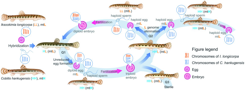
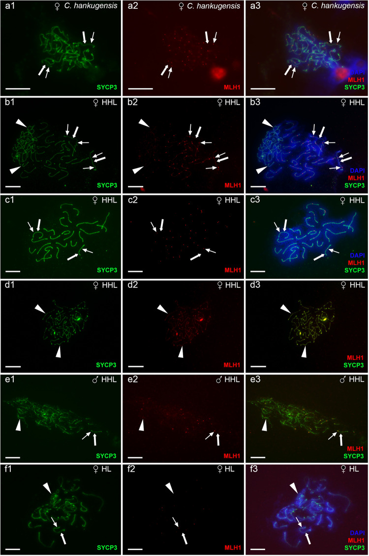
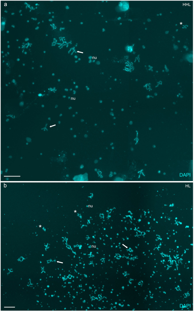
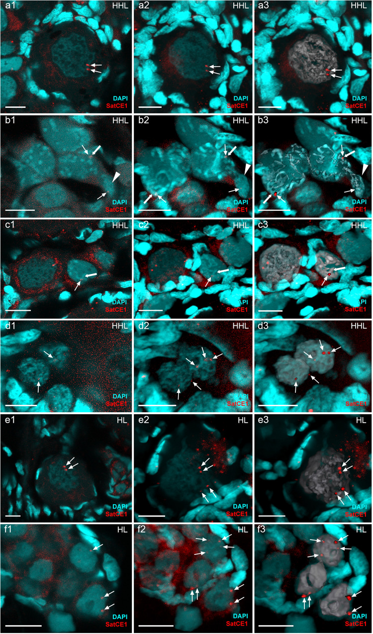
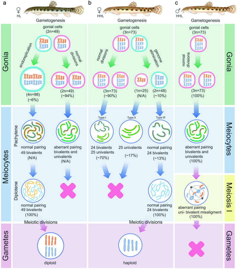
The cellular and molecular mechanisms governing sexual reproduction are conserved across eukaryotes. Nevertheless, hybridization can disrupt these mechanisms, leading to asexual reproduction, often accompanied by polyploidy. In this study, we investigate how ploidy level and ratio of parental genomes in hybrids affect their reproductive mode. We analyze the gametogenesis of sexual species and their diploid and triploid hybrids from the freshwater fish family Cobitidae, using newly developed cytogenetic markers. We find that diploid hybrid females possess oogonia and oocytes with original (diploid) and duplicated (tetraploid) ploidy. Diploid oocytes cannot progress beyond pachytene due to aberrant pairing. However, tetraploid oocytes, which emerge after premeiotic genome endoreplication, exhibit normal pairing and result in diploid gametes. Triploid hybrid females possess diploid, triploid, and haploid oogonia and oocytes. Triploid and haploid oocytes cannot progress beyond pachytene checkpoint due to aberrant chromosome pairing, while diploid oocytes have normal pairing in meiosis, resulting in haploid gametes. Diploid oocytes emerge after premeiotic elimination of a single-copied genome. Triploid hybrid males are sterile due to aberrant pairing and the failure of chromosomal segregation during meiotic divisions. Thus, changes in ploidy and genome dosage may lead to cyclical alteration of gametogenic pathways in hybrids.


Conflict of interest statement

The authors declare no competing interests.

Figures

Fig. 1
Fig. 1. Schematic overview of gametogenesis and reproduction of diploid and triploid hybrids within C. hankugensis-I. longicorpa complex (redrawn from refs. ,,).
After crosses of two parental sexual species, Cobitis hankugensis (HH, marked in blue) and Iksookimia longicorpa (LL, marked in orange), diploid hybrids (HL) are produced with the mitochondrial DNA (designated as ‘mt’) from one of the sexual species (L). Diploid hybrids form diploid clonal gametes with ‘L’ mtDNA. After fertilization of such eggs by sperm of one of the parental species, triploid hybrids with L mtDNA appear (HHL). In triploids, a single-copied genome (L) is eliminated during their gametogenesis, and the remaining haploid gametes (HH) produce haploid ‘H’ gametes with ‘L’ mtDNA. After fertilization of such gametes by sperm from C. hankugensis, diploid sexual species appear but with ‘L’ mtDNA. After fertilization of gametes produced by triploids by sperm from the other parental species, I. longicorpa, new diploid hybrids appear with ‘L’ mtDNA. Q1-3 indicates the gap of knowledge in studied asexual hybrid complex: Q1. The cytogenetic mechanisms underlying unreduced gametes formation by diploid hybrids; Q2. Gametogenic stage and mechanisms of putative genome elimination in triploid hybrid females; Q3. Gametogenic alteration underlying hybrid sterility in triploid hybrid males.
Fig. 2
Fig. 2. The analysis of pairing in pachytene oocytes (a1–d3) and spermatocytes (e1–e3) of C. hankugensis (a1–a3) and triploid HHL hybrids (b1–e3).
Synaptonemal complexes were visualized using immunostaining of lateral (SYCP3 protein, green) (a1, b1, c1, d1, and e1) and central (SYCP1 protein, red) (a2, b2, c2, d2, and e2) components. Corresponding merged figures (a3, b3, c3, d3, and e3) also include DAPI staining (blue). Accumulation of SYCP3 and SYCP1 proteins (indicated by thick arrows) allows distinguishing bivalents, while univalents accumulate only SYCP3 protein (indicated by arrowheads). Pachytene oocytes of C. hankugensis exhibit 24 fully paired bivalents (a1a3). In triploid hybrids, we observed pachytene oocytes with 24 bivalents and 25 univalents (b1b3), oocytes with 24 bivalents (c1c3), and oocytes with 25 univalents (d1d3). Triploid hybrid males exhibit pachytene oocytes only with the aberrant pairing of several bivalents and univalent (e1e3). Scale bar = 10 µm.
Fig. 3
Fig. 3. The analysis of crossover loci in pachytene oocytes (a1–d3, f1–f3) and spermatocytes (e1–e3) from gonads of C. hankugensis (a1–a3), triploid HHL hybrids (b1–e3) and diploid hybrid (f1–f3).
Crossover loci were detected by MLH1 protein (indicated by thin arrows, red) (a2, b2, c2, d2, e2, and f2) on lateral components of synaptonemal complexes (SYCP3 protein, green) (a1, b1, c1, d1, e1, and f1). Corresponding merged figures (a3, b3, c3, d3, e3, and f3) also include DAPI staining (blue). MLH1 bindings (indicated by thin arrows, red) are located on bivalents (indicated by thick arrows) and do not accumulate on univalents (indicated by arrowheads). Pachytene oocytes of C. hankugensis exhibit 24 fully with at least one crossover locus per bivalent (a1a3). In triploid hybrids, oocytes with 24 bivalents and 25 univalents have MLH1 signals only on bivalents (b1b3). Oocytes with exclusively 24 bivalents (c1c3) have recombination signals on each bivalent, while oocytes with exclusively 25 univalents (d1d3) do not have crossover locus. MLH1 immunostaining demonstrates the presence of crossover in individual bivalents formed in a triploid hybrid male (e1e3) and pachytene oocytes with an unduplicated genome (f1f3). Scale bar = 10 µm.
Fig. 4
Fig. 4. Diplotene chromosomal spreads from the individual oocytes of triploid HHL (a) and diploid HL (b) hybrid females.
A triploid hybrid’s chromosomal set of diplotene oocytes includes 24 bivalents, possibly of C. hankugensis (a). The chromosomal set of diploid hybrid includes 49 bivalents (b). Since the chromosomal spread from the individual oocyte was large, four images were merged into one in the case of (a) and (b). Chromosomes were stained with DAPI (cyan). Thick arrows indicate examples of individual bivalents; nu shows examples of extrachromosomal nucleoli (nu). Asterisks indicate enlarged bivalents in Supplementary Fig. S5c (HHL) and S5d and S5e (HL) for triploid and diploid hybrids, respectively. Scale bar = 50 µm.
Fig. 5
Fig. 5. Identification of ploidy level of cells in gonadal fragments of triploid HHL (a1–d3) and diploid hybrids (e1–f3) using whole-mount FISH with chromosome-specific SatCE1 marker.
In the diplotene oocyte of triploid HHL hybrid (a1a3), two adjacent signals are visible, suggesting the presence of two homologous chromosomes. Pachytene oocytes with bivalents and univalents (b1b3) have signals on bivalent (indicated by thick arrow) as well as on univalent (indicated by arrowhead). Pachytene oocytes only with bivalents have one signal (indicated by an arrow) on bivalent (indicated by a thick arrow) (c1c3). Diploid oogonia with two signals (indicated by arrows) and triploid oogonia with three signals (indicated by arrows) (d1d3) are shown in the ovary from the triploid HHL hybrid. In the diplotene oocyte of diploid HL hybrid (e1e3), two pairs of signals are visible, suggesting the presence of two bivalents. Diploid oogonia with two signals (indicated by arrows) and tetraploid oogonia with four signals (indicated by arrows) (f1f3) in the ovary from diploid HL hybrid. DNA is stained by DAPI (cyan). Images (a1, b1, c1, d1, e1, and f1) are single confocal sections of 0.7 µm in thickness; corresponding 3D reconstructions (a2, b2, c2, d2, e2, and f2) and 3D surface reconstructions (a3, b3, c3, d3, e3, and f3) of metaphase plates with constructed isosurfaces of the signals and cells of interest. Scale bar = 10 µm.
Fig. 6
Fig. 6. Schematic overview of gametogenesis of diploid and triploid hybrids within C. hankugensis-I. longicorpa complex.
a Diploid hybrid females (HL) have premeiotic genome endoreplication in the minor portion of oogonia. This process enables the formation of bivalents during the pachytene stage of meiosis, as each chromosome has a chromosomal copy to pair with. Afterward, such oocytes progress to diplotene, and upon completing meiosis, they form diploid gametes. b In a portion of oogonia from triploid hybrid females (on example of hybrids with HHL genome composition), genome elimination of the ‘L’ genome occurs, leading to the formation of oogonia with HH genome composition (type III) and possibly oogonia with L genome exclusively (type II). Most oogonia retain their original ploidy level without genome elimination (type I). In the pachytene stage of meiosis, type III oocytes have 24 well-paired bivalents; type I oocytes have a mixture of 24 bivalents and 25 univalents; and type II oocytes have univalents with the partial pairing of a few chromosomes. Only type III oocytes proceed beyond pachytene into the diplotene, followed by the formation of reduced haploid gametes. c In triploid hybrid males, spermatogonia retain the original ploidy level and do not undergo genome elimination or genome endoreplication. During the pachytene, spermatocytes exhibit aberrant pairings with univalent, bivalent, and multivalent formation. These aberrant spermatocytes advance beyond pachytene and proceed to the first meiotic division. During the metaphase of the first meiotic divisions, individual univalents, bivalents, and multivalents cannot properly attach to the spindle and segregate, causing the spermatocytes to become arrested at this stage of meiosis. Chromosomes of I. longicorpa are marked in orange; chromosomes of C. hankugensis are marked in blue; green indicates lateral elements in synaptonemal complexes; and red indicates central elements of synaptonemal complexes. The percentage of germ cells and oocytes with identified ploidy levels is presented in brackets; N/A indicates cells for which the percentage was not identified.

References

    1. Lenormand T, Engelstädter J, Johnston SE, Wijnker E, Haag CR. Evolutionary mysteries in meiosis. Philos. Trans. R. Soc. B: Biol. Sci. 2016;371:20160001. doi: 10.1098/rstb.2016.0001. - DOI - PMC - PubMed
    1. Otto SP, Lenormand T. Resolving the paradox of sex and recombination. Nat. Rev. Genet. 2002;3:252–261. doi: 10.1038/nrg761. - DOI - PubMed
    1. Arnold ML, Hodges SA. Are natural hybrids fit or unfit relative to their parents? Trends Ecol. Evol. 1995;10:67–71. doi: 10.1016/S0169-5347(00)88979-X. - DOI - PubMed
    1. Coyne, J. A., Orr, H. A., Coyne, J. A. & Orr, H. A. Speciation (Oxford University Press, 2004).
    1. Rieseberg LH. Chromosomal rearrangements and speciation. Trends Ecol. Evol. 2001;16:351–358. doi: 10.1016/S0169-5347(01)02187-5. - DOI - PubMed

Publication types