. 2024 Apr 8;14(1):8245.
doi: 10.1038/s41598-024-59065-9.

Bacteriophages from human skin infecting coagulase-negative Staphylococcus: diversity, novelty and host resistance

Affiliations

Bacteriophages from human skin infecting coagulase-negative Staphylococcus: diversity, novelty and host resistance

Samah E Alsaadi et al. Sci Rep. .

Abstract

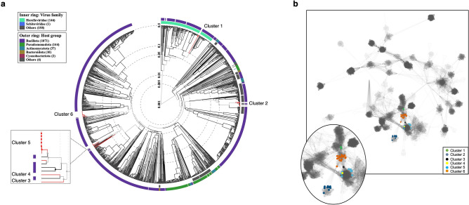
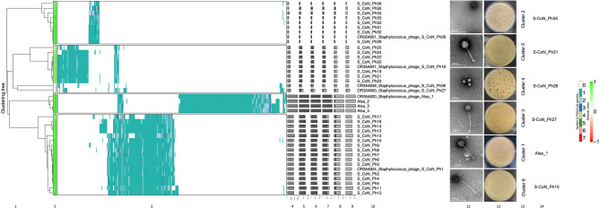
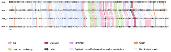
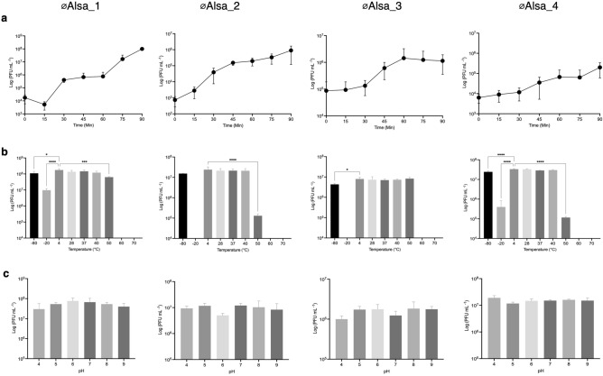
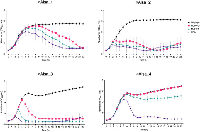
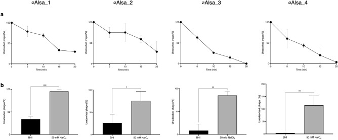
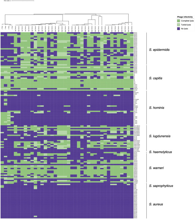
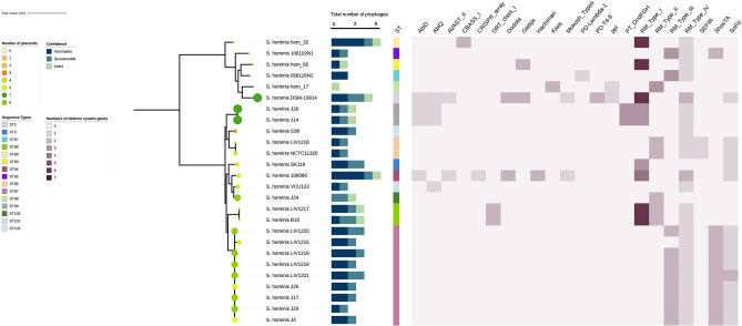
The human skin microbiome comprises diverse populations that differ temporally between body sites and individuals. The virome is a less studied component of the skin microbiome and the study of bacteriophages is required to increase knowledge of the modulation and stability of bacterial communities. Staphylococcus species are among the most abundant colonisers of skin and are associated with both health and disease yet the bacteriophages infecting the most abundant species on skin are less well studied. Here, we report the isolation and genome sequencing of 40 bacteriophages from human skin swabs that infect coagulase-negative Staphylococcus (CoNS) species, which extends our knowledge of phage diversity. Six genetic clusters of phages were identified with two clusters representing novel phages, one of which we characterise and name Alsa phage. We identified that Alsa phages have a greater ability to infect the species S. hominis that was otherwise infected less than other CoNS species by the isolated phages, indicating an undescribed barrier to phage infection that could be in part due to numerous restriction-modification systems. The extended diversity of Staphylococcus phages here enables further research to define their contribution to skin microbiome research and the mechanisms that limit phage infection.

Keywords: Bacteriophage; Comparative genomics; Defence mechanisms; Skin; Staphylococcus.


Conflict of interest statement

The authors declare no competing interests.

Figures

Figure 1
Figure 1
Phylogenetic tree and relationship of CoNS phages. (a) Staphylococcal phage genomes from this study were assigned to 6 genetic clusters (1–6) using ViPTree 3.1 that included 1332 related phage taxa. Virus families are identified on the inner ring as Herelleviridae (mint green), Schitoviridae (blue), and other unknown families (grey). Phyla of their bacterial host is indicated in the outer ring with ascribed colours shown in the key. Cluster 4 represents a novel phage genome of this study (red line) and is distinct from øIME1354_01 representing the adjacent cluster of the same node (black line). (b) Network diagram of phage genomes represented by each node, and edges are genome relatedness inferred by vConTACT. RefSeq phages (small circles) that are neighbours in the core network only are shown for clarity. Individual isolated staphylococcal phages are represented as coloured nodes and named clusters 1–6; expanded in the inset.
Figure 2
Figure 2
Hierarchical clustering of CoNS phage genomes. (1) Intergenomic distances of protein clusters were calculated using VirClust (2) and their relationship by silhouette width was examined using a hierarchical tree. The constituent viral genome clusters (VGCs) were determined based upon a 0.9 distance threshold. Similarity within each VGC is indicated by colour, with values closer to 1 being lime green. (3) Viral genome protein cluster (PCs) distribution is represented by a heatmap with rows representing individual genomes and columns as individual protein clusters together. A range of nine colours are used to indicate the number of protein clusters. Data to the right provide viral genome-specific statistics (4–9): (4) genome length (bp); (5) fraction of shared proteins (dark grey) from total proteins (light grey); (6) fraction of shared proteins within a VGC; (7) fraction of proteins specific to its own VGC; (8) proportion of proteins shared outwith a VGC; (9) the proportion of proteins shared exclusively outwith a VGC. (10) CoNS phage names from this study, provided as either their identifier or GenBank accession number, are included with reference phage genomes. (11) Electron micrographs of 6 phages, one from each cluster identified in this study [scale bar, 200 nm]. (12) Plaque morphology of each of the pictured phage and (13) their ViPTree cluster number and (14) their phage name.
Figure 3
Figure 3
Comparison of Alsa phage coding regions based on functional annotation. Genomes of øAlsa1-4 were annotated using Pharokka and HHpred and visualised with Clinker with comparison of ORF functional categories across phages indicated by colour. Grey ORFs indicate hypothetical proteins. Scale, 2.5 kb.
Figure 4
Figure 4
General characteristics of Alsa phages. One step growth curve of Alsa phages using original isolation hosts (a). Stability of phages across a range of temperatures (b) and pH (c) measured by PFU values. Statistically significant differences relative to control are indicated by asterisk: one-way ANOVA values *P < 0.05, ** P < 0.01, ***P < 0.001.
Figure 5
Figure 5
Growth kinetics of hosts with Alsa phages. Phages were cultured with their respective isolation hosts at different MOI using the absence of phage as a control. Absorbance of cells was measured over 20 h in the absence (black, closed circle) or the presence of phages at different MOI (0.01, pink square; 0.1 green triangle; 1, purple diamond) with hosts LIV1218 (øAlsa_1), LIV1220 (øAlsa_2), 104 (øAlsa_3, øAlsa_4).
Figure 6
Figure 6
Adsorption of Alsa phages and effects of periodate treatment. (a) Alsa phages were incubated with their respective isolation hosts at MOI 0.1 and free phage were titred at the indicated time periods by plaque assay. (b) The effect of sodium periodate treatment for 60 min on the proportion of phages adsorbed (MOI 0.1) over 20 min was determined with hosts LIV1218 (øAlsa_1), LIV1220 (øAlsa_2), 104 (øAlsa_3, øAlsa_4).
Figure 7
Figure 7
Host range of isolated CoNS phages. The 40 CoNS phages of this study were tested for their ability to infect 140 strains of staphylococcal species using a spot assay. Host lysis was determined visually by zone clarity with complete (dark green), turbid (light green), or no lysis (purple). The experiment was repeated a minimum of three times.
Figure 8
Figure 8
Phage defence systems and plasmid content of S. hominis. The presence of phage defence system genes in genomes of the S. hominis strains used in the host range study were detected with PADLOC. Number of defence system genes present is scored (0–6) and the total number of prophages (0–6) are indicated as either intact, incomplete, or questionable. Phylogenetic tree of S. hominis strains was generated using IQ-TREE. Sequence type (ST) of strains is indicated by vertical-coloured bars. Plasmid content (0–7 plasmids) is shown by different sized and coloured circle symbols, determined by PlasmidFinder.

Similar articles

Cited by

References

    1. Grice EA, Segre JA. The skin microbiome. Nat. Rev. Microbiol. 2011;9(4):244–253. doi: 10.1038/nrmicro2537. - DOI - PMC - PubMed
    1. Grice EA, et al. Topographical and temporal diversity of the human skin microbiome. Science. 2009;324(5931):1190–1192. doi: 10.1126/science.1171700. - DOI - PMC - PubMed
    1. Skowron K, et al. Human skin microbiome: Impact of intrinsic and extrinsic factors on skin microbiota. Microorganisms. 2021;9(3):543. doi: 10.3390/microorganisms9030543. - DOI - PMC - PubMed
    1. Flowers L, Grice EA. The skin microbiota: Balancing risk and reward. Cell Host Microbe. 2020;28(2):190–200. doi: 10.1016/j.chom.2020.06.017. - DOI - PMC - PubMed
    1. Ochlich D, Rademacher F, Drerup KA, Gläser R, Harder J. The influence of the commensal skin bacterium Staphylococcus epidermidis on the epidermal barrier and inflammation: Implications for atopic dermatitis. Exp. Dermatol. 2023;32(4):555–561. doi: 10.1111/exd.14727. - DOI - PubMed