. 2024 Aug;45(8):1660-1672.
doi: 10.1038/s41401-024-01239-2. Epub 2024 Apr 8.

Neutrophil extracellular traps promote acetaminophen-induced acute liver injury in mice via AIM2

Affiliations

Neutrophil extracellular traps promote acetaminophen-induced acute liver injury in mice via AIM2

Fan-le Zeng et al. Acta Pharmacol Sin. 2024 Aug.

Abstract

Excessive acetaminophen (APAP) can induce neutrophil activation and hepatocyte death. Along with hepatocyte dysfunction and death, NETosis (a form of neutrophil-associated inflammation) plays a vital role in the progression of acute liver injury (ALI) induced by APAP overdose. It has been shown that activated neutrophils tend to migrate towards the site of injury and participate in inflammatory processes via formation of neutrophil extracellular traps (NETs). In this study we investigated whether NETs were involved in hepatocyte injury and contributed to APAP-induced ALI progression. ALI mouse model was established by injecting overdose (350 mg/kg) of APAP. After 24 h, blood and livers were harvested for analyses. We showed that excessive APAP induced multiple programmed cell deaths of hepatocytes including pyroptosis, apoptosis and necroptosis, accompanied by significantly increased NETs markers (MPO, citH3) in the liver tissue and serum. Preinjection of DNase1 (10 U, i.p.) for two consecutive days significantly inhibited NETs formation, reduced PANoptosis and consequently alleviated excessive APAP-induced ALI. In order to clarify the communication between hepatocytes and neutrophils, we induced NETs formation in isolated neutrophils, and treated HepaRG cells with NETs. We found that NETs treatment markedly increased the activation of GSDMD, caspase-3 and MLKL, while pre-treatment with DNase1 down-regulated the expression of these proteins. Knockdown of AIM2 (a cytosolic innate immune receptor) abolished NETs-induced PANoptosis in HepaRG cells. Furthermore, excessive APAP-associated ALI was significantly attenuated in AIM2KO mice, and PANoptosis occurred less frequently. Upon restoring AIM2 expression in AIM2KO mice using AAV9 virus, both hepatic injury and PANoptosis was aggravated. In addition, we demonstrated that excessive APAP stimulated mtROS production and mitochondrial DNA (mtDNA) leakage, and mtDNA activated the TLR9 pathway to promote NETs formation. Our results uncover a novel mechanism of NETs and PANoptosis in APAP-associated ALI, which might serve as a therapeutic target.

Keywords: AIM2; APAP; NETs; PANoptosis; TLR9; acute liver injury.


Conflict of interest statement

The authors declare no competing interests.

Figures
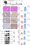
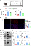
Fig. 1
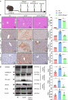
Fig. 1. Excessive acetaminophen induces liver injury and apoptosis, pyroptosis and necroptosis in mice.
a Mice were administered phosphate-buffered saline (PBS) 350 mg/kg APAP. Blood and livers were harvested after 24 h and used for various assays. b, c Representative images of hematoxylin/eosin-stained liver sections and serum alanine aminotransferase (ALT) levels. df The liver sections were stained for pyroptosis, apoptosis, and necroptosis, and representative images and quantitative histograms of GSDMD-N, cle-caspase3, and pMLKL are shown. Positive cells are marked with red arrows. g Representative immunoblots and quantitative histograms of pyroptosis markers GSDMD-N (β-actin as the loading control), apoptosis markers cle-caspase3 (β-actin as the loading control) and necroptosis markers phosphorylation forms of MLKL (MLKL as the loading control). Mean ± SEM, **P < 0.01, ***P < 0.001, and ****P < 0.0001 versus control or between the indicated groups. Similar results were obtained in five independent experiments with 6 mice per group.
Fig. 2
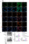
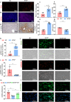
Fig. 2. Excessive acetaminophen induces hepatocyte PANoptosis via increased NETs formation in vitro.
ac Representative images of immunoflurescence images of pyroptosis markers GSDMD-N, apoptosis markers cle-caspase3 and necroptosis markers total and phosphorylation forms of MLKL. d Representative immunoblots and quantitative histograms of pyroptosis markers GSDMD-N (β-actin as the loading control), apoptosis markers cle-caspase3 (β-actin as the loading control) and necroptosis markers phosphorylation forms of MLKL (MLKL as the loading control). Mean ± SEM, **P < 0.01, ***P < 0.001, ****P < 0.0001 versus control or between the indicated groups. Similar results were obtained from five independent culture assays.
Fig. 3
Fig. 3. DNase1 treatment inhibited APAP-induced ALI as well as hepatocyte apoptosis, pyroptosis and necroptosis.
a Mice were administered phosphate-buffered saline (PBS) vehicle or DNase1 at 10 U per unit for two consecutive days, followed by PBS drug or 350 mg/kg APAP. Livers and serum were harvested after administering excess APAP 24 h for the indicated assays. b, c Representative images of areas of hepatocyte necrosis (H&E stain) and ALT levels in serum of APAP stimulation after 24 h. df Then, the liver sections were stained for pyroptosis, apoptosis and necroptosis and representative images and quantitative histograms of GSDMD-N, cle-caspase-3 and pMLKL are shown. Positive cells are marked with red arrows. g Representative immunoblots and quantitative histograms of pyroptosis markers GSDMD-N (β-actin as the loading control), and necroptosis markers phosphorylation forms of MLKL (MLKL as the loading control). Mean ± SEM. **P < 0.01, ***P < 0.001, ****P < 0.0001 versus control or between the indicated groups. Similar results were obtained in five independent experiments with 6 mice per group.
Fig. 4
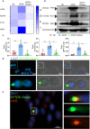
Fig. 4. Excessive acetaminophen induces the formation of PANoptosome and AIM2 pathway activation.
a cGAS, NLRP3, IFI16, and AIM2 transcripts were measured by real-time PCR. b, c Representative immunoblots and quantitative histograms of AIM2, pyrin, and ZBP1 (β-actin as the loading control). d Representative fluorescent images of NETs internalization by HepaRG. NETs were labeled with SYTOX Green and incubated with HepaRG for 6 h. The HepaRG viewed under fluorescence microscope. e Representative immunofluorescent images of co-localization with AIM2 after NETs internalization. Mean ± SEM. **P < 0.01, ***P < 0.001 versus control or between the indicated groups. Similar results were obtained in five independent experiments with 6 mice per group. Similar results were obtained from five independent culture assays.
Fig. 5
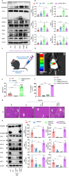
Fig. 5. Depletion of AIM2 results in reduced APAP-induced ALI and decreased formation of PANoptosome.
a, b Western blotting analyses of GSDMD, GSDMD-N, caspase-3, cle-caspase-3, phospho-MLKL, MLKL, AIM2, and ZBP1 with β-actin as a reference and quantitative histograms of these representative immunoblots in NETs-treated HepaRG cells with transfection of AIM2 siRNA. c Schematic illustrating the rescue experiment in AIM-KO mice. d, e Live imaging of small animals and real-time PCR analyses of AIM2. The results show AIM2 successful restoration in AIM2-KO mice. f, g ALT and HE. ALT show overexpressing AIM2 can aggravate liver damage in AIM2-KO mice treated with APAP. h, i Western blots analyses of GSDMD, GSDMD-N, caspase3, cle-caspase3, phospho-MLKL, MLKL, AIM2, pyrin, and ZBP1. The results show overexpressing AIM2 can aggravate liver injury and PANoptosis in AIM2-KO mice treated with APAP. Data represent the mean ± SEM. *P < 0.05, **P < 0.01, ***P < 0.001, ****P < 0.0001 versus control or between the indicated groups. Similar results were obtained in five independent experiments with 6 mice per group. Similar results were obtained from five independent culture assays.
Fig. 6
Fig. 6. mtDNA promoted the formation of NETs by TLR9-mediated signaling pathway.
a Schematic diagram of mtDNA purification by tail vein injection in mice. b Serum MPO was measured by ELISA. c Measurement of dsDNA levels in serum using Picogreen. d Representative immunofluorescence and quantitative analysis of citH3. e, f Representative immunoblots and quantitative histograms of total and phosphorylation forms of citH3, TLR9, Erk1/2, pErk1/2, p38, and p-p38. Mean ± SEM, *P < 0.05, **P < 0.01, ***P < 0.001, ****P < 0.0001 versus control or between indicated groups. Similar results were obtained in five independent experiments with 6 mice per group.
Fig. 7
Fig. 7. Excess APAP causes excess mtROS production and thus mtDNA leakage.
a Frozen sections of mouse liver tissue were stained with ROS staining solution to measure ROS changes. b The changes in mitochondrial ROS in liver tissue were detected by MitoSox staining. c Serum ROS was measured by ELISA. d, e The liver sections were stained for 8-OHdG and representative images and quantitative histograms of these representative images. f, g Real-time PCR analyses of Drp1 and MFN1 level to evaluate mitochondrial dysfunction. h Increased levels of mtDNA in serum. i mtDNA content is depleted and mtDNA integrity is impaired in the hepatocyte of APAP-induced mice in vivo. j, k HepaRG cells were stained with ROS and MitoSox staining solution to measure ROS and mtROS changes in vitro. l Immunofluorescence of 8-OHdG in vitro. m Real-time PCR analyses of mtDNA and nDNA ratios in order to indicate changes in mtDNA content in vitro. Mean ± SEM, **P < 0.01, ***P < 0.001, ****P < 0.0001 versus control or between indicated groups. Similar results were obtained in five independent experiments with 6 mice per group. Similar results were obtained from five independent culture assays.

References

    1. Jaeschke H. Acetaminophen: dose-dependent drug hepatotoxicity and acute liver failure in patients. Dig Dis. 2015;33:464–71. 10.1159/000374090 - DOI - PMC - PubMed
    1. McGill MR, Hinson JA. The development and hepatotoxicity of acetaminophen: reviewing over a century of progress. Drug Metab Rev. 2020;52:472–500. 10.1080/03602532.2020.1832112 - DOI - PMC - PubMed
    1. Ortland I, Mirjalili M, Kullak-Ublick GA, Peymani P. Drug-induced liver injury in Switzerland: an analysis of drug-related hepatic disorders in the WHO pharmacovigilance database VigiBase from 2010 to 2020. Swiss Med Wkly. 2021;151:w20503. 10.4414/smw.2021.20503 - DOI - PubMed
    1. Santos G, Figueira ERR, D’Albuquerque LAC, Lisboa PB, de Almeida MD, Filgueira NA, et al. Evaluation of drug-induced liver injury as etiology for acute liver failure in Brazil. Ann Hepatol. 2021;23:100310. 10.1016/j.aohep.2021.100310 - DOI - PubMed
    1. Darr U, Sussman NL. Drug-induced liver injury in the setting of analgesic use. Clin Liver Dis. 2020;24:121–9. 10.1016/j.cld.2019.09.008 - DOI - PubMed

MeSH terms