. 2024 Apr 8;17(1):8.
doi: 10.1186/s13039-024-00675-3.

Mosaic derivative chromosomes at chorionic villi (CV) sampling are expression of genomic instability and precursors of cryptic disease-causing rearrangements: report of further four cases

Affiliations

Mosaic derivative chromosomes at chorionic villi (CV) sampling are expression of genomic instability and precursors of cryptic disease-causing rearrangements: report of further four cases

Giulia Vitetta et al. Mol Cytogenet. .

Abstract

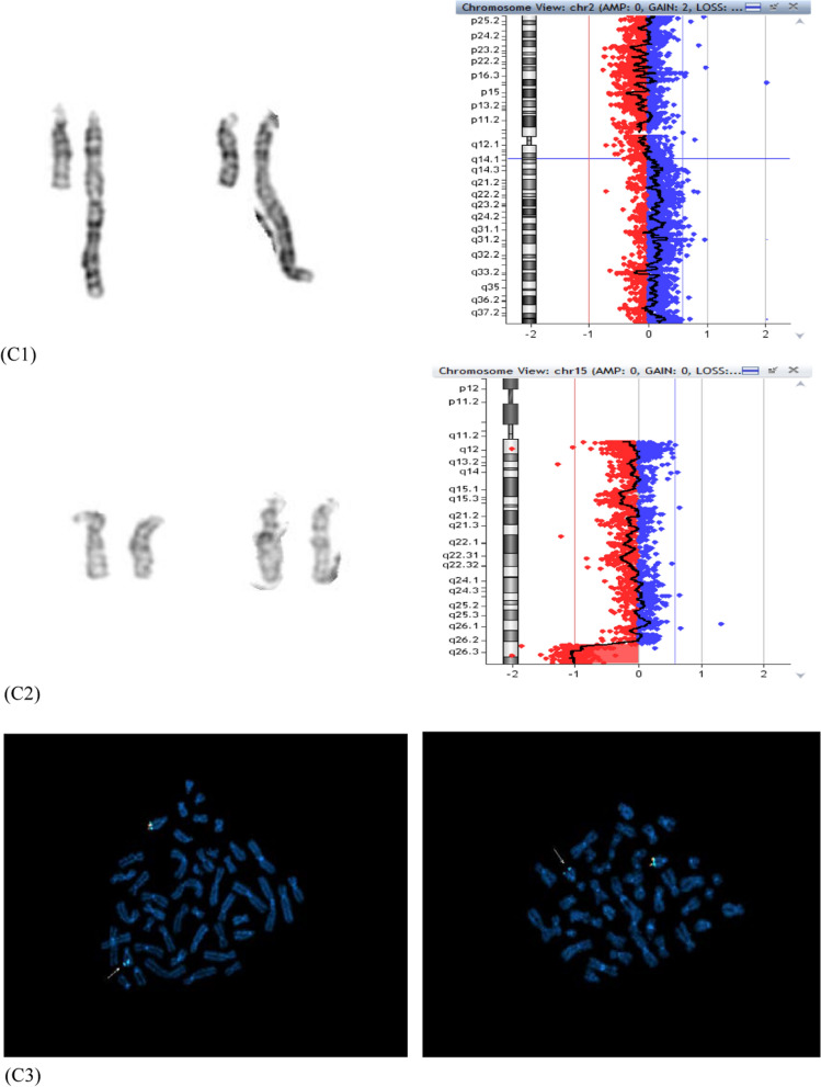
Mosaic chromosomal anomalies arising in the product of conception and the final fetal chromosomal arrangement are expression of complex biological mechanisms. The rescue of unbalanced chromosome with selection of the most viable cell line/s in the embryo and the unfavourable imbalances in placental tissues was documented in our previous paper and in the literature. We report four additional cases with mosaic derivative chromosomes in different feto-placental tissues, further showing the instability of an intermediate gross imbalance as a frequent mechanism of de novo cryptic deletions and duplications. In conclusion we underline how the extensive remodeling of unbalanced chromosomes in placental tissues represents the 'backstage' of de novo structural rearrangements, as the early phases of a long selection process that the genome undergo during embryogenesis.

Keywords: Chorionic villi; Cryptic rearrangement; Genomic instability.


Conflict of interest statement

The authors declare no competing interests.

Figures

Fig. 1
Fig. 1
Simple unbalanced translocations. The arrows indicate the abnormal chromosomes. Case1, panel A: (A1) Representative pair of Q-banded add(6)(p25) in the cytotrophoblast (left) and cryptic der(6)t(6;19) in placental mesenchymal cells (center) and in amniocytes (right) (A2) Subtelomeric FISH on cryptic derivative: the green signal marks the 6p subtelomere, with cryptic terminal deletion in the derivative (left); the red signal marks the 19q subtelomere, with cryptic 19q duplication in the derivative (right) (A3) Cryptic derivative CMA profiles: chromosome 6 with 6p25 microdeletion (left) and chromosome 19 with 19q13.43 microduplication (right). Case 2, panel B: (B1) Representative pair of Q-banded chromosomes 14: the chromosome pair with the add(14)(p11.1) (left) and the apparently normal one (right) (B2) Subtelomeric and whole-chromosome 8 painting FISH on the two cell lines: the chromosome 8 is labeled in red, the green signal marks the 8p subtelomere in the cell line with the larger duplication 8p23.3p11.1 (left) and in the one with the smaller non adjacent duplications 8p23.3p23.1 and 8p23.1p23.1 (right) (B3) chromosome 8 CMA profile from uncultured CV, showing a mosaic duplication of the entire 8p (DNA from a mixture of normal cytotrophoblast and non-mosaic abnormal mesenchyme) (left) and from amniocytes of an independent culture with only the cryptic derivative (8) that shows two discontinuous non-mosaic duplications in 8p. Case 3, panel C: (C1) Representative pairs of G-banded chromosomes 15 with the der(15)t(2q;15q) on the right (left) and chromosome2 CMA profile from CV mesenchyme with mosaic 2q14.1q37.3 duplication (right) (C2) Representative pairs of G-banded apparently normal chromosomes 15 (left) with terminal non-mosaic microdeletion 15q26.2q26.3 in CMA profile (left) (C3) Subtelomeric FISH on apparently normal cell lines: terminal microdeletion 15q26.2q26.3 with loss of the subtelomeric orange/green signal distal to light blue control probe, in cells from CV mesenchymal cells (left) and from CV direct preparation (right)
Fig. 1
Fig. 1
Simple unbalanced translocations. The arrows indicate the abnormal chromosomes. Case1, panel A: (A1) Representative pair of Q-banded add(6)(p25) in the cytotrophoblast (left) and cryptic der(6)t(6;19) in placental mesenchymal cells (center) and in amniocytes (right) (A2) Subtelomeric FISH on cryptic derivative: the green signal marks the 6p subtelomere, with cryptic terminal deletion in the derivative (left); the red signal marks the 19q subtelomere, with cryptic 19q duplication in the derivative (right) (A3) Cryptic derivative CMA profiles: chromosome 6 with 6p25 microdeletion (left) and chromosome 19 with 19q13.43 microduplication (right). Case 2, panel B: (B1) Representative pair of Q-banded chromosomes 14: the chromosome pair with the add(14)(p11.1) (left) and the apparently normal one (right) (B2) Subtelomeric and whole-chromosome 8 painting FISH on the two cell lines: the chromosome 8 is labeled in red, the green signal marks the 8p subtelomere in the cell line with the larger duplication 8p23.3p11.1 (left) and in the one with the smaller non adjacent duplications 8p23.3p23.1 and 8p23.1p23.1 (right) (B3) chromosome 8 CMA profile from uncultured CV, showing a mosaic duplication of the entire 8p (DNA from a mixture of normal cytotrophoblast and non-mosaic abnormal mesenchyme) (left) and from amniocytes of an independent culture with only the cryptic derivative (8) that shows two discontinuous non-mosaic duplications in 8p. Case 3, panel C: (C1) Representative pairs of G-banded chromosomes 15 with the der(15)t(2q;15q) on the right (left) and chromosome2 CMA profile from CV mesenchyme with mosaic 2q14.1q37.3 duplication (right) (C2) Representative pairs of G-banded apparently normal chromosomes 15 (left) with terminal non-mosaic microdeletion 15q26.2q26.3 in CMA profile (left) (C3) Subtelomeric FISH on apparently normal cell lines: terminal microdeletion 15q26.2q26.3 with loss of the subtelomeric orange/green signal distal to light blue control probe, in cells from CV mesenchymal cells (left) and from CV direct preparation (right)
Fig. 2
Fig. 2
Inv-dup del translocation. A Representative pairs of G-banded chromosome 5 (derivative 5p on the right): inv-dup del t(2;5) derivative in direct preparation (left) and inv-dup del(5) derivative in long-term culture (right) Inv-dup del t(2;5) CMA profiles in CV direct preparation, with mosaic duplication 2p25.3p21 (log2 ratio + 0.35) (left) and non-mosaic terminal microdeletion 5p15.33 adjacent to duplication 5p15.33p13.1 (right). C Inv-dup del(5) CMA profile in CV long-term culture, showing the absence of 2p25.3p21 duplication (a normal profile for chromosome 2 on the left) and the remaining non-mosaic derivative inv-dup del(5) (right)
Fig. 3
Fig. 3
Schematic representation of mechanisms leading to gross simple unbalanced translocations in case 1, 2 and 3. Donor chromosome pair is in yellow, recipient chromosome pair is in blue, the telomeric sequences are in grey, the red line indicates the breakage event, the sticky terminal portion of deleted chromosome that will stabilize in different ways is indicated in red. Mechanism A (upper sequence): The trisomy (A1) normalizes by anaphase lagging with chromothripsis of the supernumerary chromosome in the micronucleus (A2, right); a breakage event in the recipient chromosome (either in p or in q arm) with loss of the terminal portion (A3), the deleted chromosome stabilizes by telomere (p or q) capture from the fragmented chromosome leading to the simple unbalanced translocation (A4). This mechanism presupposes a second chromosomal breakage event at the same initial breakpoint in the recipient chromosome (A4) for the onset of the second cryptic derivative chromosome, either by a second chromothripsis event and smaller segments recapture (case 1 and 2) or by loss of the translocated portion and neo-telomere formation (case 3) (see the text for the details). Mechanism B (lower sequence): a breakage event in the recipient chromosome (either in p or in q arm) with loss of the terminal portion leads to the deleted chromosome (B1) which stabilizes in different independent ways in the two daughter cells during early embryonic development, by telomere (p or q) capture from donor chromosome in one cell (B2, the result is the simple unbalanced translocation) and by de novo telomere synthesis in the other cell (B3, the result is the cryptic derivative)
Fig. 4
Fig. 4
Schematic representation of mechanisms leading to inv-dup del translocation (2p;5p) and inv-dup del(5) in case 4. The red line indicates the breakage event, the deletion on chromosome 5 is in blue, the portion of donor chromosome 2 is in yellow, the telomeric sequences are in grey, the neo-telomere is in red. A breakage event on chromosome 5p A leads to the deleted chromosome B that stabilizes its sticky end by forming an unstable intermediate dicentric C whose asymmetric breakage results into an inv-dup del and a deleted chromosome D; the inv-dup del stabilizes by capturing a large portion of chromosome 2p containing the telomeric sequence, leading to the inv-dup del translocation (2p;5p) E, G. Two mechanism may be considered to explain also the presence of the smaller inv-dup del (5): the mechanism 1 (upper) presupposes the loss of the translocated chromosome 2 portion with stabilization of the inv-dup del (5) by de novo telomere synthesis F, the mechanism 2 assumes two independent repair events in the two daughter cells of the mitotic division, which stabilize the inv-dup del either by translocation with 2p in one cell daughter (G left) or by neo-telomere formation in the other ones (H left)

References

    1. Akera T, Lampson MA. Chromosome segregation: poor supervision in the early stage of life. Curr Biol. 2019;29:R156–R158. doi: 10.1016/j.cub.2019.01.036. - DOI - PubMed
    1. Baker NE, Li W. Cell competition and its possible relation to cancer. Cancer Res. 2008;68:5505–5507. doi: 10.1158/0008-5472.CAN-07-6348. - DOI - PubMed
    1. Benbouchta Y. 15q26 deletion in a patient with congenital heart defect, growth restriction and intellectual disability: case report and literature review. Ital J Pediatr. 2021;47(1):188. doi: 10.1186/s13052-021-01121-5. - DOI - PMC - PubMed
    1. Bolton H, Graham SJL, Van der Aa N, Kumar P, Theunis K, Gallardo EF, Voet T, Zernicka-Goetz M. Mouse model of chromosome mosaicism reveals lineage-specific depletion of aneuploid cells and normal developmental potential. Nat Commun. 2016;7:11165. doi: 10.1038/ncomms11165. - DOI - PMC - PubMed
    1. Bonaglia MC, Kurtas NE, Errichiello E, Bertuzzo S, Beri S, Mehrjouy MM, Provenzano A, Vergani D, Pecile V, Novara F, Reho P, Di Giacomo MC, Discepoli G, Giorda R, Aldred MA, Santos-Rebouças CB, Goncalves AP, Abuelo DN, Giglio S, Ricca I, Franchi F, Patsalis P, Sismani C, Morí MA, Nevado J, Tommerup N, Zuffardi O. De novo unbalanced translocations have a complex history/aetiology. Hum Genet. 2018;137:817–829. doi: 10.1007/s00439-018-1941-9. - DOI - PubMed

LinkOut - more resources