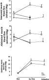
Respiratory and locomotor muscle blood flow measurements using near-infrared spectroscopy and indocyanine green dye in health and disease
- PMID: 38590151
- PMCID: PMC11003331
- DOI: 10.1177/14799731241246802
Respiratory and locomotor muscle blood flow measurements using near-infrared spectroscopy and indocyanine green dye in health and disease
Abstract
Measuring respiratory and locomotor muscle blood flow during exercise is pivotal for understanding the factors limiting exercise tolerance in health and disease. Traditional methods to measure muscle blood flow present limitations for exercise testing. This article reviews a method utilising near-infrared spectroscopy (NIRS) in combination with the light-absorbing tracer indocyanine green dye (ICG) to simultaneously assess respiratory and locomotor muscle blood flow during exercise in health and disease. NIRS provides high spatiotemporal resolution and can detect chromophore concentrations. Intravenously administered ICG binds to albumin and undergoes rapid metabolism, making it suitable for repeated measurements. NIRS-ICG allows calculation of local muscle blood flow based on the rate of ICG accumulation in the muscle over time. Studies presented in this review provide evidence of the technical and clinical validity of the NIRS-ICG method in quantifying respiratory and locomotor muscle blood flow. Over the past decade, use of this method during exercise has provided insights into respiratory and locomotor muscle blood flow competition theory and the effect of ergogenic aids and pharmacological agents on local muscle blood flow distribution in COPD. Originally, arterial blood sampling was required via a photodensitometer, though the method has subsequently been adapted to provide a local muscle blood flow index using venous cannulation. In summary, the significance of the NIRS-ICG method is that it provides a minimally invasive tool to simultaneously assess respiratory and locomotor muscle blood flow at rest and during exercise in health and disease to better appreciate the impact of ergogenic aids or pharmacological treatments.
Keywords: Muscle blood flow; chronic obstructive pulmonary disease; near-infrared spectroscopy.
Conflict of interest statement
Declaration of conflicting interestsThe author(s) declared no potential conflicts of interest with respect to the research, authorship, and/or publication of this article.
Figures


Similar articles
-
Near-infrared spectroscopy using indocyanine green dye for minimally invasive measurement of respiratory and leg muscle blood flow in patients with COPD.J Appl Physiol (1985). 2018 Sep 1;125(3):947-959. doi: 10.1152/japplphysiol.00959.2017. Epub 2018 Jun 21. J Appl Physiol (1985). 2018. PMID: 29927736
-
Near-infrared spectroscopy and indocyanine green derived blood flow index for noninvasive measurement of muscle perfusion during exercise.J Appl Physiol (1985). 2010 Apr;108(4):962-7. doi: 10.1152/japplphysiol.01269.2009. Epub 2010 Jan 28. J Appl Physiol (1985). 2010. PMID: 20110542
-
Blood flow index using near-infrared spectroscopy and indocyanine green as a minimally invasive tool to assess respiratory muscle blood flow in humans.Am J Physiol Regul Integr Comp Physiol. 2011 Apr;300(4):R984-92. doi: 10.1152/ajpregu.00739.2010. Epub 2011 Feb 2. Am J Physiol Regul Integr Comp Physiol. 2011. PMID: 21289237
-
Respiratory and locomotor muscle blood flow during exercise in health and chronic obstructive pulmonary disease.Exp Physiol. 2020 Dec;105(12):1990-1996. doi: 10.1113/EP088104. Epub 2020 Mar 29. Exp Physiol. 2020. PMID: 32103536 Review.
-
Monitoring tissue oxygen availability with near infrared spectroscopy (NIRS) in health and disease.Scand J Med Sci Sports. 2001 Aug;11(4):213-22. doi: 10.1034/j.1600-0838.2001.110404.x. Scand J Med Sci Sports. 2001. PMID: 11476426 Review.
Cited by
-
Application value of indocyanine green fluorescence imaging in assessing blood supply during laparoscopic radical resection of rectal cancer.Exp Ther Med. 2025 Aug 12;30(4):194. doi: 10.3892/etm.2025.12944. eCollection 2025 Oct. Exp Ther Med. 2025. PMID: 40901040 Free PMC article.
References
-
- Zakynthinos SG, Vogiatzis I. The major limitation to exercise performance in COPD is inadequate energy supply to the respiratory and locomotor muscles vs. lower limb muscle dysfunction vs. dynamic hyperinflation. Exercise intolerance in COPD: putting the pieces of the puzzle together. J Appl Physiol (1985) 2008; 105(2): 760. - PubMed
-
- Lunardi AC, Marques da Silva CC, Rodrigues Mendes FA, et al. Musculoskeletal dysfunction and pain in adults with asthma. J Asthma 2011; 48(1): 105–110. - PubMed
-
- Aubier M, Murciano D, Menu Y, et al. Dopamine effects on diaphragmatic strength during acute respiratory failure in chronic obstructive pulmonary disease. Ann Intern Med 1989; 110(1): 17–23. - PubMed
Publication types
MeSH terms
Substances
LinkOut - more resources
Full Text Sources
Miscellaneous

